Bilateral superficial brachial artery – a case report
Neelamjit Kaur1*, Patnaik V. V. Gopichand1, Rajan Kumar Singla2
1Department of Anatomy, Maharishi Markandeshwar Institute of Medical Sciences and Research, Mullana, Ambala, India.
2Department of Anatomy, Government Medical College, Amritsar, Punjab, India.
- *Corresponding Author:
- Dr. Neelamjit Kaur, MS Anatomy
Assistant Professor, Department of Anatomy, Maharishi Markandeshwar Institute of Medical Sciences and Research Mullana, Ambala, India.
Tel: +91 172 5092902
E-mail: neelamjit@yahoo.co.in
Date of Received: September 13th, 2010
Date of Accepted: March 11th, 2011
Published Online: December 21th, 2011
© IJAV. 2011; 4: 207–210.
[ft_below_content] =>Keywords
superficial brachial artery, vascular surgery, arterial pattern, brachial artery
Introduction
Variations in the arterial pattern of upper limb are common and have been reported by several investigators [1–3]. The great variability of this arterial pattern may be attributed to the failure of regression of some paths of embryonic arterial trunks [2,3]. The presence of superficial brachial artery and the usual pattern of its branching in the upper arm or forearm have also been reported with prevalence rate varying from 0.2–25% but mostly unilaterally [3–6]. A rare case of bilateral superficial brachial artery in an adult female cadaver was observed during our routine dissection studies in the Department of Anatomy, Govt. Medical College, Amritsar, India. Since a bilateral presentation is extremely rare, that gave us an impetus to report this case.
Case Report
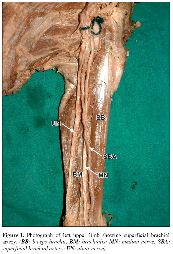
During a routine undergraduate dissection of 60-year-old female cadaver, superficial brachial artery was observed in both the upper limbs. On the left side, the artery was crossing superficial to median nerve from medial to lateral side in the middle 1/3rd of the arm. It descended to the cubital fossa lateral to median nerve and terminated by dividing into radial and ulnar arteries (Figure 1).
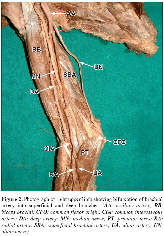
In the right arm of the same cadaver, the brachial artery after its usual origin, bifurcated into superficial and deep branches in upper 1/3rd of arm, which coursed superficial and deep to median nerve, respectively. The former reached the cubital fossa and divided into radial and ulnar arteries whereas the latter continued as common interrosseous artery in the forearm further dividing into anterior and posterior interrosseous arteries. Ulnar artery coming from superficial brachial artery in this limb coursed superficial to superficial head of pronator teres muscle (Figure 2). No other vascular variation was found in either of the limbs.
Figure 2: Photograph of right upper limb showing bifurcation of brachial artery into superficial and deep branches. (AA: axillary artery; BB: biceps brachii; CFO: common flexor origin; CIA: common interosseous artery; DA: deep artery; MN: median nerve; PT: pronator teres; RA: radial artery; SBA: superficial brachial artery; UA: ulnar artery; UN: ulnar nerve)
Discussion
Superficial brachial artery is named so because it runs superficial to median nerve and replaces the main trunk [7]. It has also been reasoned out to be an atavistic condition since a main brachial artery crossing superficial to median nerve is said to be the usual arrangement in the primates [8] or a persisting embryonic vessel [9]. A pattern similar to the one found in left limb of the present case, i.e. brachial artery passing superficial to median nerve and bifurcating normally into radial and ulnar artery has been reported earlier by Keen [5] in 3.6% and Lengele and Dhem [10] in 0.8% of population. On the other hand, the one in the right limb of the present specimen has been encountered in 1% of cases by Fuss et al. [1], 1.25% by Baeza et al. [3] and 0.3% by Skopakoff [5].
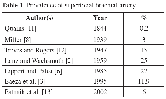
However, majority of authors have given a prevalence of superficial brachial artery varying from 0.2-25% but without specifying its pattern (Table 1). Similarly all of them are silent about its bilateral occurrence except a few sporadic cases [11].
Ontogeny
Superficial brachial artery emerges from the axial artery during stage IV of upper limb vascular development (embryo of 21 mm crown-rump length). During the next stage of embryonic development it is joined to the axial artery by means of several anastomotic branches at the level of axilla, upper arm and elbow [9]. The resulting arterial pattern thus acquires a plexiform appearance. Further it is the choice of unusual paths in primitive vascular plexus, persistence of vessels normally obliterated, disappearance of vessels normally retained and incomplete development of vessels that gives rise to anomalous blood vessels [11].
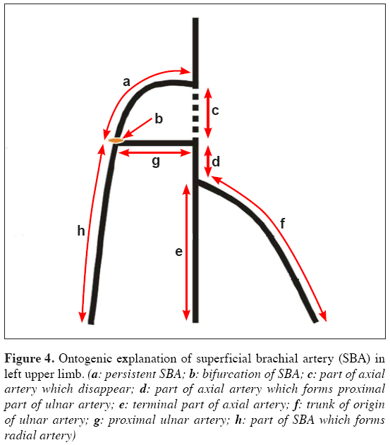
Development of superficial brachial artery in left limb of the present case can be easily made out if we look at the stages of development of axial artery of upper limb as described by Singer [9] (Figure 4). Accordingly, it seems that in Singer stage V, after development of communicating branch (g) between superficial brachial artery and main brachial artery, instead of proximal part of superficial brachial artery (a), it is the proximal part of main brachial artery (c) which retrogressed and severed its communication with common interosseous artery. The superficial brachial artery continued to supply the radial artery. The communication which usually forms proximal part of radial artery now forms proximal part of ulnar artery (g), its middle part being formed by the middle part of brachial artery (d) and terminal part by ulnar artery proper (f). The common interosseous artery (e) came as a branch of ulnar artery. The bifurcation of brachial artery which is usually seen at ‘h’ is now seen at ‘b’.
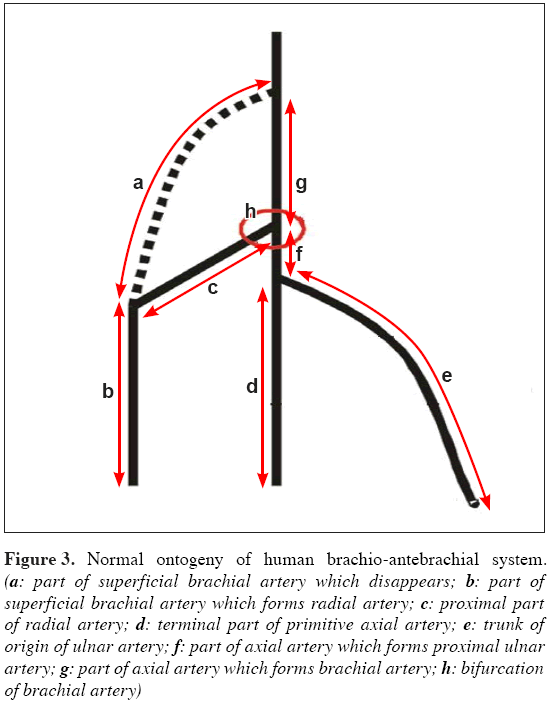
Figure 3: Normal ontogeny of human brachio-antebrachial system. (a: part of superficial brachial artery which disappears; b: part of superficial brachial artery which forms radial artery; c: proximal part of radial artery; d: terminal part of primitive axial artery; e: trunk of origin of ulnar artery; f: part of axial artery which forms proximal ulnar artery; g: part of axial artery which forms brachial artery; h: bifurcation of brachial artery)
Figure 4: Ontogenic explanation of superficial brachial artery (SBA) in left upper limb. (a: persistent SBA; b: bifurcation of SBA; c: part of axial artery which disappear; d: part of axial artery which forms proximal part of ulnar artery; e: terminal part of axial artery; f: trunk of origin of ulnar artery; g: proximal ulnar artery; h: part of SBA which forms radial artery)
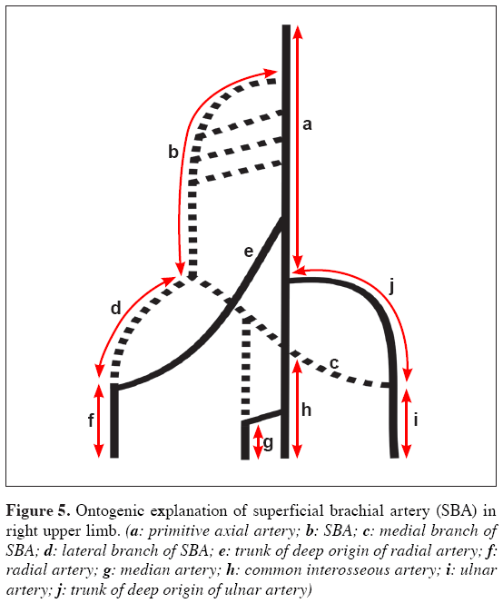
In the right limb, the development of superficial brachial artery can be explained by referring to the modifications made by Rodriguez-Baeza at al. [3] in the ontogeny of human brachio-antebrachial system. According to these, normally superficial brachial artery has 2 terminal branches, a lateral (the distal part of which is destined to become the distal part of radial artery) and a medial one (which has 2 terminal branches, the median and ulnar arteries). The radial artery and ulnar artery anastomose with the corresponding branch of primitive axial artery (trunks of deep origin; Figure 5, e, j). In the subsequent stages the trunk with deep origin (shown as solid lines in Figure 5) prevail hemodynamically and the proximal part of medial and lateral branches of superficial brachial artery begin to involute (open lines).
Figure 5: Ontogenic explanation of superficial brachial artery (SBA) in right upper limb. (a: primitive axial artery; b: SBA; c: medial branch of SBA; d: lateral branch of SBA; e: trunk of deep origin of radial artery; f: radial artery; g: median artery; h: common interosseous artery; i: ulnar artery; j: trunk of deep origin of ulnar artery)
In the present case, it seems that proximal trunk of superficial origin of radial artery (d in Figure 5) and of ulnar artery (c in Figure 5) took hemodynamic predominance and failed to retrogress. As a result deep trunks of origin of these vessels (e for radial artery; j for ulnar artery) retrogressed, giving rise to a superficial brachial artery dividing into radial and ulnar arteries. Similarly both superficial brachial artery (b) and deep brachial artery (a) persisted and later continued as axial artery and thence common interosseous artery.
Clinical Applications
It is important for a clinician to be aware of a superficial brachial artery because its position not only makes it more vulnerable to trauma but also makes it accessible to cannulation, if needed. In case of intravenous injection of drugs, care must be taken to avoid a superficially placed artery because it may lead to gangrene or loss of hand [2]. One way of avoiding this complication is routine examination of forearm for unusual pulses before any injection of drugs is undertaken.
References
- Fuss FK, Matula CW, Tschabitscher M. Die Arteria brachialis superficialis. Anat Anz. 1985; 160: 285–294. (German)
- Tountas CP, Bergman RA. Anatomic Variations of the Upper Extremity. New York, Churchill Livingstone. 1993; 196–210.
- Rodriguez-Baeza A, Nebot J, Ferreira B, Reina F, Perez J, Sanudo JR, Roig M. An anatomical study and ontogenetic explanation of 23 cases with variations in the main pattern of the human brachio-antebrachial arteries. J Anat. 1995; 187: 473–479.
- Mc Cormack LJ, Cauldwell EW, Anson BJ. Brachial and antebrachial arterial patterns; a study of 750 extremities. Surg Gynecol Obstet. 1953; 96: 43–54.
- Keen JA. A study of the arterial variations in the limbs, with special reference to symmetry of vascular patterns. Am J Anat. 1961; 108: 245–261.
- Lippert H, Pabst R. Arterial Variations in Man. New York: Springer; 1985; 68–73.
- Adachi B. Arteriensystem der Japaner. Kyoto, Verlag der Kaiserlich-Japanischen Universitat zu Kyoto. 1928; 205–210.
- Miller RA. Observations upon the arrangement of the axillary artery and brachial plexus. Am J of Anat. 1939; 64: 143–163.
- Singer E. Embryological patterns persisting in the arteries of the arm. Anat Rec. 1933; 55: 406–413.
- Lengele B, Dhem A. Unusual variations of the vasculonervous elements of the human axilla. Report of three cases. Arch Anat Histol Embryol. 1989; 72: 57,67.
- Sharma T, Singla RK, Sachdeva K. Bilateral superficial brachial artery. Kathmandu Univ Med J (KUMJ). 2009; 7: 426–428.
- Treves FB, Rogers L. Surgical Applied Anatomy. 11th Ed., Cassell & Co. Ltd, London Toronto, Melbourne and Sydney. 1947; 230–266.
- Patnaik VVG, Kalsey G, Singla RK. Branching pattern of brachial artery – a morphological study. J Anat Soc India. 2002; 51: 176–186.
Neelamjit Kaur1*, Patnaik V. V. Gopichand1, Rajan Kumar Singla2
1Department of Anatomy, Maharishi Markandeshwar Institute of Medical Sciences and Research, Mullana, Ambala, India.
2Department of Anatomy, Government Medical College, Amritsar, Punjab, India.
- *Corresponding Author:
- Dr. Neelamjit Kaur, MS Anatomy
Assistant Professor, Department of Anatomy, Maharishi Markandeshwar Institute of Medical Sciences and Research Mullana, Ambala, India.
Tel: +91 172 5092902
E-mail: neelamjit@yahoo.co.in
Date of Received: September 13th, 2010
Date of Accepted: March 11th, 2011
Published Online: December 21th, 2011
© IJAV. 2011; 4: 207–210.
Abstract
Though variations in the arterial pattern of upper limb are fairly common, a bilateral presentation as encountered in this case, i.e. superficial brachial artery seen bilaterally in an adult female cadaver is extremely rare. Its origin, course and termination were different in both the limbs. On the left side, the brachial artery in the middle 1/3rd of arm, coursed superficial to the median nerve and on the right side it divided in upper 1/3rd of arm into superficial and deep arteries. Former coursed superficial to median nerve and terminated in the cubital fossa by dividing into radial and ulnar artery while the latter coursed deep to median nerve and continued as common interosseous artery. Superficial brachial artery is reported earlier by different authors in frequency range of 0.2–25%, but mostly unilaterally. Further ontogeny and clinical implications of the variant are explained in detail.
-Keywords
superficial brachial artery, vascular surgery, arterial pattern, brachial artery
Introduction
Variations in the arterial pattern of upper limb are common and have been reported by several investigators [1–3]. The great variability of this arterial pattern may be attributed to the failure of regression of some paths of embryonic arterial trunks [2,3]. The presence of superficial brachial artery and the usual pattern of its branching in the upper arm or forearm have also been reported with prevalence rate varying from 0.2–25% but mostly unilaterally [3–6]. A rare case of bilateral superficial brachial artery in an adult female cadaver was observed during our routine dissection studies in the Department of Anatomy, Govt. Medical College, Amritsar, India. Since a bilateral presentation is extremely rare, that gave us an impetus to report this case.
Case Report
During a routine undergraduate dissection of 60-year-old female cadaver, superficial brachial artery was observed in both the upper limbs. On the left side, the artery was crossing superficial to median nerve from medial to lateral side in the middle 1/3rd of the arm. It descended to the cubital fossa lateral to median nerve and terminated by dividing into radial and ulnar arteries (Figure 1).
In the right arm of the same cadaver, the brachial artery after its usual origin, bifurcated into superficial and deep branches in upper 1/3rd of arm, which coursed superficial and deep to median nerve, respectively. The former reached the cubital fossa and divided into radial and ulnar arteries whereas the latter continued as common interrosseous artery in the forearm further dividing into anterior and posterior interrosseous arteries. Ulnar artery coming from superficial brachial artery in this limb coursed superficial to superficial head of pronator teres muscle (Figure 2). No other vascular variation was found in either of the limbs.
Figure 2: Photograph of right upper limb showing bifurcation of brachial artery into superficial and deep branches. (AA: axillary artery; BB: biceps brachii; CFO: common flexor origin; CIA: common interosseous artery; DA: deep artery; MN: median nerve; PT: pronator teres; RA: radial artery; SBA: superficial brachial artery; UA: ulnar artery; UN: ulnar nerve)
Discussion
Superficial brachial artery is named so because it runs superficial to median nerve and replaces the main trunk [7]. It has also been reasoned out to be an atavistic condition since a main brachial artery crossing superficial to median nerve is said to be the usual arrangement in the primates [8] or a persisting embryonic vessel [9]. A pattern similar to the one found in left limb of the present case, i.e. brachial artery passing superficial to median nerve and bifurcating normally into radial and ulnar artery has been reported earlier by Keen [5] in 3.6% and Lengele and Dhem [10] in 0.8% of population. On the other hand, the one in the right limb of the present specimen has been encountered in 1% of cases by Fuss et al. [1], 1.25% by Baeza et al. [3] and 0.3% by Skopakoff [5].
However, majority of authors have given a prevalence of superficial brachial artery varying from 0.2-25% but without specifying its pattern (Table 1). Similarly all of them are silent about its bilateral occurrence except a few sporadic cases [11].
Ontogeny
Superficial brachial artery emerges from the axial artery during stage IV of upper limb vascular development (embryo of 21 mm crown-rump length). During the next stage of embryonic development it is joined to the axial artery by means of several anastomotic branches at the level of axilla, upper arm and elbow [9]. The resulting arterial pattern thus acquires a plexiform appearance. Further it is the choice of unusual paths in primitive vascular plexus, persistence of vessels normally obliterated, disappearance of vessels normally retained and incomplete development of vessels that gives rise to anomalous blood vessels [11].
Development of superficial brachial artery in left limb of the present case can be easily made out if we look at the stages of development of axial artery of upper limb as described by Singer [9] (Figure 4). Accordingly, it seems that in Singer stage V, after development of communicating branch (g) between superficial brachial artery and main brachial artery, instead of proximal part of superficial brachial artery (a), it is the proximal part of main brachial artery (c) which retrogressed and severed its communication with common interosseous artery. The superficial brachial artery continued to supply the radial artery. The communication which usually forms proximal part of radial artery now forms proximal part of ulnar artery (g), its middle part being formed by the middle part of brachial artery (d) and terminal part by ulnar artery proper (f). The common interosseous artery (e) came as a branch of ulnar artery. The bifurcation of brachial artery which is usually seen at ‘h’ is now seen at ‘b’.
Figure 3: Normal ontogeny of human brachio-antebrachial system. (a: part of superficial brachial artery which disappears; b: part of superficial brachial artery which forms radial artery; c: proximal part of radial artery; d: terminal part of primitive axial artery; e: trunk of origin of ulnar artery; f: part of axial artery which forms proximal ulnar artery; g: part of axial artery which forms brachial artery; h: bifurcation of brachial artery)
Figure 4: Ontogenic explanation of superficial brachial artery (SBA) in left upper limb. (a: persistent SBA; b: bifurcation of SBA; c: part of axial artery which disappear; d: part of axial artery which forms proximal part of ulnar artery; e: terminal part of axial artery; f: trunk of origin of ulnar artery; g: proximal ulnar artery; h: part of SBA which forms radial artery)
In the right limb, the development of superficial brachial artery can be explained by referring to the modifications made by Rodriguez-Baeza at al. [3] in the ontogeny of human brachio-antebrachial system. According to these, normally superficial brachial artery has 2 terminal branches, a lateral (the distal part of which is destined to become the distal part of radial artery) and a medial one (which has 2 terminal branches, the median and ulnar arteries). The radial artery and ulnar artery anastomose with the corresponding branch of primitive axial artery (trunks of deep origin; Figure 5, e, j). In the subsequent stages the trunk with deep origin (shown as solid lines in Figure 5) prevail hemodynamically and the proximal part of medial and lateral branches of superficial brachial artery begin to involute (open lines).
Figure 5: Ontogenic explanation of superficial brachial artery (SBA) in right upper limb. (a: primitive axial artery; b: SBA; c: medial branch of SBA; d: lateral branch of SBA; e: trunk of deep origin of radial artery; f: radial artery; g: median artery; h: common interosseous artery; i: ulnar artery; j: trunk of deep origin of ulnar artery)
In the present case, it seems that proximal trunk of superficial origin of radial artery (d in Figure 5) and of ulnar artery (c in Figure 5) took hemodynamic predominance and failed to retrogress. As a result deep trunks of origin of these vessels (e for radial artery; j for ulnar artery) retrogressed, giving rise to a superficial brachial artery dividing into radial and ulnar arteries. Similarly both superficial brachial artery (b) and deep brachial artery (a) persisted and later continued as axial artery and thence common interosseous artery.
Clinical Applications
It is important for a clinician to be aware of a superficial brachial artery because its position not only makes it more vulnerable to trauma but also makes it accessible to cannulation, if needed. In case of intravenous injection of drugs, care must be taken to avoid a superficially placed artery because it may lead to gangrene or loss of hand [2]. One way of avoiding this complication is routine examination of forearm for unusual pulses before any injection of drugs is undertaken.
References
- Fuss FK, Matula CW, Tschabitscher M. Die Arteria brachialis superficialis. Anat Anz. 1985; 160: 285–294. (German)
- Tountas CP, Bergman RA. Anatomic Variations of the Upper Extremity. New York, Churchill Livingstone. 1993; 196–210.
- Rodriguez-Baeza A, Nebot J, Ferreira B, Reina F, Perez J, Sanudo JR, Roig M. An anatomical study and ontogenetic explanation of 23 cases with variations in the main pattern of the human brachio-antebrachial arteries. J Anat. 1995; 187: 473–479.
- Mc Cormack LJ, Cauldwell EW, Anson BJ. Brachial and antebrachial arterial patterns; a study of 750 extremities. Surg Gynecol Obstet. 1953; 96: 43–54.
- Keen JA. A study of the arterial variations in the limbs, with special reference to symmetry of vascular patterns. Am J Anat. 1961; 108: 245–261.
- Lippert H, Pabst R. Arterial Variations in Man. New York: Springer; 1985; 68–73.
- Adachi B. Arteriensystem der Japaner. Kyoto, Verlag der Kaiserlich-Japanischen Universitat zu Kyoto. 1928; 205–210.
- Miller RA. Observations upon the arrangement of the axillary artery and brachial plexus. Am J of Anat. 1939; 64: 143–163.
- Singer E. Embryological patterns persisting in the arteries of the arm. Anat Rec. 1933; 55: 406–413.
- Lengele B, Dhem A. Unusual variations of the vasculonervous elements of the human axilla. Report of three cases. Arch Anat Histol Embryol. 1989; 72: 57,67.
- Sharma T, Singla RK, Sachdeva K. Bilateral superficial brachial artery. Kathmandu Univ Med J (KUMJ). 2009; 7: 426–428.
- Treves FB, Rogers L. Surgical Applied Anatomy. 11th Ed., Cassell & Co. Ltd, London Toronto, Melbourne and Sydney. 1947; 230–266.
- Patnaik VVG, Kalsey G, Singla RK. Branching pattern of brachial artery – a morphological study. J Anat Soc India. 2002; 51: 176–186.


