Multiple variations of the azygos venous system: a case report.
1Department of Anatomy, Sri Guru Ram Das Institute of Medical Sciences and Research, Vallah, India.
2ESI Hospital, Amritsar, India
- *Corresponding Author:
- Dr. Seema
Associate Professor, Department of Anatomy, SGRD Institute of Medical, Sciences and Research, Vallah, India.
Tel: +91 991 4754354
E-mail: drseema16@gmail.com
Date of Received: February 7th, 2012
Date of Accepted: September 29th, 2012
Published Online: February 14th, 2013
© Int J Anat Var (IJAV). 2013; 6: 34–35.
[ft_below_content] =>Keywords
azygos vein, hemiazygos vein, accessory hemiazygos vein, posterior mediastinum
Introduction
Variations related to the azygos system are not rare [1]. They vary much in their mode of origin, course, tributaries, anastomosis and termination. Azygos veins are important cava-caval and porto-caval junctions, thus forming collateral circulation in caval vein occlusion and in portal hypertension [2]. The azygos venous system develops on the basis of multiple transformations of the subcardinal veins [3]. Therefore it is helpful to recognize the presence of these variations in order to properly interpret CT and MRI scans. The present study showed a rare and special variation of the azygos venous system in a human cadaver.
Case Report
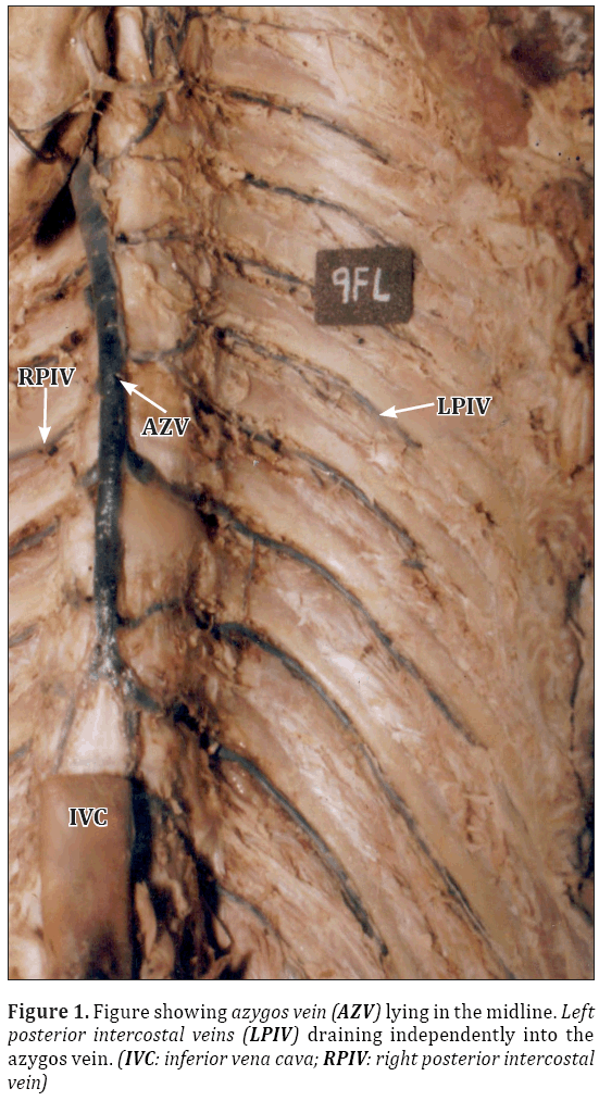
During the dissection of the posterior mediastinum of a 50-year-old male cadaver, the formation, course and the drainage of the azygos vein was found in the midline. The second to fifth left posterior intercostal veins joined to form a common trunk which crossed the midline and drained in the azygos vein. This channel represented the left superior intercostal vein. The sixth left posterior intercostal vein drained independently into the azygos vein. The sixth posterior intercostal vein tributary along with the seventh left posterior intercostal vein formed a common trunk draining into the azygos vein. The eighth left posterior intercostal vein drained into the common trunk formed by the sixth and the seventh left posterior intercostal vein. The ninth and the tenth left posterior intercostal veins drained independently into the azygos vein. The hemiazygos vein after its formation in the abdomen, entered the thorax, received the eleventh left posterior intercostal vein and ultimately drained into the azygos vein. A small connecting vein was found between the ninth and the tenth left posterior intercostal veins.
Discussion
Azygos venous system embryologically generates from subcardinal veins. The right subcardinal vein forms azygos vein and the left subcardinal vein forms hemiazygos vein. The left superior intercostal vein and accessory hemiazygos vein are derived from the left posterior cardinal vein and this vein simultaneously forms the upper part of the azygos vein. The part that connects hemiazygos vein to azygos vein is actually remainder of the anastomosis between the left and the right posterior cardinal vein [4].
In the present case, there were underdeveloped left azygos venous line on the left side, and the left intersegmental veins reached directly to the azygos vein. Different aspects of the azygos venous system have been reported. Ozbek et al reported a case in which hemiazygos vein was absent [5]. Fifth and sixth left posterior intercostal veins with third and fourth veins opened to left superior intercostal vein. Kocabiyik et al found a case in which there was no complete accessory hemiazygos vein in the azygos vein system, and posterior intercostal veins drained bilaterally to the azygos vein [6].The variant azygos venous system may easily be confused with aneurysm, lymphadenopathy and other conditions like tumor [7,8]. Mahato reported the variant accessory hemiazygos system with persistent cranial segment of posterior cardinal vein [9]. It is important to keep in mind this kind of variations in the mediastinal operations or surgery of large vessels.
Conclusion
It is important to report and document the different variations of the azygos venous system because especially in CT and MRI scans. Some variations of the azygos venous system can easily be confused with pathological conditions such as aneurysms, tumors and enlarged lymph nodes. Finally, it is of utmost importance for the surgeons during the mediastinal operations of the possibility of a variation of the azygos venous system.
References
- Mezzogiorno A, Passiatore C. An atypic pattern of azygos venous system in man. Anat Anz. 1988; 165: 277–281.
- Arslan G, Cubuk M, Ozkaynak C, Sindel T, Luleci E. Absence of the azygos vein. Clin Imaging. 2000; 24: 157–158.
- Caggiati A, Barberini F. Partial agenesis of the azygos vein: a case report. Ann Anat. 1996; 178: 273–275.
- Standring S, ed. Gray’s Anatomy. The Anatomical Basis of Clinical Practice. 40th Ed., New York, Churchill Livingstone. 2008: 207–208.
- Ozbek A, Dalcik C, Colak T, Dalcik H. Multiple variations of the azygos venous system. Surg Radiol Anat. 1999; 21: 83–85.
- Kocabiyik N, Kutoglu T, Albay S, Yalcin B, Ozan H. Azygos vein system abnormality: case report. Gulhane Med J. 2006; 48: 180–182.
- Sieunarine K, May J, White GH, Harris JP. Anomalous Azygos Vein: A potential danger during endoscopic thoracic sympathectomy. Aust N Z J Surg. 1997; 67: 578–579.
- Takasugi JE, Godwin JD. CT appearance of the retroaortic anastomoses of the azygos system. AJR Am J Roentgenol. 1990; 154: 41–44.
- Mahato NK. Anomalous accessory hemi-azygos system with persistent cranial segment of posterior cardinal vein - A case report. Braz J Morphol Sci. 2009; 26: 177–179.
1Department of Anatomy, Sri Guru Ram Das Institute of Medical Sciences and Research, Vallah, India.
2ESI Hospital, Amritsar, India
- *Corresponding Author:
- Dr. Seema
Associate Professor, Department of Anatomy, SGRD Institute of Medical, Sciences and Research, Vallah, India.
Tel: +91 991 4754354
E-mail: drseema16@gmail.com
Date of Received: February 7th, 2012
Date of Accepted: September 29th, 2012
Published Online: February 14th, 2013
© Int J Anat Var (IJAV). 2013; 6: 34–35.
Abstract
The venous system variations are generally explained on the basis of their embryological development. Tributaries of the azygos venous system varies greatly. Variations of azygos venous system and especially of the hemiazygos veins are not clearly described in the literature. In this case, the azygos vein instead of lying on the right side of the vertebral column was in the midline of posterior mediastinum. The left azygos venous line was ill defined and lower left posterior intercostal veins were opening independently into the azygos vein. The left superior intercostal vein was opening into the azygos vein. The accessory hemiazygos vein was ill defined. The origin and termination of the azygos venous system was found normal. These variations are discussed in view of its embryological development. Clinically these variations should be kept in mind while doing mediastinal surgery of large vessels.
-Keywords
azygos vein, hemiazygos vein, accessory hemiazygos vein, posterior mediastinum
Introduction
Variations related to the azygos system are not rare [1]. They vary much in their mode of origin, course, tributaries, anastomosis and termination. Azygos veins are important cava-caval and porto-caval junctions, thus forming collateral circulation in caval vein occlusion and in portal hypertension [2]. The azygos venous system develops on the basis of multiple transformations of the subcardinal veins [3]. Therefore it is helpful to recognize the presence of these variations in order to properly interpret CT and MRI scans. The present study showed a rare and special variation of the azygos venous system in a human cadaver.
Case Report
During the dissection of the posterior mediastinum of a 50-year-old male cadaver, the formation, course and the drainage of the azygos vein was found in the midline. The second to fifth left posterior intercostal veins joined to form a common trunk which crossed the midline and drained in the azygos vein. This channel represented the left superior intercostal vein. The sixth left posterior intercostal vein drained independently into the azygos vein. The sixth posterior intercostal vein tributary along with the seventh left posterior intercostal vein formed a common trunk draining into the azygos vein. The eighth left posterior intercostal vein drained into the common trunk formed by the sixth and the seventh left posterior intercostal vein. The ninth and the tenth left posterior intercostal veins drained independently into the azygos vein. The hemiazygos vein after its formation in the abdomen, entered the thorax, received the eleventh left posterior intercostal vein and ultimately drained into the azygos vein. A small connecting vein was found between the ninth and the tenth left posterior intercostal veins.
Discussion
Azygos venous system embryologically generates from subcardinal veins. The right subcardinal vein forms azygos vein and the left subcardinal vein forms hemiazygos vein. The left superior intercostal vein and accessory hemiazygos vein are derived from the left posterior cardinal vein and this vein simultaneously forms the upper part of the azygos vein. The part that connects hemiazygos vein to azygos vein is actually remainder of the anastomosis between the left and the right posterior cardinal vein [4].
In the present case, there were underdeveloped left azygos venous line on the left side, and the left intersegmental veins reached directly to the azygos vein. Different aspects of the azygos venous system have been reported. Ozbek et al reported a case in which hemiazygos vein was absent [5]. Fifth and sixth left posterior intercostal veins with third and fourth veins opened to left superior intercostal vein. Kocabiyik et al found a case in which there was no complete accessory hemiazygos vein in the azygos vein system, and posterior intercostal veins drained bilaterally to the azygos vein [6].The variant azygos venous system may easily be confused with aneurysm, lymphadenopathy and other conditions like tumor [7,8]. Mahato reported the variant accessory hemiazygos system with persistent cranial segment of posterior cardinal vein [9]. It is important to keep in mind this kind of variations in the mediastinal operations or surgery of large vessels.
Conclusion
It is important to report and document the different variations of the azygos venous system because especially in CT and MRI scans. Some variations of the azygos venous system can easily be confused with pathological conditions such as aneurysms, tumors and enlarged lymph nodes. Finally, it is of utmost importance for the surgeons during the mediastinal operations of the possibility of a variation of the azygos venous system.
References
- Mezzogiorno A, Passiatore C. An atypic pattern of azygos venous system in man. Anat Anz. 1988; 165: 277–281.
- Arslan G, Cubuk M, Ozkaynak C, Sindel T, Luleci E. Absence of the azygos vein. Clin Imaging. 2000; 24: 157–158.
- Caggiati A, Barberini F. Partial agenesis of the azygos vein: a case report. Ann Anat. 1996; 178: 273–275.
- Standring S, ed. Gray’s Anatomy. The Anatomical Basis of Clinical Practice. 40th Ed., New York, Churchill Livingstone. 2008: 207–208.
- Ozbek A, Dalcik C, Colak T, Dalcik H. Multiple variations of the azygos venous system. Surg Radiol Anat. 1999; 21: 83–85.
- Kocabiyik N, Kutoglu T, Albay S, Yalcin B, Ozan H. Azygos vein system abnormality: case report. Gulhane Med J. 2006; 48: 180–182.
- Sieunarine K, May J, White GH, Harris JP. Anomalous Azygos Vein: A potential danger during endoscopic thoracic sympathectomy. Aust N Z J Surg. 1997; 67: 578–579.
- Takasugi JE, Godwin JD. CT appearance of the retroaortic anastomoses of the azygos system. AJR Am J Roentgenol. 1990; 154: 41–44.
- Mahato NK. Anomalous accessory hemi-azygos system with persistent cranial segment of posterior cardinal vein - A case report. Braz J Morphol Sci. 2009; 26: 177–179.


