Variant origin of left vertebral arteryin
Rajan Kumar Singla, Tripta Sharma and Kanika Sachdeva*
Department of Anatomy, Government Medical College, Amritsar, Punjab, India
- *Corresponding Author:
- Dr. Kanika Sachdeva, MBBS
Department of Anatomy, Government Medical College, Amritsar, 143001, Punjab, India
Tel: +91 183 2401291
E-mail: kanikadr.sarang@yahoo.com
Date of Received: May 7th, 2010
Date of Accepted: June 30th, 2010
Published Online: July 30th, 2010
© IJAV. 2010; 3: 97–99.
[ft_below_content] =>Keywords
vertebral artery, subclavian artery,arch of aorta
Introduction
In anatomy, normality embraces a range of morphologies. It includes those that are more common and others called variations which are less frequent but not considered abnormal [1]. Arterial derangements within the thorax are common, complex and can assume many diverse forms. These derangements in origin and course of main vessels occurring either individually or in combination with other cardiovascular defects are mostly explainable on ontogenic basis, which can thus be blamed for a myriad of clinically relevant anomalies [2].
The major branches of arch of aorta are the great ways for blood supply to the head and upper limb, and are of particular interest in clinical angiography. The proximal segment of these branches and of the aortic arch is common sites for atherosclerosis with clinical consequences for blood supply to brain [3]. Anomalies of origin and distribution of these branches can cause changes in cerebral haemodynamics that may lead to cerebral abnormalities [4].
An understanding of the variability of vertebral artery remains most important in angiography and surgical procedures where an incompatible knowledge of anatomy can lead to complications [5]. According to the standard textbooks of anatomy, vertebral artery is the largest and most constant stem of subclavian artery, both in origin and distribution [6], arising from postero-superior aspect of its first part [7]. Rarely, it may arise directly from the arch of aorta. One such case was found in a male cadaver, arteryin the Department of Anatomy, Government Medical College, Amritsar, Punjab, India, which is being reported here.
Case Report
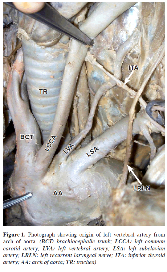
During routine undergraduate dissections on a 60-year-old male cadaver, left vertebral artery was seen arising from aortic arch, between the origins of left common carotid and left subclavian arteries. After its origin, the first part of the artery followed its normal course to enter the foramen transversarium of the sixth cervical vertebra (Figure 1). However, the right vertebral artery arose from the posterosuperior aspect of first part of right subclavian artery, as usual.
Figure 1: Photograph showing origin of left vertebral artery from arch of aorta. (BCT: brachiocephalic trunk; LCCA: left common carotid artery; LVA: left vertebral artery; LSA: left subclavian artery; LRLN: left recurrent laryngeal nerve; ITA: inferior thyroid artery; AA: arch of aorta; TR: trachea)
Discussion
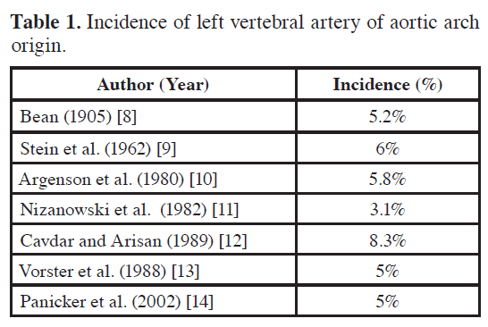
A vertebral artery of aortic arch origin has been earlier described by different authors in the range of 3.1-8.3 % (Table I).
It is universally agreed upon that the anatomical and morphological variations of vertebral artery are significant for diagnostic and surgical procedures in head and neck region, where an incompatible knowledge can lead to complications.
A variant origin of vertebral artery of this kind may favor cerebral disorders because of alterations in cerebral haemodynamics [4]. Although overall incidence of variant origin of prevertebral segment of vertebral artery is very low, it is extremely important to be aware of these complications in such patients [14].
The most frequent pathology affecting the extracranial vertebral artery is atherosclerosis [15]. The commonest site of which, according to Fischer et al. is at the origin of vessel from subclavian artery [16]. They further underlined its importance during the evaluation of vertebro-basilar insufficiency thought to be due to atherosclerotic disease, and during cannulation of vertebral artery for endovascular procedures in such patients. Vitte et al. stressed to keep the variant origin of vertebral artery in mind during its manual compression used routinely for positional haemodynamics vertebro-basilar insufficiency and cautioned to take a special care in these cases [17].
Ontogeny
Arey is of the view that the anomalous blood vessels may be due to (i) the choice of unusual paths in the primitive vascular plexus, (ii) the persistence of vessels normally obliterated, (iii) the disappearance of vessels normally retained, (iv) incomplete development, and (v) fusions and absorption of the parts usually distinct [18].
While discussing development of the vascular system in any part of the body, two fundamental questions arise [19]:
1. What is the origin of blood vessels in the body of embryo?
2. What is the primitive form of vessels in any area, and the manner of changes from this to that of the adult?
Vorster et al. believe that there are two factors that influence the development of the branches of subclavian artery [13]. First, ability of the blood to follow the longitudinal channels offering the least resistance and second, the tension on the vessels; resulting from the caudal shifting of the heart and aorta. Further citing the work of Congdon [20], they opined that vertebral artery develops from longitudinal arterial channels of intersegmental anastomosis. Due to caudal shifting of aorta, the proximal parts of these segmental arteries are exposed to longitudinal tension and bending with the resulting retarded blood flow. It must have resulted in abnormal connections between the longitudinal channels and the aortic arch.
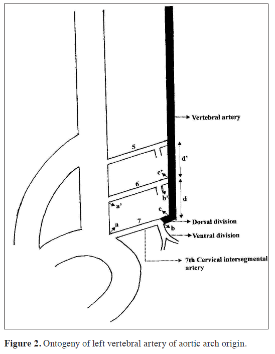
However, Panicker et al. [14], acknowledging the views of Vorster et al. [13], opined that a left vertebral artery of aortic arch origin may be because of persistence of dorsal division of left 6th intersegmental artery as 1st part of vertebral artery instead of that of left 7th dorsal intersegmental artery.
Normally, 1st part of vertebral artery develops from dorsal division (segment bc) of 7th intersegmental artery (segment ab) which itself forms proximal part of left subclavian artery. The 6th intersegmental artery (segment a′b′) and its dorsal division (segment b′c′) usually disappear as does the segment aa’ of dorsal aorta (Figure 2).
In cases, where vertebral artery arises from aortic arch, we feel, that dorsal branch of 6th intersegmental artery (segment b′c′), 6th intersegmental artery itself (segment a′b′) and segment aa′ of dorsal aorta fail to disappear, so blood flow through these persist forming a vertebral artery of aortic arch origin. As a preferential blood flows to this persistent channel (a-a′-b′-c′), the blood flow through channel (b-c-d) decreases which ultimately disappears.
References
- Willan PL, Humpherson JR. Concepts of variation and normality in morphology: Important issues at risk of neglect in modern undergraduate medical courses. Clin Anat. 1999; 12: 186–190.
- Nathan H, Seidel MR. The association of a retroesophageal right subclavian artery, a right-sided terminating thoracic duct, and a left vertebral artery of aortic origin: anatomical and clinical considerations. Acta Anat (Basel). 1983; 117: 362–373.
- Zamir M, Sinclair P. Origin of brachiocephalic trunk, left carotid and left subclavian arteries from the arch of human aorta. Invest Radiol. 1991; 26: 128–133.
- Bernardi L, Dettori P. Angiographic study of a rare anomalous origin of vertebral artery. Neuroradiol. 1975; 9: 43–47.
- Wasserman BA, Milkulis DJ, Mananzione JV. Origin of the right vertebral artery from the left side of the aortic arch proximal to the origin of the left subclavian artery. AJNR Am J Neuroradiol. 1992; 13: 355–358.
- Hollinshead WH. Arteries: The Neck. In: Anatomy for Surgeons. Vol. I, The Head & Neck. New York, Paul B Hoeber, Inc, Medical Book Department of Harpers & Brothers. 1954; 467–474.
- Williams, PL, Bannister, LH, Berry MM, Collins P, Dyson M, Dussek JE, Ferguson MWJ, eds. Gray’s Anatomy. 38th Ed., New York, London, Churchill Livingstone. 1995; 1529–1536.
- Bean RB. A composite study of the subclavian artery in man. Am J Anat. 1905; 4: 303–328.
- Stein BM, McCormick WF, Rodriguez JN, Taveras JM. Postmortom angiography of cerebral vascular system. Arch Neurol. 1962; 7: 545–559.
- Argenson C, Francke JP. Sylla S, Dintimille H, Papasian S, diMarino V. The vertebral arteries (segments V1 and V2). Anat Clin. 1980; 2: 29–41.
- Nizanowski C, Noczynski L, Suder E. Variability of the origin of ramifications of the subclavian artery in humans (studies on the Polish population). Folia Morphol (Warsz). 1982; 41: 281–294.
- Cavdar S, Arisan E. Variations in the extracranial origin of the human vertebral artery. Acta Anat (Basel). 1989; 135: 236–238.
- Vorster W, Du Plooy PT, Meiring JH. Abnormal origin of internal thoracic and vertebral arteries. Clin Anat. 1998; 11: 33–37.
- Panicker HK, Tarnekar A, Dhawane V, Ghosh SK. Anomalous origin of left vertebral artery – embryological basis and applied aspects – A case report. J Anat. Soc. India. 2002; 51: 234–235.
- Matula C, Trattnig S, Tschabitscher M, Day JD, Koos WT. The course of the prevertebral segment of the vertebral artery: anatomy and clinical significance. Surg Neurol. 1997; 48: 125–131.
- Fisher CM, Gore I, Obake N, White PD. Atherosclerosis of the carotid and arteries – extracranial and intracranial. J Neuropathol Exp Neurol 1965; 24: 244-245
- Vitte E, Feron JM, Guerin-Surville H, Koskas F. Anatomical study of digital compression of the vertebral artery at its origin at the suboccipital triangle. Anat Clin. 1985; 7: 77–82.
- Arey LB. Development of arteries. The vascular system. In: Developmental Anatomy. A Textbook and Laboratory Manual of Embryology. 6th Ed., Philadelphia and London, WB Saunders Company. 1957; 367–373.
- Keibel FN, Mall FP. Development of the vascular system. In: Manual of Human Embryology. Vol II, Philadelphia and London, J. B. Lippincott Company. 1912: 659–667.
- Congdon ED. Transformation of the aortic arch system during during the development of human embryo. Contrib Embryol Carnegie Inst. 1922; 68: 47–110.
Rajan Kumar Singla, Tripta Sharma and Kanika Sachdeva*
Department of Anatomy, Government Medical College, Amritsar, Punjab, India
- *Corresponding Author:
- Dr. Kanika Sachdeva, MBBS
Department of Anatomy, Government Medical College, Amritsar, 143001, Punjab, India
Tel: +91 183 2401291
E-mail: kanikadr.sarang@yahoo.com
Date of Received: May 7th, 2010
Date of Accepted: June 30th, 2010
Published Online: July 30th, 2010
© IJAV. 2010; 3: 97–99.
Abstract
Arterial derangements within the thorax are common, complex and can assume many diverse forms. Accurate knowledge of the normal and variant arterial anatomy of the vertebral artery is important for clinical procedures and vascular radiology. In the present paper, a rare variation of vertebral artery is being reported. The artery on left side, took origin directly from the arch of aorta. Earlier, vertebral artery of aortic arch origin has been described by different authors in the range of 3.1-8.3%. Further its ontogeny and clinical implications are discussed in detail. The present report should be of interest for the clinician with regard to vascular variations in the neck and thoracic region, and may give insight into elucidating the developmental mechanism of angiogenesis.
-Keywords
vertebral artery, subclavian artery,arch of aorta
Introduction
In anatomy, normality embraces a range of morphologies. It includes those that are more common and others called variations which are less frequent but not considered abnormal [1]. Arterial derangements within the thorax are common, complex and can assume many diverse forms. These derangements in origin and course of main vessels occurring either individually or in combination with other cardiovascular defects are mostly explainable on ontogenic basis, which can thus be blamed for a myriad of clinically relevant anomalies [2].
The major branches of arch of aorta are the great ways for blood supply to the head and upper limb, and are of particular interest in clinical angiography. The proximal segment of these branches and of the aortic arch is common sites for atherosclerosis with clinical consequences for blood supply to brain [3]. Anomalies of origin and distribution of these branches can cause changes in cerebral haemodynamics that may lead to cerebral abnormalities [4].
An understanding of the variability of vertebral artery remains most important in angiography and surgical procedures where an incompatible knowledge of anatomy can lead to complications [5]. According to the standard textbooks of anatomy, vertebral artery is the largest and most constant stem of subclavian artery, both in origin and distribution [6], arising from postero-superior aspect of its first part [7]. Rarely, it may arise directly from the arch of aorta. One such case was found in a male cadaver, arteryin the Department of Anatomy, Government Medical College, Amritsar, Punjab, India, which is being reported here.
Case Report
During routine undergraduate dissections on a 60-year-old male cadaver, left vertebral artery was seen arising from aortic arch, between the origins of left common carotid and left subclavian arteries. After its origin, the first part of the artery followed its normal course to enter the foramen transversarium of the sixth cervical vertebra (Figure 1). However, the right vertebral artery arose from the posterosuperior aspect of first part of right subclavian artery, as usual.
Figure 1: Photograph showing origin of left vertebral artery from arch of aorta. (BCT: brachiocephalic trunk; LCCA: left common carotid artery; LVA: left vertebral artery; LSA: left subclavian artery; LRLN: left recurrent laryngeal nerve; ITA: inferior thyroid artery; AA: arch of aorta; TR: trachea)
Discussion
A vertebral artery of aortic arch origin has been earlier described by different authors in the range of 3.1-8.3 % (Table I).
It is universally agreed upon that the anatomical and morphological variations of vertebral artery are significant for diagnostic and surgical procedures in head and neck region, where an incompatible knowledge can lead to complications.
A variant origin of vertebral artery of this kind may favor cerebral disorders because of alterations in cerebral haemodynamics [4]. Although overall incidence of variant origin of prevertebral segment of vertebral artery is very low, it is extremely important to be aware of these complications in such patients [14].
The most frequent pathology affecting the extracranial vertebral artery is atherosclerosis [15]. The commonest site of which, according to Fischer et al. is at the origin of vessel from subclavian artery [16]. They further underlined its importance during the evaluation of vertebro-basilar insufficiency thought to be due to atherosclerotic disease, and during cannulation of vertebral artery for endovascular procedures in such patients. Vitte et al. stressed to keep the variant origin of vertebral artery in mind during its manual compression used routinely for positional haemodynamics vertebro-basilar insufficiency and cautioned to take a special care in these cases [17].
Ontogeny
Arey is of the view that the anomalous blood vessels may be due to (i) the choice of unusual paths in the primitive vascular plexus, (ii) the persistence of vessels normally obliterated, (iii) the disappearance of vessels normally retained, (iv) incomplete development, and (v) fusions and absorption of the parts usually distinct [18].
While discussing development of the vascular system in any part of the body, two fundamental questions arise [19]:
1. What is the origin of blood vessels in the body of embryo?
2. What is the primitive form of vessels in any area, and the manner of changes from this to that of the adult?
Vorster et al. believe that there are two factors that influence the development of the branches of subclavian artery [13]. First, ability of the blood to follow the longitudinal channels offering the least resistance and second, the tension on the vessels; resulting from the caudal shifting of the heart and aorta. Further citing the work of Congdon [20], they opined that vertebral artery develops from longitudinal arterial channels of intersegmental anastomosis. Due to caudal shifting of aorta, the proximal parts of these segmental arteries are exposed to longitudinal tension and bending with the resulting retarded blood flow. It must have resulted in abnormal connections between the longitudinal channels and the aortic arch.
However, Panicker et al. [14], acknowledging the views of Vorster et al. [13], opined that a left vertebral artery of aortic arch origin may be because of persistence of dorsal division of left 6th intersegmental artery as 1st part of vertebral artery instead of that of left 7th dorsal intersegmental artery.
Normally, 1st part of vertebral artery develops from dorsal division (segment bc) of 7th intersegmental artery (segment ab) which itself forms proximal part of left subclavian artery. The 6th intersegmental artery (segment a′b′) and its dorsal division (segment b′c′) usually disappear as does the segment aa’ of dorsal aorta (Figure 2).
In cases, where vertebral artery arises from aortic arch, we feel, that dorsal branch of 6th intersegmental artery (segment b′c′), 6th intersegmental artery itself (segment a′b′) and segment aa′ of dorsal aorta fail to disappear, so blood flow through these persist forming a vertebral artery of aortic arch origin. As a preferential blood flows to this persistent channel (a-a′-b′-c′), the blood flow through channel (b-c-d) decreases which ultimately disappears.
References
- Willan PL, Humpherson JR. Concepts of variation and normality in morphology: Important issues at risk of neglect in modern undergraduate medical courses. Clin Anat. 1999; 12: 186–190.
- Nathan H, Seidel MR. The association of a retroesophageal right subclavian artery, a right-sided terminating thoracic duct, and a left vertebral artery of aortic origin: anatomical and clinical considerations. Acta Anat (Basel). 1983; 117: 362–373.
- Zamir M, Sinclair P. Origin of brachiocephalic trunk, left carotid and left subclavian arteries from the arch of human aorta. Invest Radiol. 1991; 26: 128–133.
- Bernardi L, Dettori P. Angiographic study of a rare anomalous origin of vertebral artery. Neuroradiol. 1975; 9: 43–47.
- Wasserman BA, Milkulis DJ, Mananzione JV. Origin of the right vertebral artery from the left side of the aortic arch proximal to the origin of the left subclavian artery. AJNR Am J Neuroradiol. 1992; 13: 355–358.
- Hollinshead WH. Arteries: The Neck. In: Anatomy for Surgeons. Vol. I, The Head & Neck. New York, Paul B Hoeber, Inc, Medical Book Department of Harpers & Brothers. 1954; 467–474.
- Williams, PL, Bannister, LH, Berry MM, Collins P, Dyson M, Dussek JE, Ferguson MWJ, eds. Gray’s Anatomy. 38th Ed., New York, London, Churchill Livingstone. 1995; 1529–1536.
- Bean RB. A composite study of the subclavian artery in man. Am J Anat. 1905; 4: 303–328.
- Stein BM, McCormick WF, Rodriguez JN, Taveras JM. Postmortom angiography of cerebral vascular system. Arch Neurol. 1962; 7: 545–559.
- Argenson C, Francke JP. Sylla S, Dintimille H, Papasian S, diMarino V. The vertebral arteries (segments V1 and V2). Anat Clin. 1980; 2: 29–41.
- Nizanowski C, Noczynski L, Suder E. Variability of the origin of ramifications of the subclavian artery in humans (studies on the Polish population). Folia Morphol (Warsz). 1982; 41: 281–294.
- Cavdar S, Arisan E. Variations in the extracranial origin of the human vertebral artery. Acta Anat (Basel). 1989; 135: 236–238.
- Vorster W, Du Plooy PT, Meiring JH. Abnormal origin of internal thoracic and vertebral arteries. Clin Anat. 1998; 11: 33–37.
- Panicker HK, Tarnekar A, Dhawane V, Ghosh SK. Anomalous origin of left vertebral artery – embryological basis and applied aspects – A case report. J Anat. Soc. India. 2002; 51: 234–235.
- Matula C, Trattnig S, Tschabitscher M, Day JD, Koos WT. The course of the prevertebral segment of the vertebral artery: anatomy and clinical significance. Surg Neurol. 1997; 48: 125–131.
- Fisher CM, Gore I, Obake N, White PD. Atherosclerosis of the carotid and arteries – extracranial and intracranial. J Neuropathol Exp Neurol 1965; 24: 244-245
- Vitte E, Feron JM, Guerin-Surville H, Koskas F. Anatomical study of digital compression of the vertebral artery at its origin at the suboccipital triangle. Anat Clin. 1985; 7: 77–82.
- Arey LB. Development of arteries. The vascular system. In: Developmental Anatomy. A Textbook and Laboratory Manual of Embryology. 6th Ed., Philadelphia and London, WB Saunders Company. 1957; 367–373.
- Keibel FN, Mall FP. Development of the vascular system. In: Manual of Human Embryology. Vol II, Philadelphia and London, J. B. Lippincott Company. 1912: 659–667.
- Congdon ED. Transformation of the aortic arch system during during the development of human embryo. Contrib Embryol Carnegie Inst. 1922; 68: 47–110.


