A case of bilateral double renal veins
Sudeshna Majumdar1*, Sibani Mazumder1, Hironmoy Roy2, Asis Kumar Ghoshal3
1Department of Anatomy, Calcutta National Medical College, Kolkata, India.
2Department of Anatomy, Murshidabad Medical College, Murshidabad, India.
3Department of Anatomy, Institute of Post Graduate Medical Education and Research, Kolkata, India.
- *Corresponding Author:
- Dr. Sudeshna Majumdar
Associate Professor, Department of Anatomy, Calcutta National Medical College, Kolkata, West Bengal, India.
Tel: +91 (943) 3007363
E-mail: sudeshnamajumdar.2007@rediffmail.com
Date of Received: March 8th, 2012
Date of Accepted: October 21st, 2012
Published Online: April 21st, 2013
© Int J Anat Var (IJAV). 2013; 6: 61–63.
[ft_below_content] =>Keywords
right and left renal veins, double renal veins, hilar structures of kidney
Introduction
The large renal veins lie anterior to the renal arteries and open into the inferior vena cava almost at right angles. The left is three times of the right in length (7.5 cm and 2.5 cm). The left renal vein runs from its origin in the renal hilum, posterior to the splenic vein and the body of pancreas, and then across the anterior aspect of aorta. The left testicular or ovarian vein enters the left renal vein from below, and the suprarenal vein usually receiving one of the left inferior phrenic veins, enters it above. The right renal vein is behind the descending part of duodenum. The right testicular or ovarian vein and the right suprarenal vein enter the inferior vena cava directly. The left renal vein may be double, one vein passing posterior, the other anterior to the aorta before joining the inferior vena cava. This is sometimes referred to as the persistence of the ‘renal collar’. Because of its close relationship to the aorta, the left renal vein may be ligated during aortic aneurysm [1]. Several other variations of renal veins have also been reported.
Case Report
While doing the routine dissection for the undergraduate students in Calcutta National Medical College, in a 70-year-old old male cadaver few variations in the hilar blood vessels of both the kidneys were noted. The vessels concerned were dissected properly in the posterior abdominal wall, painted and relevant photographs were taken. Variations in the hilar structures of both the kidneys were noted minutely.
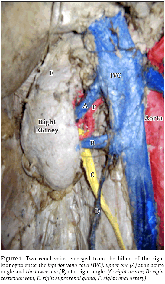
It was found that the right kidney had two renal veins. The veins emerged from the hilum to enter the inferior vena cava separately – upper one at an acute angle and the lower one at a right angle. The right testicular vein drained into the inferior vena cava at an acute angle at the point of entry of the right renal vein into the inferior vena cava (Figure 1).
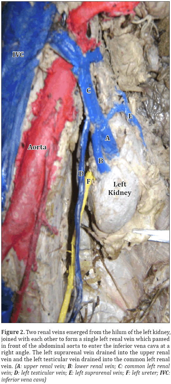
Left kidney was also found to have two renal veins which emerged from the hilum, passed medially and joined with each other to form a single left renal vein. This common renal vein passed in front of the abdominal aorta to enter the inferior vena cava at a right angle. The left suprarenal vein drained into the upper renal vein and the left testicular vein drained into the common renal vein (Figure 2).
Figure 2: Two renal veins emerged from the hilum of the left kidney, joined with each other to form a single left renal vein which passed in front of the abdominal aorta to enter the inferior vena cava at a right angle. The left suprarenal vein drained into the upper renal vein and the left testicular vein drained into the common left renal vein. (A: upper renal vein; B: lower renal vein; C: common left renal vein; D: left testicular vein; E: left suprarenal vein; F: left ureter; IVC: inferior vena cava)
In case of both the kidneys, the renal veins were passing in front of the corresponding renal artery.
Discussion
Ontogenic review
In the embryo, on each side the anterior and posterior cardinal veins (drain the body wall) join with each other and form the right and left common cardinal veins before entering the respective sinus horn of the developing heart (in the 4th week of intrauterine life). During the 5th to 7th week of the embryo, a number of additional veins are formed, among them the supracardinal veins drain the body wall by the way of the intercostal veins; the azygos veins and the subcardinal veins drain the mesonephros and connected to the postcardinal veins [1–2]. With the regression of the major parts of postcardinal veins, the subcardinal veins are connected to the supracardinal veins. Along with the enlargement of the metanephros, the venous drainage of the mesonephric ridge is assumed by subcardinal veins [3]. The two subcardinal veins are connected to each other via the pre-aortic anastomotic plexus forming a part of the left renal vein. The subcardinal veins mainly drain the kidneys and partly disappear in the caudal part [4].
Previously Recorded Cases
In an extensive study, Pick and Anson found that 40.5% of all kidneys examined, had more than two vessels, that was, more than a single artery and a single vein [5]. For most part of the body, variations or anomalies of veins are far more frequent than those of arteries, but this is not true of the vascular pedicle of kidney. Supernumerary veins are present in 14.4% and supernumerary arteries are present in 32.25% of the kidneys [5].
Dhar reported a case with segmental branches of right renal artery being sandwiched between two right renal veins and the emergence of two renal veins at the hilum of right kidney which drained separately into inferior vena cava [6]. This case had similarity with the right-sided variation of our case. Toda et al. reported about two patients with double left renal veins and abdominal aortic aneurysm [7].
According to Pick and Anson most of the multiple renal veins are present on the right side; in their series 27.8% additional veins were on right side and 1% on the left side [5]. Satyapal stated that the incidence of additional renal vein was 26% on the right side as compared to 2.6% on the left side [8].
This vascular variation shows a major significance in renal surgery, in partial or total nephrectomy and in renal transplant [9]. The renal hilum should be examined properly prior to any surgical procedure in this region by radiographic examination [7,10], which is also important for the post-operative management [2]. Contrast-enhanced CT scan can provide enough information about venous variations of kidney, which is important especially before any abdominal operation like the operation of abdominal aortic aneurysm [7].
Conclusion
Multiple vascular variations near the hilum of the kidney are present in seemingly normal patients. A sound knowledge of possible variations of the hilar vessels is important for radiologists, urologists and surgeons [10]. This variation will also enhance our knowledge in gross anatomy.
Acknowledgement
We express our heartiest gratitude to all the faculty members and other staff of Department of Anatomy, Calcutta National Medical College, Kolkata, India, for their cordial help to complete this case report.
References
- Standring S, Jeremiah HC, Glass J, Collins P, Wigley C, eds. Gray’s Anatomy. The Anatomical Basis of Clinical Practice. 39th Ed., London, New York, Oxford, Philadelphia, St. Louis, Sydney, Toronto, Elsevier Churchill Livingstone. 2005; 1276–1277.
- Sadler TW. Langman’s Medical Embryology. 11th Ed., Baltimore, Philadelphia, New Delhi, Lippincott Williams and Wilkins. 2009; 193–196.
- Williams PL, Bannister HL, Martin MB, Collins P, Dyson M, Dussek EJ, Ferguson WJM, Glabella G, eds. Gray’s Anatomy. 38th Ed., New York, Edinburgh, London, Madrid, Tokyo and Melbourne, Churchill Livingstone. 1995; 324–325, 1600 1602.
- McClure CFW, Butler EG. The development of the vena cava inferior in man. Am J Anat. 1925; 35: 331–383.
- Pick JW, Anson BJ. The renal vascular pedicle. An anatomical study of 430 bodyhalves. J Urol. 1940; 44: 411–434.
- Dhar P. An additional renal vein. Clin Anat. 2002; 15: 64–66.
- Toda R, Iguro Y, Moriyama Y, Hisashi Y, Masuda H, Sakata R. Double renal vein associated with abdominal aortic aneurysm. Ann Thorac Cardiovasc Surg. 2001; 7: 113–115.
- Satyapal KS, Rambiritch V, Pillai G. Additional renal veins: incidence and morphometry. Clin Anat. 1995; 8: 51–55.
- Dhar P, Lal K. Main and accessory renal arteries--a morphological study. Ital J Anat Embryol. 2005; 110: 101–110.
- Soni S, Wadhwa A. Multiple Variations in the paired arteries of abdominal aorta – clinical implications. J Clin Diagn Res. 2010; 4: 2622–2625.
Sudeshna Majumdar1*, Sibani Mazumder1, Hironmoy Roy2, Asis Kumar Ghoshal3
1Department of Anatomy, Calcutta National Medical College, Kolkata, India.
2Department of Anatomy, Murshidabad Medical College, Murshidabad, India.
3Department of Anatomy, Institute of Post Graduate Medical Education and Research, Kolkata, India.
- *Corresponding Author:
- Dr. Sudeshna Majumdar
Associate Professor, Department of Anatomy, Calcutta National Medical College, Kolkata, West Bengal, India.
Tel: +91 (943) 3007363
E-mail: sudeshnamajumdar.2007@rediffmail.com
Date of Received: March 8th, 2012
Date of Accepted: October 21st, 2012
Published Online: April 21st, 2013
© Int J Anat Var (IJAV). 2013; 6: 61–63.
Abstract
Variations in the number and arrangement of renal vessels are common. There may be multiple renal arteries and renal veins which are important in different investigative and surgical procedures, especially during renal transplantation. While doing the routine dissection for the undergraduate students in Calcutta National Medical College, in one male cadaver of seventy years, double renal veins were found on both sides. This case report, regarding the renal veins may have importance not only in clinical anatomy, but also in the field of gross anatomy.
-Keywords
right and left renal veins, double renal veins, hilar structures of kidney
Introduction
The large renal veins lie anterior to the renal arteries and open into the inferior vena cava almost at right angles. The left is three times of the right in length (7.5 cm and 2.5 cm). The left renal vein runs from its origin in the renal hilum, posterior to the splenic vein and the body of pancreas, and then across the anterior aspect of aorta. The left testicular or ovarian vein enters the left renal vein from below, and the suprarenal vein usually receiving one of the left inferior phrenic veins, enters it above. The right renal vein is behind the descending part of duodenum. The right testicular or ovarian vein and the right suprarenal vein enter the inferior vena cava directly. The left renal vein may be double, one vein passing posterior, the other anterior to the aorta before joining the inferior vena cava. This is sometimes referred to as the persistence of the ‘renal collar’. Because of its close relationship to the aorta, the left renal vein may be ligated during aortic aneurysm [1]. Several other variations of renal veins have also been reported.
Case Report
While doing the routine dissection for the undergraduate students in Calcutta National Medical College, in a 70-year-old old male cadaver few variations in the hilar blood vessels of both the kidneys were noted. The vessels concerned were dissected properly in the posterior abdominal wall, painted and relevant photographs were taken. Variations in the hilar structures of both the kidneys were noted minutely.
It was found that the right kidney had two renal veins. The veins emerged from the hilum to enter the inferior vena cava separately – upper one at an acute angle and the lower one at a right angle. The right testicular vein drained into the inferior vena cava at an acute angle at the point of entry of the right renal vein into the inferior vena cava (Figure 1).
Left kidney was also found to have two renal veins which emerged from the hilum, passed medially and joined with each other to form a single left renal vein. This common renal vein passed in front of the abdominal aorta to enter the inferior vena cava at a right angle. The left suprarenal vein drained into the upper renal vein and the left testicular vein drained into the common renal vein (Figure 2).
Figure 2: Two renal veins emerged from the hilum of the left kidney, joined with each other to form a single left renal vein which passed in front of the abdominal aorta to enter the inferior vena cava at a right angle. The left suprarenal vein drained into the upper renal vein and the left testicular vein drained into the common left renal vein. (A: upper renal vein; B: lower renal vein; C: common left renal vein; D: left testicular vein; E: left suprarenal vein; F: left ureter; IVC: inferior vena cava)
In case of both the kidneys, the renal veins were passing in front of the corresponding renal artery.
Discussion
Ontogenic review
In the embryo, on each side the anterior and posterior cardinal veins (drain the body wall) join with each other and form the right and left common cardinal veins before entering the respective sinus horn of the developing heart (in the 4th week of intrauterine life). During the 5th to 7th week of the embryo, a number of additional veins are formed, among them the supracardinal veins drain the body wall by the way of the intercostal veins; the azygos veins and the subcardinal veins drain the mesonephros and connected to the postcardinal veins [1–2]. With the regression of the major parts of postcardinal veins, the subcardinal veins are connected to the supracardinal veins. Along with the enlargement of the metanephros, the venous drainage of the mesonephric ridge is assumed by subcardinal veins [3]. The two subcardinal veins are connected to each other via the pre-aortic anastomotic plexus forming a part of the left renal vein. The subcardinal veins mainly drain the kidneys and partly disappear in the caudal part [4].
Previously Recorded Cases
In an extensive study, Pick and Anson found that 40.5% of all kidneys examined, had more than two vessels, that was, more than a single artery and a single vein [5]. For most part of the body, variations or anomalies of veins are far more frequent than those of arteries, but this is not true of the vascular pedicle of kidney. Supernumerary veins are present in 14.4% and supernumerary arteries are present in 32.25% of the kidneys [5].
Dhar reported a case with segmental branches of right renal artery being sandwiched between two right renal veins and the emergence of two renal veins at the hilum of right kidney which drained separately into inferior vena cava [6]. This case had similarity with the right-sided variation of our case. Toda et al. reported about two patients with double left renal veins and abdominal aortic aneurysm [7].
According to Pick and Anson most of the multiple renal veins are present on the right side; in their series 27.8% additional veins were on right side and 1% on the left side [5]. Satyapal stated that the incidence of additional renal vein was 26% on the right side as compared to 2.6% on the left side [8].
This vascular variation shows a major significance in renal surgery, in partial or total nephrectomy and in renal transplant [9]. The renal hilum should be examined properly prior to any surgical procedure in this region by radiographic examination [7,10], which is also important for the post-operative management [2]. Contrast-enhanced CT scan can provide enough information about venous variations of kidney, which is important especially before any abdominal operation like the operation of abdominal aortic aneurysm [7].
Conclusion
Multiple vascular variations near the hilum of the kidney are present in seemingly normal patients. A sound knowledge of possible variations of the hilar vessels is important for radiologists, urologists and surgeons [10]. This variation will also enhance our knowledge in gross anatomy.
Acknowledgement
We express our heartiest gratitude to all the faculty members and other staff of Department of Anatomy, Calcutta National Medical College, Kolkata, India, for their cordial help to complete this case report.
References
- Standring S, Jeremiah HC, Glass J, Collins P, Wigley C, eds. Gray’s Anatomy. The Anatomical Basis of Clinical Practice. 39th Ed., London, New York, Oxford, Philadelphia, St. Louis, Sydney, Toronto, Elsevier Churchill Livingstone. 2005; 1276–1277.
- Sadler TW. Langman’s Medical Embryology. 11th Ed., Baltimore, Philadelphia, New Delhi, Lippincott Williams and Wilkins. 2009; 193–196.
- Williams PL, Bannister HL, Martin MB, Collins P, Dyson M, Dussek EJ, Ferguson WJM, Glabella G, eds. Gray’s Anatomy. 38th Ed., New York, Edinburgh, London, Madrid, Tokyo and Melbourne, Churchill Livingstone. 1995; 324–325, 1600 1602.
- McClure CFW, Butler EG. The development of the vena cava inferior in man. Am J Anat. 1925; 35: 331–383.
- Pick JW, Anson BJ. The renal vascular pedicle. An anatomical study of 430 bodyhalves. J Urol. 1940; 44: 411–434.
- Dhar P. An additional renal vein. Clin Anat. 2002; 15: 64–66.
- Toda R, Iguro Y, Moriyama Y, Hisashi Y, Masuda H, Sakata R. Double renal vein associated with abdominal aortic aneurysm. Ann Thorac Cardiovasc Surg. 2001; 7: 113–115.
- Satyapal KS, Rambiritch V, Pillai G. Additional renal veins: incidence and morphometry. Clin Anat. 1995; 8: 51–55.
- Dhar P, Lal K. Main and accessory renal arteries--a morphological study. Ital J Anat Embryol. 2005; 110: 101–110.
- Soni S, Wadhwa A. Multiple Variations in the paired arteries of abdominal aorta – clinical implications. J Clin Diagn Res. 2010; 4: 2622–2625.


