A case report: double renal arteries
Patel Shashikala*, Wanjari Anjali, Naik Anshuman and Deshpande Jayshree
Department of Anatomy, Jawaharlal Nehru Medical College, Sawangi (Meghe) Wardha, Maharashtra, India
- *Corresponding Author:
- Dr. Patel Shashikala, MBBS, MD
Department of Anatomy, Jawaharlal Nehru Medical College, Sawangi (Meghe) Wardha 442004, M.S., India
Tel: +91 9970499132
E-mail: shashi_patel27@rediffmail.com
Date of Received: June 2nd, 2011
Date of Accepted: April 16th, 2012
Published Online: July 8th, 2012
© Int J Anat Var (IJAV). 2012; 5: 22–24.
[ft_below_content] =>Keywords
kidney, renal artery, anatomical variation, double, unilateral
Introduction
The renal arteries usually arise from the anterolateral or lateral aspect of the abdominal aorta just below the origin of the superior mesenteric artery [1]. Near the hilum of the kidney, each renal artery divides into anterior and posterior branch, which in turn divides into a number of segmental arteries supplying the different renal segments [2]. Classically, a single renal artery supplies each kidney [3]. Among renal morphological variations, those most often encountered are variations in the number of the renal arteries, of which double renal arteries are the most frequent [4]. Explanation for individual or combined variations of renal arteries had been related to the embryological development of vessels from the lateral mesonephric branches of the dorsal aorta [5]. Knowledge of the variations of renal vascular anatomy has importance in exploration and treatment of renal trauma, renal transplantation, renovascular hypertension, renal artery embolization, angioplasty or vascular reconstruction for congenital and acquired lesions, surgery for abdominal aortic aneurysm and conservative or radical renal surgery [6]. The objective of this case report and review of literature is to bring awareness to clinicians about the variations in the blood supply of the kidney, especially for those who are performing invasive procedures and vascular surgeries on kidney.
Case Report
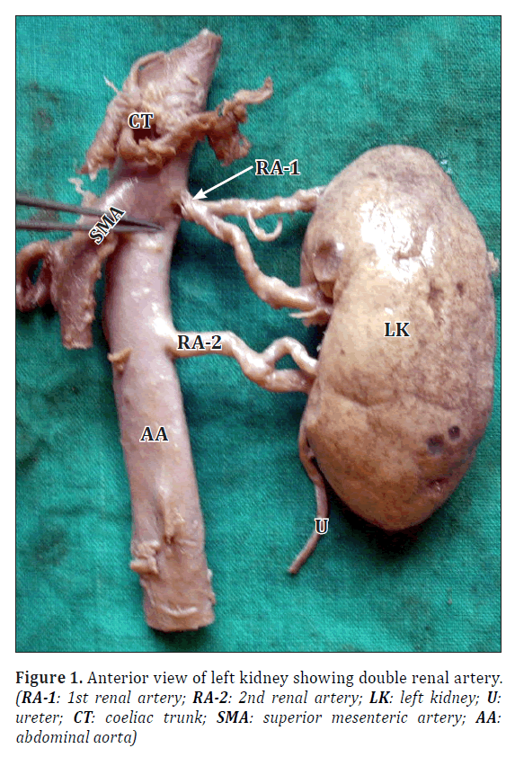
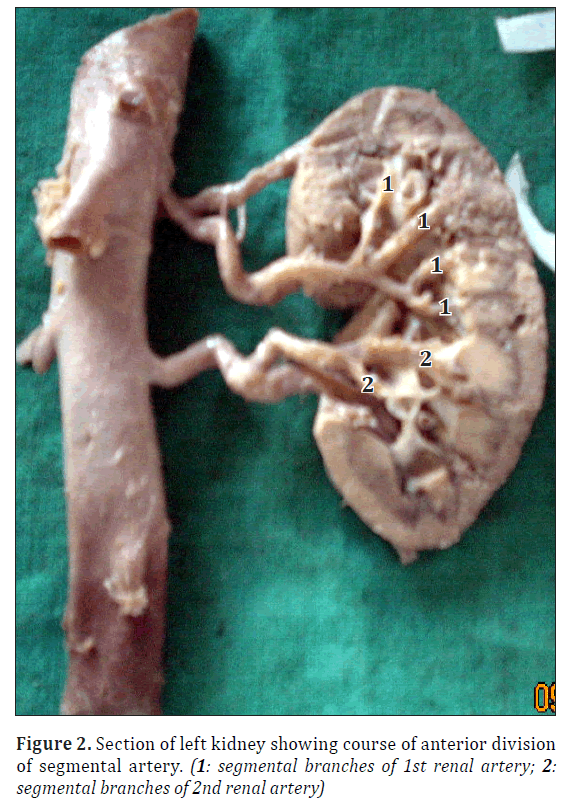
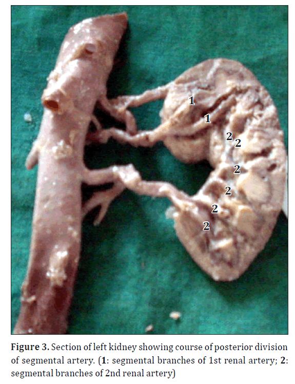
The case showed unilateral left double renal artery in 54-year-old male cadaver during routine dissection of abdomen in Department of Anatomy Jawaharlal Nehru Medical College Sawangi (Meghe) Wardha. First renal artery (RA) arose from aorta at the level of L1 vertebra, whereas 2nd renal artery arose from same 5 cm below to the first one. Both RA ran laterally and entered the kidney through the hilum with their anterior and posterior divisions. One branch (superior polar artery) of 1st renal artery arose from 0.5 cm away from main origin, ran supero-laterally to reach the upper pole of the kidney and supplied it. Anterior division of 1st RA divided into 4 segmental arteries (1 apical, 1 upper, 2 middle) having intrarenal course, whereas 2nd RA gave 2 segmental arteries (1 middle and 1 lower). Posterior division of 1st RA gave 2 (apical) whereas that of 2nd gave 6 (2 upper, 2 middle, 2 lower) segmental arteries.
Discussion
Double renal arteries with an aortic origin are frequent vascular variations, representing the persistence of the embryonic vessels, the lateral branches of the mesonephros, within the renal ascent [7]. The nomenclature of the variations of the renal arteries is still not clear, as different authors described them as additional, accessory, hilar, inferior and superior polar arteries. We named our renal arteries as double renal arteries [1]. Double renal arteries usually arise from the abdominal aorta, that entered the kidney through the hilum, the one with a larger caliber and thus supplying a larger renal territory, was designated the main renal artery and the other as the hilar supplementary renal artery [4]. In recent years, interest in the surgical and medical aspects of double renal arteries has been high because during renal surgical procedures, besides hemorrhage and loss of renal parenchyma, arterial lesions may induce segmental ischemia followed by hypertension. The presence of double renal arteries increases the complexity of renal transplantation; kidneys with double arterial supply being involved in a higher percentage of transplant failures than kidneys showing no variation [4].
Ashken stated that the incidence of double renal arteries in hypertensive subjects was 40%, compared with 20% of normotensive subjects [8].
Our results are quite similar with those of Bergman et al. and Bulic et al. [9,10]. Bergman et al. observed double renal and testicular arteries in a well-developed 69-year-old Caucasian male. The right kidney had two renal arteries, one at its usual midorgan (hilar) position and one inferior polar [9]. Bulic et al. reported a right kidney receiving two renal arteries from the aorta that were similar in diameter, both entering through the hilum [10]. Satyapal et al. described double renal arteries in 31.3% of the African population in their study, 30.9% of the white population, 18.5% of the half-caste population and 13.5% of the Indian population [11]. Rusu reported bilateral doubled renal arteries on the right side as superior hilar and inferior hilar renal arteries, and on the left side as superior hilar and inferior polar renal arteries [12]. Bordei et al. found 54 double renal arteries originating from the aorta in 272 kidneys (20%); six of them were bilateral (2,2%) [4].
With the increasing demand for kidney transplantation, living donor grafts has become the major source for maintaining the donor pool, and successful allograft with double arteries has become a necessity. Nevertheless, to plan the adequate surgical procedure and to avoid any vascular complication, arteriography should be performed prior to every nephrectomy [3]. The variations described in the current observation present a unique pattern of congenital renal vascular variants having surgical and radiological importance.
Acknowledgements
I (Patel Shashikala) am grateful to God, who always with me. The special thank goes to my helpful guide Dr. Anjali Wanjari, the supervision and support that she gave truly help the progression and smoothness of the research paper. The co-operation is much indeed appreciated. Last but not least I would like to thank my lovely husband Dr. Anshuman Naik, who encourage, support and help me in completing this paper successfully.
References
- Patasi B, Boozary A. A case report: accessory right renal artery. Int J Anat Var (IJAV). 2009; 2: 119–121.
- Gupta V, Kotgirwar S, Trivedi S, Deopujari R, Singh V. Bilateral variations in renal vasculature. Int J Anat Var (IJAV). 2010; 3: 53–55.
- Gurses IA, Kale A, Gayretli O, Bayraktar B, Usta A, Kayaalp ME, Ari Z. Bilateral variations of renal and testicular arteries. Int J Anat Var (IJAV). 2009; 2: 45–47.
- Bordei P, Sapte E, Iliescu D. Double renal arteries originating from the aorta. Surg Radiol Anat. 2004; 26: 474–479.
- Felix W. Mesonephric arteries (aa. mesonephricae). In: Kiebel F, Mall FP, eds. Manual of Human Embryology. Vol 2, Philadelphia, Lippincott. 1912; 820–825.
- Fernandes RMP, Conte FHP, Favorito La, Abidu-Figueiredo M, Babinski MA. Triple right renal vein: an uncommon variation. Int J Morphol. 2005; 23: 231–233.
- Langman J, Sadler TW. Embryologie Médicale. Paris, Pradel. 1996; 301.
- Ashken MH. A study of the renal vascular patterns in hypertension, chronic pyelonephritis and other diseases. Hunterian Lecture delivered at the Royal College of Surgeons of England on 24th February 1966. Ann R Coll Surg Engl. 1967; 40: 82–99.
- Bergman RA, Cassell MD, Sahinoglu K, Heidger PM Jr. Human doubled renal and testicular arteries. Ann Anat. 1992; 174: 313–315.
- Bulic K, Ivkic G, Pavic T. A case of duplicated right renal artery and triplicated left renal artery. Ann Anat. 1996; 178: 281–283.
- Satyapal KS, Haffejee AA, Singh B, Ramsaroop L, Robbs JV, Kalideen JM. Additional renal arteries: incidence and morphometry. Surg Radiol Anat. 2001; 23: 33–38.
- Rusu MC. Human bilateral doubled renal and testicular arteries with a left testicular arterial arch around the left renal vein. Rom J Morphol Embryol. 2006; 47: 197–200.11222222
Patel Shashikala*, Wanjari Anjali, Naik Anshuman and Deshpande Jayshree
Department of Anatomy, Jawaharlal Nehru Medical College, Sawangi (Meghe) Wardha, Maharashtra, India
- *Corresponding Author:
- Dr. Patel Shashikala, MBBS, MD
Department of Anatomy, Jawaharlal Nehru Medical College, Sawangi (Meghe) Wardha 442004, M.S., India
Tel: +91 9970499132
E-mail: shashi_patel27@rediffmail.com
Date of Received: June 2nd, 2011
Date of Accepted: April 16th, 2012
Published Online: July 8th, 2012
© Int J Anat Var (IJAV). 2012; 5: 22–24.
Abstract
Proper knowledge of variations of arteries supplying the kidney is essential not only to the anatomists but also to the surgeons. The present case shows unilateral left double renal artery in 54-year-old male cadaver during routine dissection of abdomen. First renal artery was arising from aorta at the level of L1 vertebra whereas second renal artery was arising from same 5 cm below it. Details are discussed.
-Keywords
kidney, renal artery, anatomical variation, double, unilateral
Introduction
The renal arteries usually arise from the anterolateral or lateral aspect of the abdominal aorta just below the origin of the superior mesenteric artery [1]. Near the hilum of the kidney, each renal artery divides into anterior and posterior branch, which in turn divides into a number of segmental arteries supplying the different renal segments [2]. Classically, a single renal artery supplies each kidney [3]. Among renal morphological variations, those most often encountered are variations in the number of the renal arteries, of which double renal arteries are the most frequent [4]. Explanation for individual or combined variations of renal arteries had been related to the embryological development of vessels from the lateral mesonephric branches of the dorsal aorta [5]. Knowledge of the variations of renal vascular anatomy has importance in exploration and treatment of renal trauma, renal transplantation, renovascular hypertension, renal artery embolization, angioplasty or vascular reconstruction for congenital and acquired lesions, surgery for abdominal aortic aneurysm and conservative or radical renal surgery [6]. The objective of this case report and review of literature is to bring awareness to clinicians about the variations in the blood supply of the kidney, especially for those who are performing invasive procedures and vascular surgeries on kidney.
Case Report
The case showed unilateral left double renal artery in 54-year-old male cadaver during routine dissection of abdomen in Department of Anatomy Jawaharlal Nehru Medical College Sawangi (Meghe) Wardha. First renal artery (RA) arose from aorta at the level of L1 vertebra, whereas 2nd renal artery arose from same 5 cm below to the first one. Both RA ran laterally and entered the kidney through the hilum with their anterior and posterior divisions. One branch (superior polar artery) of 1st renal artery arose from 0.5 cm away from main origin, ran supero-laterally to reach the upper pole of the kidney and supplied it. Anterior division of 1st RA divided into 4 segmental arteries (1 apical, 1 upper, 2 middle) having intrarenal course, whereas 2nd RA gave 2 segmental arteries (1 middle and 1 lower). Posterior division of 1st RA gave 2 (apical) whereas that of 2nd gave 6 (2 upper, 2 middle, 2 lower) segmental arteries.
Discussion
Double renal arteries with an aortic origin are frequent vascular variations, representing the persistence of the embryonic vessels, the lateral branches of the mesonephros, within the renal ascent [7]. The nomenclature of the variations of the renal arteries is still not clear, as different authors described them as additional, accessory, hilar, inferior and superior polar arteries. We named our renal arteries as double renal arteries [1]. Double renal arteries usually arise from the abdominal aorta, that entered the kidney through the hilum, the one with a larger caliber and thus supplying a larger renal territory, was designated the main renal artery and the other as the hilar supplementary renal artery [4]. In recent years, interest in the surgical and medical aspects of double renal arteries has been high because during renal surgical procedures, besides hemorrhage and loss of renal parenchyma, arterial lesions may induce segmental ischemia followed by hypertension. The presence of double renal arteries increases the complexity of renal transplantation; kidneys with double arterial supply being involved in a higher percentage of transplant failures than kidneys showing no variation [4].
Ashken stated that the incidence of double renal arteries in hypertensive subjects was 40%, compared with 20% of normotensive subjects [8].
Our results are quite similar with those of Bergman et al. and Bulic et al. [9,10]. Bergman et al. observed double renal and testicular arteries in a well-developed 69-year-old Caucasian male. The right kidney had two renal arteries, one at its usual midorgan (hilar) position and one inferior polar [9]. Bulic et al. reported a right kidney receiving two renal arteries from the aorta that were similar in diameter, both entering through the hilum [10]. Satyapal et al. described double renal arteries in 31.3% of the African population in their study, 30.9% of the white population, 18.5% of the half-caste population and 13.5% of the Indian population [11]. Rusu reported bilateral doubled renal arteries on the right side as superior hilar and inferior hilar renal arteries, and on the left side as superior hilar and inferior polar renal arteries [12]. Bordei et al. found 54 double renal arteries originating from the aorta in 272 kidneys (20%); six of them were bilateral (2,2%) [4].
With the increasing demand for kidney transplantation, living donor grafts has become the major source for maintaining the donor pool, and successful allograft with double arteries has become a necessity. Nevertheless, to plan the adequate surgical procedure and to avoid any vascular complication, arteriography should be performed prior to every nephrectomy [3]. The variations described in the current observation present a unique pattern of congenital renal vascular variants having surgical and radiological importance.
Acknowledgements
I (Patel Shashikala) am grateful to God, who always with me. The special thank goes to my helpful guide Dr. Anjali Wanjari, the supervision and support that she gave truly help the progression and smoothness of the research paper. The co-operation is much indeed appreciated. Last but not least I would like to thank my lovely husband Dr. Anshuman Naik, who encourage, support and help me in completing this paper successfully.
References
- Patasi B, Boozary A. A case report: accessory right renal artery. Int J Anat Var (IJAV). 2009; 2: 119–121.
- Gupta V, Kotgirwar S, Trivedi S, Deopujari R, Singh V. Bilateral variations in renal vasculature. Int J Anat Var (IJAV). 2010; 3: 53–55.
- Gurses IA, Kale A, Gayretli O, Bayraktar B, Usta A, Kayaalp ME, Ari Z. Bilateral variations of renal and testicular arteries. Int J Anat Var (IJAV). 2009; 2: 45–47.
- Bordei P, Sapte E, Iliescu D. Double renal arteries originating from the aorta. Surg Radiol Anat. 2004; 26: 474–479.
- Felix W. Mesonephric arteries (aa. mesonephricae). In: Kiebel F, Mall FP, eds. Manual of Human Embryology. Vol 2, Philadelphia, Lippincott. 1912; 820–825.
- Fernandes RMP, Conte FHP, Favorito La, Abidu-Figueiredo M, Babinski MA. Triple right renal vein: an uncommon variation. Int J Morphol. 2005; 23: 231–233.
- Langman J, Sadler TW. Embryologie Médicale. Paris, Pradel. 1996; 301.
- Ashken MH. A study of the renal vascular patterns in hypertension, chronic pyelonephritis and other diseases. Hunterian Lecture delivered at the Royal College of Surgeons of England on 24th February 1966. Ann R Coll Surg Engl. 1967; 40: 82–99.
- Bergman RA, Cassell MD, Sahinoglu K, Heidger PM Jr. Human doubled renal and testicular arteries. Ann Anat. 1992; 174: 313–315.
- Bulic K, Ivkic G, Pavic T. A case of duplicated right renal artery and triplicated left renal artery. Ann Anat. 1996; 178: 281–283.
- Satyapal KS, Haffejee AA, Singh B, Ramsaroop L, Robbs JV, Kalideen JM. Additional renal arteries: incidence and morphometry. Surg Radiol Anat. 2001; 23: 33–38.
- Rusu MC. Human bilateral doubled renal and testicular arteries with a left testicular arterial arch around the left renal vein. Rom J Morphol Embryol. 2006; 47: 197–200.11222222


