A Case Report on IAN damage and Accurate Pre-operative Visualization
Received: 03-Oct-2022, Manuscript No. ijav-22-5516; Editor assigned: 05-Oct-2022, Pre QC No. ijav-22-5516 (PQ); Accepted Date: Oct 24, 2022; Reviewed: 19-Oct-2022 QC No. ijav-22-5516; Revised: 24-Oct-2022, Manuscript No. ijav-22-5516 (R); Published: 31-Oct-2022, DOI: 10.37532/1308-4038.15(10).223
Citation: Hvizdosova N. A Case Report on IAN damage and Accurate Pre-operative Visualization. Int J Anat Var. 2022;15(10):230-230.
This open-access article is distributed under the terms of the Creative Commons Attribution Non-Commercial License (CC BY-NC) (http://creativecommons.org/licenses/by-nc/4.0/), which permits reuse, distribution and reproduction of the article, provided that the original work is properly cited and the reuse is restricted to noncommercial purposes. For commercial reuse, contact reprints@pulsus.com
Abstract
This case report associating a permanent IAN shows an affected third molar with an associated Dentiger cyst, with the IAN pointing laterally and running along the lateral side of the mandible. In this case the limitations of standard radiographic techniques such as orthopantomograms are repeated.
Keywords
Cone beam computed tomography; Impacted third molars; Maxillofacial anatomy; Inferior alveolar nerve
INTRODUCTION
No specific number of IAN violations is expected Root canal treatment with these complications Relatively rare use of thermoplastic ends Dental filling materials are becoming more and more popular practitioners who practice endodontic, i.e. Nerve injury may occur more frequently [1- 2]. Additionally after reviewing many articles on IAN violations extrusion of other root canal filling materials Mandibular teeth material nerve clinically, IAN injury manifests as a sensory deficit Paresthesia, Anesthesia, Hypoesthesia etc. Oral hyperesthesia and his IAN innervation skin area.
CASE REPORT
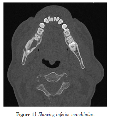
A middle-aged woman who was experiencing pain and swelling in her left upper jaw was evaluated in a clinic. Approximately two years prior, she had similar problems in the upper jaw’s left premolar area and had visited an oral surgeon. The patient had apisectomy on their first and second premolars as well as a root canal retreatment. She visited our clinic after consulting a second oral surgeon for advice after experiencing discomfort for an additional two years. During a thorough examination, it was found that the patient had right side numbness in the brain region that developed 10 years ago during root canal therapy on 47 teeth [3]. Ten years after receiving treatment, she stated that the discomfort was gradually getting better but the numbness had not changed. Her 3rd quadrant teeth felt like “wooden”. She complained that it was difficult to drool and paint her lips Tick. The patient had no history of systemic disease. Dental 3D cone-beam CT imaging was performed. This showed radiopaque material in the lower region Alveolar bone extends posteriorly the tip of the mesial root of tooth 47 (Figure 1). No one else Specific details of endodontic treatment or used I had a filling.
DISCUSSION
Examination of our patient showed evidence of change there is a feeling of redness from the midline to the lower right of the upper lip. Commissures are Vermilion from the lower lip to the lower edge of the mandible compared to the contralateral side. She significantly reduced overall coldness, pinprick, light touch detection and her two-point discrimination. Problem field intraoral examination showed normal Gums and fullness of tongue and tongue Anesthesia of the labial gingiva of the mandible Right second premolar to midline. Cranial nerve nothing else stood out. Was a physical examination also inconspicuous? According to the patient the feel of your lips will not change after root canal treatment [4-5].
CONCLUSION
Contact with nerves or toxic overfilled endodontic agents material. In most cases the damage is reversible. Necessary conservative or surgical treatment is applied Will be on time. However, in the above cases, no treatment is given It could be beneficial. That is the most important lesson from this case there is a prevention of this type of nerve damage. Dentists and endodontists should be aware that sequence of overstretching or periapical extrusion of endodontic filling material.
ACKNOWLEDGEMENT
None
REFERENCES
- Jones AV. Craig GT. Franklin CD. Range and demographics of odontogenic cysts diagnosed in the UK over a 30 year period. J Oral Pathol Med. 35(8):500-700.
- Rood JP, Shehab N. The radiological prediction of inferior alveolar nerve injury during third molar surgery. Br J Oral Maxillofac Surg. 28(1):20-25.
- Sarikov R, Juodzbalys G. Inferior alveolar nerve injury after mandibular third molar extraction: a literature review. J Oral Maxillofac Res. 29; 5(4):e1.
- Tantanapornkul W, Kiyoshi O, Yoshikuni F, Masashi Y, et al. A comparative study of cone-beam computed tomography and conventional panoramic radiography in assessing the topographic relationship between the mandibular canal and impacted third molars. Oral Surg Oral Med Oral Pathol Oral Radiol Endod. 103(2):253-259.
- Maegawa H, Sano K, Kitagawa Y. Preoperative assessment of the relationship between the mandibular third molar and mandibular canal by axial computed tomography. Oral Surg Oral Med Oral Pathol Oral Radiol Endod. 96(5):639-46.
Indexed at, Google Scholar, Crossref
Indexed at, Google Scholar, Crossref
Indexed at, Google Scholar, Crossref
Indexed at, Google Scholar, Crossref


