A Case Report on Radiological Examination of Maxilla Mandibular Fixation
Received: 02-Jan-2023, Manuscript No. ijav-23-6077; Editor assigned: 05-Jan-2023, Pre QC No. ijav-23-6077 (PQ); Accepted Date: Jan 24, 2023; Reviewed: 19-Jan-2023 QC No. ijav-23-6077; Revised: 24-Jan-2023, Manuscript No. ijav-23-6077 (R); Published: 31-Jan-2023, DOI: I:10.37532/1308-4038.16(1).239
Citation: Karl M. A Case Report on Radiological Examination of Maxilla Mandibular Fixation. Int J Anat Var. 2023;16(1):252-252.
This open-access article is distributed under the terms of the Creative Commons Attribution Non-Commercial License (CC BY-NC) (http://creativecommons.org/licenses/by-nc/4.0/), which permits reuse, distribution and reproduction of the article, provided that the original work is properly cited and the reuse is restricted to noncommercial purposes. For commercial reuse, contact reprints@pulsus.com
Abstract
A review of the literature, was performed which bared no former clinical cases reported but a prevalence of 1.2 of triadic foramina after probing dry craniums or radiographic. The discussion stresses the significance of acceptable preoperative radiological examination in the clinical situation especially when unrestricted surgery is planned.
Keywords
Maxilla mandibular fixation; Triadic foramina; Radiological examination
INTRODUCTION
The internal jitters, is primarily a sensitive whim-whams and innervate after leaving the foramen the lower CA nines and premolars and thus play an important part in procedures in this area similar as administration of original anesthesia and surgical intervention. The absence and variation of appurtenant internal foramina has been reported in dry mortal bills and on radiographs preliminarily, and can range from (0.2) to (10.6) on one side [1- 3]. A double internal foramen appears in roughly 1 on the left side in Egyptian and Polynesian populations and in 1.1 on the right side of a Melanesian group.
CASE REPORT
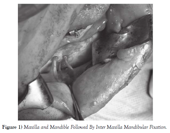
A 28- time-old Male was involved in a road accident performing in bilateral condylar neck fractures of the beak and an oblique fracture at the region of first molar on the right hand side. Under general anesthesia bow bars were applied in the nib and beak followed by inter maxilla mandibular fixation (MMF) in order to achieve a correct occlusion. A vestibular gash was also placed from the lower central incisor to the area distal of the first molar on the right hand side. The immediate postoperative course and the primary mending were uneventful. The case was reviewed with regard to fracture stability, occlusion and any other possible complications similar as sensitive or motor disturbance but was without any complaints (Figure 1).
DISCUSSION
After reviewing the literature to the stylish of our knowledge, this is the first clinical case proved and reported with a triadic internal foramina discovered during surgical treatment of a beak fracture [4]. While treating a mandibular fracture with open reduction and ostheo conflation we anticipated to and one but set up three foramina. We considered only two to be of significance due to the small periphery of the third whim-whams but took care not to damage any of the jitters. Postoperatively reviewing the preoperative reckoned tomography the first foramen was easily depicted distally of the only being premolar and the alternate foramen deposited apical of the mesial root of first molar. Eventually the third minor foramen was observed in between the two. On the postoperative panoramicx-ray only two foramina could be detected. This confirms CT as is formerly well established, to be the system of choice, to probe and describe deconstruction. This is incompletely due to that CT has a low average deformation of 1.8, compared to panoramic which has 23.5 and periapical radiographs 14 which is essential to be apprehensive of when relating delicate anatomical structures [5]. The mindfulness of the possibility of the actuality of anatomical variations in this region which is apparent from reviewing the literature makes it essential to have access to preoperative high quality radiology and good capability to interpret the x-ray while planning surgery.
CONCLUSION
It is essential to be aware of the possibility of these anatomical variations already when planning surgery and when viewing the pre-operative radiological examination. Thus we consider that it is important to report on the risk of anatomical variation of mental foramina, in order to avoid nerve damage in connection with surgical procedures.
ACKNOWLEDGEMENT
None.
CONFLICTS OF INTEREST
None.
REFERENCES
- Kay LW. Some anthropologic investigations of interest to oral surgeons. Int J Oral Surg. 1974; 3(6):363-79.
- Agthong S, Huanmanop T, Chentanez V. Anatomical variation of the supraorbital, infraorbital, and mental foramina related to gender and side. J Oral Maxillofac Surg. 2005; 63(6):800-804.
- Katakami K, Mishima A, Shiozaki K, Shimoda S, et al. Characteristics of accessory mental foramina observed on limited cone-beam computed tomography images. J Endod. 2008; 34(12):1441-1445.
- Sonick M, Abrahams J, Faiella RA. A comparison of the accuracy of periapical, panoramic, and computerized tomographic radiographs in locating the mandibular canal. Int J Oral Maxil-lofac Implants. 1994; 9:455-468.
- Naitoh M, Hiraiwa Y, Aimiya H, Gotoh K, et al. Accessory mental foramen assessment using cone-beam computed tomography. Oral Surg Oral Med Oral Pathol Oral Radiol Endod. 2009; 107(2):289-294.
Indexed at, Google Scholar, Crossref
Indexed at, Google Scholar, Crossref
Indexed at, Google Scholar, Crossref


