A Case Report on the Dorsoepitrochlearis Muscle
2 Department of Human Anatomy, Mount Kenya University, Nairobi, Kenya
Received: 03-Apr-2023, Manuscript No. ijav-23-6344; Editor assigned: 04-Apr-2023, Pre QC No. ijav-23-6344 (PQ); Accepted Date: Apr 21, 2023; Reviewed: 18-Apr-2023 QC No. ijav-23-6344; Revised: 21-Apr-2023, Manuscript No. ijav-23-6344 (R); Published: 28-Apr-2023, DOI: 10.37532/1308-4038.16(4).257
Citation: Nurani MK, Kenneth M, Gakure JN et al. A Case Report on the Dorsoepitrochlearis Muscle. Int J Anat Var. 2023;16(4):290-291.
This open-access article is distributed under the terms of the Creative Commons Attribution Non-Commercial License (CC BY-NC) (http://creativecommons.org/licenses/by-nc/4.0/), which permits reuse, distribution and reproduction of the article, provided that the original work is properly cited and the reuse is restricted to noncommercial purposes. For commercial reuse, contact reprints@pulsus.com
Abstract
Anatomic variations in the upper limb are of great significance as they pose implications on surgical techniques and raise the risk of intraoperative injury. The presence of dorsoepitrochlearis muscle was discovered during routine dissection of a male cadaver at the Department of Human Anatomy. This variant muscle was found in the right axillary region, originating as a single strip from latissimus dorsi and attaching onto the medial epicondyle of the humerus.
Dorsoepitrochlearis is a rare anatomical variation whose spectrum of clinical manifestation may extend from asymptomatic to muscular paresthesia and atrophy as well as motion restriction. A rich basis of knowledge of anatomical variations is crucial in diagnosis and management.
Keywords
Musculus dorsoepitrochlearis; Latissimus dorsi
INTRODUCTION
Anatomic variations in the upper limb are of great significance as they pose implications on surgical techniques and raise the risk of intraoperative injury [1]. Muscular variations, in particular, have been reported previously in literature, and extend from those of supernumerary heads of biceps brachii, panniculus carnosus, and to those noting the presence of the dorsoepitrochlearis muscle [2].
The dorsoepitrochlearis muscle, or latissimo-condyloideus muscle (also referred to as latissimo-epitrochlearis, dorsoepicondylar medial muscle, appendix of latissimus dorsi, tensor fasciae antebrachii, acessorius latissimus dorsi and dorso-antebrachialis), is a longitudinal supernumerary muscle of which arises from the latissimus dorsi muscle [3]. With a prevalence of about 1.9% [4], it arises as a muscular or fibro muscular slip from the antero-inferior border of latissimus dorsi, passing over the axilla and crossing the brachial plexus to attach onto the medial epicondyle of humerus. Muscular slips from dorsoepitrochlearis may also arise and insert onto various structures including the greater tubercle of the humerus or the coracoid process [3]. When present, the dorsoepitrochlearis muscle derives its innervation from the thoracodorsal nerve [4].
Presence of dorsoepitrochlearis may have numerous implications. It has been reported to manifest clinically by limiting the shoulder range of motion and causing axillary deformity [5] and, in other instances, to cause nerve compression in the axilla [4]. In some cases, however, it remains present asymptomatically and becomes an incidental finding during cadaveric dissections [6]. With respect to this, the present study aims to study and report the dorsoepitrochlearis muscle whose presence may be of anatomical, clinical and surgical interest.
CASE REPORT
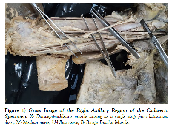
We report the presence of a complete form of dorsoepitrochlearis, which is a muscular variation in the arm, found during upper limb dissection of a male cadaver at the Department of Human Anatomy. This atypical muscle was discovered in the right axillary area arising as a single strip from latissimus dorsi, and crossing the arm to insert onto the medial epicondyle of the humerus, interacting with muscular, neural and vascular elements of the arm as it traversed. There were no other anomalies noted in the cadaveric specimen [Figure 1].
DISCUSSION
Previous studies have reported the presence of the dorsoepitrochlearis muscle giving significance to its rare yet implicative nature [1,4,5,7]. Farfan-C et al. (2019) reported it as a cadaveric finding originating from three fascicles. The upper fascicle as an extension of pectoral fascia and the middle and lower one emerging from latissimus dorsi these three united into a thin muscular belly that inserted onto the medial epicondyle [1]. On the other hand, Natsis et al. (2012) reported dorsoepitrochlearis as a finding on Magnetic Resonance Imaging arising from latissimus dorsi muscle and coursing towards the medial surface of the humerus [5]. In addition, Haninec et al. (2009) reported four cases of dorsoepitrochlearis, where muscular strips originated from the anteroinferior border of latissimus dorsi, crossed the axilla under the axillary fascia and joined the deep surface of the tendon of pectoralis major muscle to insert on the greater tubercle of humerus [4].
The presence and morphology of the dorsoepitrochlearis muscle can be described by latissimus dorsi development [7]. Latissimus dorsi initially appears as a cranio-caudal strip on the lateral side of the body, extending from the 11th rib and neighboring vertebrae to the humerus, the latter being its point of insertion [8]. It then fans out to achieve the definitive position of its attachments on the vertebral spines and on the iliac crest. As it develops, the arch of the muscular primordium that crosses the axilla is interrupted by apoptosis. However, muscular remnants of the arch may persist and constitute the dorsoepitrochlearis muscle [1,7]. This may be complete, in the case where the insertion of dorsoepitrochlearis is on the medial epicondyle, or incomplete, where insertion is more proximal [4].
When it comes to comparative anatomy, dorsoeptrochlearis is reported as being found in majority of mammals and as being common to all quadruped mammals [4]. Similarly, a homologous muscle has been described in amphibians and reptilians. In birds, latissimus dorsi has the same cranial and caudal parts, and the typically formed dorsoepitrochlearis muscle is called metapatagial latissimus dorsi. As such, dorsoepitrochlearis is considered a phylogenetically ancient structure whose morphogenetic mechanism is fixed in the mammalian genome [4].
In terms of mechanics and functional implications, dorsoepitrochlearis could function as a weak adductor at the shoulder given its attachments and fiber orientation [1], or, as a restrictor of movement around the glenohumeral joint [5]. It may also have compressive effects on neurovascular structures within the axilla. This compression could generate clinical symptoms such as numbness, tingling, pain and paresthesia, whose etiology may not be picked promptly as is the case in rare muscular variations [4,9]. As such, it is crucial to keep note of muscular variations and their implications on adjacent neurovasculature [1].
In conclusion, the dorsoepitrochlearis muscle is a rare anatomical variation whose spectrum of clinical manifestation may extend from asymptomatic to muscular paresthesia and atrophy as well as motion restriction. A rich basis of knowledge of anatomical variations is crucial in diagnosis and management
CONFLICTS OF INTEREST
No conflict of interest among authors.
REFERENCES
- Farfán-CE, Inzunza-HO, Echeverría-MM, Inostroza-RV. The Dorsoepicondylar Medial Muscle a Clinically Relevant Anatomical Variation. Int J Morphol. 2019; 37:600-605.
- Tubbs RS, Shoja MM, Loukas M. Bergman’s Comprehensive Encyclopedia of Human Anatomic Variation. Wiley. 2016.
- Boyle EK, Mahon V, Diogo R. Muscles Lost in Our Adult Primate Ancestors Still Imprint in Us: on Muscle Evolution, Development, Variations, and Pathologies. Curr Mol Bio Rep. 2020; 6:32-50.
- Haninec P , Tomás R, Kaiser R, Cihák R. Development and clinical significance of the musculus dorsoepitrochlearis in men. Clin Anat. 2009; 22:481-488.
- Natsis K, Totlis T, Vlasis K, Sofidis G, Lazaridis N, et al. Dorsoepitrochlearis muscle: an unknown cause of shoulder motion limitation and axilla deformity. J Orthop Sci. 2012; 17(2):186-188.
- Matsen FA. Rockwood and Matsen’s the shoulder. 6th ed. Philadelphia: Elsevier. 2021.
- Shah IP, Yadav A, Mehta R, Thatte M. Variation of the latissimus dorsi. Indian J Plast Surg. 2014; 47(3):453-5.
- Moore KL, Persaud TVN. The Developing Human: Clinically Oriented Embryology. Saunders. 1998.
- Salgado G, Cantín M, Inzunza O, Muñoz A, Saez J, et al. Bilateral reversed palmaris longus muscle: a rare anatomical variation. Folia Morphol (Warsz). 2012; 71(1):52-5.
Indexed at, Google Scholar, Crossref
Indexed at, Google Scholar, Crossref
Indexed at, Google Scholar, Crossref
Indexed at, Google Scholar, Crossref
Indexed at, Google Scholar, Crossref
Indexed at, Google Scholar, Crossref


