A case report on unilateral high up origin of a testicular artery with embryological review
Pranab Mukherjee1*, Hironmoy Roy2 and Amit Mallik2
1Department of Anatomy, R.G. Kar Medical College, Kolkata, WB, India
2Department of Anatomy, North Bengal Medical College, Siliguri, WB, India
- *Corresponding Author:
- Dr. Pranab Mukherjee
Associate Professor, Department of Anatomy, R.G. Kar Medical College Mahamaya Road, P.O. Hijalpukur P.S. Habra, Dist. (N) 24 pgs WB, 734271, India
Tel: +91 943 4568024
E-mail: pranabanatomy@gmail.com
Date of Received: April 17th, 2011
Date of Accepted: August 10th, 2011
Published Online: August 15th, 2011
© Int J Anat Var (IJAV). 2011; 4: 147–148.
[ft_below_content] =>Keywords
testicular artery, descent of testis, ascent of kidney, left renal vein
Introduction
Variations of anatomical structures are always appreciated by the anatomists not only to help clinicians, but also to enrich own subject. Testicular arteries are the paired lateral splanchnic braches of abdominal aorta usually arise just below the origin of renal arteries proximal to the inferior mesenteric artery quite in the level of second lumbar vertebra. With a long slender oblique retroperitoneal course over the paravertebral psoas major muscles they reach the deep inguinal ring, where they crosses the ureter anteriorly, join with the ductus deferens to pass through the inguinal canal to reach the testis [1].
Case Report
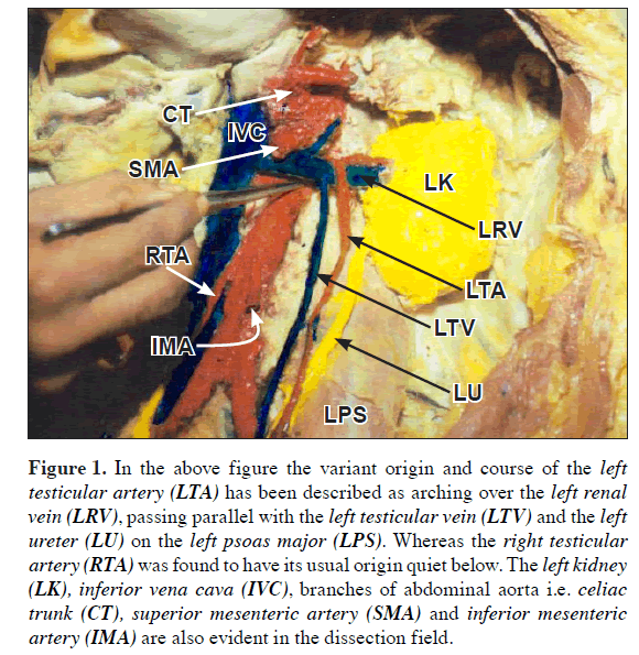
In course of the routine dissection of a 62-year-old male cadaver, asymmetrical origins of the testicular arteries were noted. On right side the artery has taken origin from its usual location and coursed below normally, on the other hand in the left side it has originated from a point just ventral to left renal artery. In the early course it has ascended upward behind the left renal vein to arch over it distal to the draining point of the left suprarenal vein close to the hilum of the left kidney, and then coursed downwards lying on the left psoas major, subsequently crossed the ureter superficially to attain more lateral position at the level of bifurcation of aorta to reach the deep inguinal ring (Figure 1). It is important to note that ipsilateral kidney as well as the testis grossly showed no difference compared to the other side.
Figure 1: In the above figure the variant origin and course of the left testicular artery (LTA) has been described as arching over the left renal vein (LRV), passing parallel with the left testicular vein (LTV) and the left ureter (LU) on the left psoas major (LPS). Whereas the right testicular artery (RTA) was found to have its usual origin quiet below. The left kidney (LK), inferior vena cava (IVC), branches of abdominal aorta i.e. celiac trunk (CT), superior mesenteric artery (SMA) and inferior mesenteric artery (IMA) are also evident in the dissection field.
Discussion
Genital ridge gets its blood supply from the corresponding segments of abdominal aorta which persists throughout the life even after the descent of the testis [2]. The site of origin of the blood vessel represents the embryological origin of it [3]. Kidney develops in the pelvic cavity and in due course of time ascends in the adult position. During such migration, the blood vessels communicate the developing organ from the respective segments of the abdominal aorta or its branches and in parallel the veins go to drain in respective segments of inferior vena cava [4].
During the ascent of the kidney to its prescheduled position left renal vein having a long course across the abdominal aorta, can elevate the free flowing course testicular artery. This happens when the arterial courses of the ascending kidney and descending testis somewhere crosses each other.
Notkovich in 1956 described three types of gonadal arteries based on the anatomical relation with the renal vein, where he has vividly mentioned the category-III gonadal artery, where it arises below and arches over the renal vein. Secondly, preponderance of such variants was found more in males than in females [5].
Discussion
Arching of the both right and left gonadal arteries over the left renal vein were also highlighted by Nathan et. al in 1976 [6]. Bilateral doubled renal and testicular arteries with left testicular arteries arching over the left renal veins have been reported by Rusu in 2006 [7]. Similar to present case i.e. left testicular artery arching over the left renal vein was also reported earlier from southern part of India by Mamatha et al. in 2010 [8].
High origin of the gonadal artery is developmentally cranial mesonephric arteries and in the early courses such arteries pass anterior to subcardinal anastomoses [9]. This may be the explanation of high origin and unusual course of testicular artery.
Conclusion
Unusual origin and arched course of the left testicular artery definitely carries a clinico-surgical importance. This can be explained in the light of elevation of the left testicular artery by the mesonephric vein connection the left kidney and inferior vena cava at the subcardinal segment. Surgeons dealing with the renal pedicle during renal surgery can provide an unintentional knot over the arching gonadal artery. Secondly, compressive effect of the renal vein can precipitate the ‘nut-craker syndrome’ [10]. Last but not least to mention that ipsilateral gonad also may functionally jeopardize due to obstructed blood flow in its artery.
Acknowledgement
The authors are to acknowledge the Principal of NB Medical College and the colleague faculties of the Department of Anatomy for their kind guidance and cooperation.
References
- Gabella G. Testicular artery. Anterolateral visceral arteries. In: Williams PL, Bannister LH, Berry MM, Collins P, Dyson M, Dussek JE, Ferguson MW, eds. Gray’s Anatomy. 38th Ed., Edinburgh, Churchill Livingstone. 1995; 318, 1557–1558.
- Agur AMR, Lee MJ. Grant’s Atlas of Anatomy. 10th Ed., Philadelphia, Lippincott’s Williams and Wilkins. 1999: 143–145.
- Hamilton WJ, Mossman HW. Human Embryology. 4th Ed., Williams and Wilkins. 1976; 430–431.
- Vize PD, Woolf AS, Bard JBL. The Kidney. From Normal Development to Congenital Disease. Amsterdam, Academic Press. 2003: 251–266.
- Notkovich H. Variation of testicular or ovarian arteries in relation to renal pedicle. Surg Gynecol Obstet. 1956; 103; 487–495.
- Nathan H, Tobias PV, Wellsted MD. An unusual case of right and left testicular arteries arching over the left renal vein. Br J Urol. 1976; 48: 135–138.
- Rusu Mc. Human bilateral doubled renal and testicular arteries with a left testicular arterial arch around the left renal vein. Rom J Morphol Embryol. 2006; 47: 197–200.
- Mamatha Y, Prakash BS, Latha PK, Ramesh BR. Variant course of left gonadal artery. International Journal of Anatomical Variations (IJAV). 2010; 3: 132–133.
- Keibel F, Mall FP. Manual of Human Embryology. Vol 2., Philadelphia, Lippincott, 1912: 820–825.
- Rudloff U, Holmes RJ, Prem JT, Faust GR, Moldwin R, Siegel D. Mesoaortic compression of the left renal vein (nutcracker syndrome): case reports and review of the literature. Ann Vasc Surg. 2006; 20: 120–129.
Pranab Mukherjee1*, Hironmoy Roy2 and Amit Mallik2
1Department of Anatomy, R.G. Kar Medical College, Kolkata, WB, India
2Department of Anatomy, North Bengal Medical College, Siliguri, WB, India
- *Corresponding Author:
- Dr. Pranab Mukherjee
Associate Professor, Department of Anatomy, R.G. Kar Medical College Mahamaya Road, P.O. Hijalpukur P.S. Habra, Dist. (N) 24 pgs WB, 734271, India
Tel: +91 943 4568024
E-mail: pranabanatomy@gmail.com
Date of Received: April 17th, 2011
Date of Accepted: August 10th, 2011
Published Online: August 15th, 2011
© Int J Anat Var (IJAV). 2011; 4: 147–148.
Abstract
High up origin of the left testicular artery was noted during routine dissection of an elderly male cadaver. Just after origin it was found to ascend upwards behind the left renal vein to arch over it, distal to the left suprarenal vein close to the hilum of the left kidney. Such an extensive unusual course of testicular artery seems to posses an immense anatomical, embryological and clinical relevance to study to help surgeons to avoid unintentional injury during renal operation. Compression of renal vein by such an artery can produce renal congestion with vice-versa effect on testicular atrophy. Simultaneous descent of testis with ascent of kidney can enlighten such a variation as detailed below.
-Keywords
testicular artery, descent of testis, ascent of kidney, left renal vein
Introduction
Variations of anatomical structures are always appreciated by the anatomists not only to help clinicians, but also to enrich own subject. Testicular arteries are the paired lateral splanchnic braches of abdominal aorta usually arise just below the origin of renal arteries proximal to the inferior mesenteric artery quite in the level of second lumbar vertebra. With a long slender oblique retroperitoneal course over the paravertebral psoas major muscles they reach the deep inguinal ring, where they crosses the ureter anteriorly, join with the ductus deferens to pass through the inguinal canal to reach the testis [1].
Case Report
In course of the routine dissection of a 62-year-old male cadaver, asymmetrical origins of the testicular arteries were noted. On right side the artery has taken origin from its usual location and coursed below normally, on the other hand in the left side it has originated from a point just ventral to left renal artery. In the early course it has ascended upward behind the left renal vein to arch over it distal to the draining point of the left suprarenal vein close to the hilum of the left kidney, and then coursed downwards lying on the left psoas major, subsequently crossed the ureter superficially to attain more lateral position at the level of bifurcation of aorta to reach the deep inguinal ring (Figure 1). It is important to note that ipsilateral kidney as well as the testis grossly showed no difference compared to the other side.
Figure 1: In the above figure the variant origin and course of the left testicular artery (LTA) has been described as arching over the left renal vein (LRV), passing parallel with the left testicular vein (LTV) and the left ureter (LU) on the left psoas major (LPS). Whereas the right testicular artery (RTA) was found to have its usual origin quiet below. The left kidney (LK), inferior vena cava (IVC), branches of abdominal aorta i.e. celiac trunk (CT), superior mesenteric artery (SMA) and inferior mesenteric artery (IMA) are also evident in the dissection field.
Discussion
Genital ridge gets its blood supply from the corresponding segments of abdominal aorta which persists throughout the life even after the descent of the testis [2]. The site of origin of the blood vessel represents the embryological origin of it [3]. Kidney develops in the pelvic cavity and in due course of time ascends in the adult position. During such migration, the blood vessels communicate the developing organ from the respective segments of the abdominal aorta or its branches and in parallel the veins go to drain in respective segments of inferior vena cava [4].
During the ascent of the kidney to its prescheduled position left renal vein having a long course across the abdominal aorta, can elevate the free flowing course testicular artery. This happens when the arterial courses of the ascending kidney and descending testis somewhere crosses each other.
Notkovich in 1956 described three types of gonadal arteries based on the anatomical relation with the renal vein, where he has vividly mentioned the category-III gonadal artery, where it arises below and arches over the renal vein. Secondly, preponderance of such variants was found more in males than in females [5].
Discussion
Arching of the both right and left gonadal arteries over the left renal vein were also highlighted by Nathan et. al in 1976 [6]. Bilateral doubled renal and testicular arteries with left testicular arteries arching over the left renal veins have been reported by Rusu in 2006 [7]. Similar to present case i.e. left testicular artery arching over the left renal vein was also reported earlier from southern part of India by Mamatha et al. in 2010 [8].
High origin of the gonadal artery is developmentally cranial mesonephric arteries and in the early courses such arteries pass anterior to subcardinal anastomoses [9]. This may be the explanation of high origin and unusual course of testicular artery.
Conclusion
Unusual origin and arched course of the left testicular artery definitely carries a clinico-surgical importance. This can be explained in the light of elevation of the left testicular artery by the mesonephric vein connection the left kidney and inferior vena cava at the subcardinal segment. Surgeons dealing with the renal pedicle during renal surgery can provide an unintentional knot over the arching gonadal artery. Secondly, compressive effect of the renal vein can precipitate the ‘nut-craker syndrome’ [10]. Last but not least to mention that ipsilateral gonad also may functionally jeopardize due to obstructed blood flow in its artery.
Acknowledgement
The authors are to acknowledge the Principal of NB Medical College and the colleague faculties of the Department of Anatomy for their kind guidance and cooperation.
References
- Gabella G. Testicular artery. Anterolateral visceral arteries. In: Williams PL, Bannister LH, Berry MM, Collins P, Dyson M, Dussek JE, Ferguson MW, eds. Gray’s Anatomy. 38th Ed., Edinburgh, Churchill Livingstone. 1995; 318, 1557–1558.
- Agur AMR, Lee MJ. Grant’s Atlas of Anatomy. 10th Ed., Philadelphia, Lippincott’s Williams and Wilkins. 1999: 143–145.
- Hamilton WJ, Mossman HW. Human Embryology. 4th Ed., Williams and Wilkins. 1976; 430–431.
- Vize PD, Woolf AS, Bard JBL. The Kidney. From Normal Development to Congenital Disease. Amsterdam, Academic Press. 2003: 251–266.
- Notkovich H. Variation of testicular or ovarian arteries in relation to renal pedicle. Surg Gynecol Obstet. 1956; 103; 487–495.
- Nathan H, Tobias PV, Wellsted MD. An unusual case of right and left testicular arteries arching over the left renal vein. Br J Urol. 1976; 48: 135–138.
- Rusu Mc. Human bilateral doubled renal and testicular arteries with a left testicular arterial arch around the left renal vein. Rom J Morphol Embryol. 2006; 47: 197–200.
- Mamatha Y, Prakash BS, Latha PK, Ramesh BR. Variant course of left gonadal artery. International Journal of Anatomical Variations (IJAV). 2010; 3: 132–133.
- Keibel F, Mall FP. Manual of Human Embryology. Vol 2., Philadelphia, Lippincott, 1912: 820–825.
- Rudloff U, Holmes RJ, Prem JT, Faust GR, Moldwin R, Siegel D. Mesoaortic compression of the left renal vein (nutcracker syndrome): case reports and review of the literature. Ann Vasc Surg. 2006; 20: 120–129.


