A common insertion of thoracoepicondylaris and an axillary arch of Langer: a case report
Polly Lama* and Binod Kumar Tamang
Department of Anatomy, Sikkim Manipal University, Gangtok, Sikkim, India
- *Corresponding Author:
- Polly Lama,MSc, (PhD scholar)
Lecturer
Department of Anatomy
Sikkim Manipal University
Sikkim, India
Tel: +91 954 7591771
E-mail: polly_1in@yahoo.co.in
Date of Received: September 18th, 2009
Date of Accepted: February 11th, 2010
Published Online: April 27th, 2010
© Int J Anat Var (IJAV). 2010; 3: 63–64.
[ft_below_content] =>Keywords
thoracoepicondylaris, chondroepitrochlearis, dorsoepitrochlearis, costohumeralis, costoepitrochlearis
Introduction
An axillary arch is considered to be an variant muscle or tendinous slip that arises form the lower border of latissimus dorsi to join the tendon of pectoralis major, coracobrachalis, or fascia over the biceps. It is present in about 7% of the cases and may be multiple [1]. An axillary arch is thought to be a vestige of panniculus carnosus muscle [2].
Thoracoepicondylaris is a very rare muscular variant which arises as an accessory slip from pectoralis major muscle, crosses the axilla and inserts onto the medial epicondyle of the humerus and medial brachial intermuscular septum [3]; according to Loukas et al., the term thoracoepicondylaris more accurately reflects the origin and insertion of the variant muscular slip.
Many cases has been observed in the past about the occurrences of axillary arches, chondroepitrochlearis, dorsoepitrochlearis, costoepitrochlearis and costohumeralis but variations in relation to the common insertion of pectoralis major and latissimus dorsi muscle are rare and maybe important for diagnosis of unexpected and uncommon clinical conditions.
It has been reported that such rare muscular slips may produce neurovascular complications as it arches over the axillary nerves and vessels [4]. Considering the frequency of procedures in this area, the knowledge of anatomical variation in the axilla is important for any surgical intervention [5].
Case Report
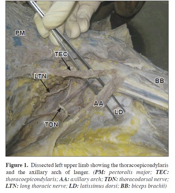
During a routine dissection for teaching medical students the left side upper limb of a 38-year-old male cadaver reportwas dissected. The skin, superficial fascia and deep fascia was removed to expose the pectoral region and the flexor compartment of the arm. The pectoralis major muscle along with a variant muscular slip was noted. The variant muscular slip arose as separate fibers from the inferior aspect of the bulky pectoralis major muscle. The muscular slip was identified as thoracoepicondylaris, and it measured approximately about 18 cm in length and 1.5 cm in width. After carefully removing the axillary group of lymph nodes and the axillary fascia, another variant muscular slip was observed arising from the inferolateral aspect of the latissimus dorsi muscle which measured approximately 7 cm in length and 2.5 cm in thickness, these variant muscle was identified as “axillary arch of Langer”. The most unique feature lied at the insertion, both the muscular variants coursed superficial through the axillary vessels and the nerves of the brachial plexus; and finally inserted at common site into the fascia covering the biceps brachii (Figure 1). The right side of the axilla was also dissected; but the arrangement and attachment of the pectoralis major and latissimus dorsi muscles were as usual and the variable muscular slip was not present.
Discussion
Muscular variations of the upper limb in relation to the pectoralis major and latissimus dorsi muscle have been reported before, but yet it remains important to continue describing rare variations as they arise.
An axilla or the armpit is the space between the upper arm and the side of the thorax, bounded in front by the pectoralis major muscle and behind by the subscapularis and latissimus dorsi, medially by the serratus anterior muscle and contains neurovascular bundle and lymph node for the upper limb and side wall of thorax [6]. Considering the pectoralis major muscle fibers, muscular variations may be present in the form of chondroepitrochlearis, costohumeralis, costoepitrochlearis [5]. Latissimus dorsi also presents an aberrant muscular slip which inserts either around the coracoid process of the scapula or along with the tendon of pectoralis major [7]. This unusual muscle bundles are commonly referred to as “Axillary arch Muscle” [8]. Such muscular variant is surgically very important, since it arches superficial to the neurovascular bundle within the axilla. Thus, variant muscular fibers may not only restrict the abduction of the arm but may also be the cause axillary vein entrapment syndrome [9], and ulnar nerve entrapment [10].
Previous studies have been reported on variant slips coursing along the inferior border of pectoralis major and said to have been originated from ribs or the costal cartilages [5,10]. Samuel and Vollola reported an accessory muscular slip originating from the pectoralis major and inserting on to the medial epicondyle of the humerus and medial intermuscular septum [11]. Variation of the latissimus dorsi muscle fibers as “axillary arch of Langer” is considerable and has been reported many times before [12]. Muscular slip arising from the latissimus dorsi or pectoralis major muscle inserting into many structures including the fascia and the flexor muscles of the arm [13], coracobrachalis, biceps brachii and long head of the triceps brachii [14], teres major [12], the coracoid process of the scapula [15], and medial condyle of the humerus as chondroepitrochlearis muscle [16] have been reported earlier. Chiba et al. suggested that chondroepitrochlearis is always associated with axillary arch muscle and is present in 7 to 13% of the population [1]. Voto and Weiner reported a clinical case of an infant with a contracture of chondroepitrochlearis in whom the axillary arch muscle was absent [17].
The present case discussed here confirms with previous findings; however, it has never been reported before an insertion of variant muscular slips from pectoralis major and latissimus dorsi muscle into a common site.
To conclude, this muscular variant of the pectoralis major and latissimus dorsi holds a great deal of importance as it is rare and can explain the loss of ability to elevate the shoulders. Idiopathic contracture resulting from the unusual slips of pectoralis major and latissimus dorsi may require surgical interventions. Aberrant slips may also cause axillary vein entrapment and has been suggested to have a role in development of lymphedema of the upper limb. The variant therefore attracts clinical attention and because of its potential to cause significant defects it may be of particular interests to orthopedic surgeons, neurologist and cosmetic surgeons.
References
- Chiba S, Suzuki T, Kasai T. A rare anomaly of the pectoralis major-the chondroepitrochlearis. Okajimas Folia Anat Jpn. 1983; 60: 175–186.
- http://www.mondofacto.com/facts/dictionary?query=axillary+arch&action=look+it+up (accessed February 11th, 2010).
- Loukas M, Louis RG Jr, Kwiatkowska M. Chondroepitrochlearis muscle, a case report and a suggested revision of the current nomenclature. Surg Radiol Anat. 2005; 27: 354–356.
- Spinner RJ, Carmichael SW, Spinner M. Infraclavicular ulnar nerve entrapment due to chondroepitrochlearis muscle. J Hand Surg Br. 1991; 16: 315–317.
- Rao RT, Shetty P, Rao S. Additional slip of pectoralis major muscle-the costohumeralis. International Journal of Anatomical Variations (IJAV). 2009; 2: 35–37.
- McMinn RMH, Ed. Last’s Anatomy Regional and Applied. 9th Ed., Edinburgh, Churchill-Livingstone. 1997; 63–65.
- Dharap A. An unusually medial axillary arch muscle. J Anat. 1994; 184: 639–641.
- Gardner E, Gray DJ, O Rahilly R. Anatomy: A regional study of human structure. 4th Ed., Philadelphia, W.B. Saunders. 1975; 105.
- Daniels IR, della Rovere GQ. The axillary arch of Langer--the most common muscular variation in the axilla. Breast Cancer Res Treat. 2000; 59: 77–80.
- Jaijesh P. Unilateral appearance of chondro-epitrochlearis muscle – a case report. Indian J Plast Surg. 2005; 2: 164–166.
- Samuel VP, Vollala VR. Unusual pectoralis major muscle: the chondroepitrochlearis. Anat Sci Int. 2008; 83: 277–279.
- Bergman RA, Thompson SA, Afifi AK. Catalog of Human Variation. 8th Ed., Baltimore, Urban and Schwarzenberg. 1988; 26–27.
- Hollinshead WH. Anatomy for Surgeons. 33rd Ed., Philadelphia, Harper and Row. 1966; 280–281.
- Davies DV, Davies F, ed. Gray’s Anatomy, Descriptive and Applied. 33rd Ed., London, Longmans. 1962; 638.
- Vare AM, Indurkar GM. Some anomalous findings in the axillary musculature. J Anat Soc India. 1965; 14: 478–484.
- Landry SO Jr. The phylogenetic significance of the chondroepitrochlearis muscle and its accompanying pectoral abnormalities. J Anat. 1958; 92: 57–61.
- Voto SJ, Weiner DS. The chondroepitrochlearis muscle. J Pediatr Orthop. 1987; 7: 213–214.
- Kasai T, Chiba S. True nature of the muscular arch of the axilla and its nerve supply. Kaibogaku Zashi. 1977; 52: 309–336.
Polly Lama* and Binod Kumar Tamang
Department of Anatomy, Sikkim Manipal University, Gangtok, Sikkim, India
- *Corresponding Author:
- Polly Lama,MSc, (PhD scholar)
Lecturer
Department of Anatomy
Sikkim Manipal University
Sikkim, India
Tel: +91 954 7591771
E-mail: polly_1in@yahoo.co.in
Date of Received: September 18th, 2009
Date of Accepted: February 11th, 2010
Published Online: April 27th, 2010
© Int J Anat Var (IJAV). 2010; 3: 63–64.
Abstract
Variant muscle slips arising from the pectoralis major or the latissimus dorsi muscle have been described before. Here in these report, we present a rare case of aberrant muscular slip originating from the pectoralis major called as the “thoracoepicondylaris” and an unusual slip from the latissimus dorsi called as “axillary arch of Langer” to have a common insertion after arching superficial to the axillary neurovascular bundle into the fascia covering the biceps brachii. Clinical consideration of such a variation is discussed since the knowledge of the muscle is important for differential diagnosis.
-Keywords
thoracoepicondylaris, chondroepitrochlearis, dorsoepitrochlearis, costohumeralis, costoepitrochlearis
Introduction
An axillary arch is considered to be an variant muscle or tendinous slip that arises form the lower border of latissimus dorsi to join the tendon of pectoralis major, coracobrachalis, or fascia over the biceps. It is present in about 7% of the cases and may be multiple [1]. An axillary arch is thought to be a vestige of panniculus carnosus muscle [2].
Thoracoepicondylaris is a very rare muscular variant which arises as an accessory slip from pectoralis major muscle, crosses the axilla and inserts onto the medial epicondyle of the humerus and medial brachial intermuscular septum [3]; according to Loukas et al., the term thoracoepicondylaris more accurately reflects the origin and insertion of the variant muscular slip.
Many cases has been observed in the past about the occurrences of axillary arches, chondroepitrochlearis, dorsoepitrochlearis, costoepitrochlearis and costohumeralis but variations in relation to the common insertion of pectoralis major and latissimus dorsi muscle are rare and maybe important for diagnosis of unexpected and uncommon clinical conditions.
It has been reported that such rare muscular slips may produce neurovascular complications as it arches over the axillary nerves and vessels [4]. Considering the frequency of procedures in this area, the knowledge of anatomical variation in the axilla is important for any surgical intervention [5].
Case Report
During a routine dissection for teaching medical students the left side upper limb of a 38-year-old male cadaver reportwas dissected. The skin, superficial fascia and deep fascia was removed to expose the pectoral region and the flexor compartment of the arm. The pectoralis major muscle along with a variant muscular slip was noted. The variant muscular slip arose as separate fibers from the inferior aspect of the bulky pectoralis major muscle. The muscular slip was identified as thoracoepicondylaris, and it measured approximately about 18 cm in length and 1.5 cm in width. After carefully removing the axillary group of lymph nodes and the axillary fascia, another variant muscular slip was observed arising from the inferolateral aspect of the latissimus dorsi muscle which measured approximately 7 cm in length and 2.5 cm in thickness, these variant muscle was identified as “axillary arch of Langer”. The most unique feature lied at the insertion, both the muscular variants coursed superficial through the axillary vessels and the nerves of the brachial plexus; and finally inserted at common site into the fascia covering the biceps brachii (Figure 1). The right side of the axilla was also dissected; but the arrangement and attachment of the pectoralis major and latissimus dorsi muscles were as usual and the variable muscular slip was not present.
Discussion
Muscular variations of the upper limb in relation to the pectoralis major and latissimus dorsi muscle have been reported before, but yet it remains important to continue describing rare variations as they arise.
An axilla or the armpit is the space between the upper arm and the side of the thorax, bounded in front by the pectoralis major muscle and behind by the subscapularis and latissimus dorsi, medially by the serratus anterior muscle and contains neurovascular bundle and lymph node for the upper limb and side wall of thorax [6]. Considering the pectoralis major muscle fibers, muscular variations may be present in the form of chondroepitrochlearis, costohumeralis, costoepitrochlearis [5]. Latissimus dorsi also presents an aberrant muscular slip which inserts either around the coracoid process of the scapula or along with the tendon of pectoralis major [7]. This unusual muscle bundles are commonly referred to as “Axillary arch Muscle” [8]. Such muscular variant is surgically very important, since it arches superficial to the neurovascular bundle within the axilla. Thus, variant muscular fibers may not only restrict the abduction of the arm but may also be the cause axillary vein entrapment syndrome [9], and ulnar nerve entrapment [10].
Previous studies have been reported on variant slips coursing along the inferior border of pectoralis major and said to have been originated from ribs or the costal cartilages [5,10]. Samuel and Vollola reported an accessory muscular slip originating from the pectoralis major and inserting on to the medial epicondyle of the humerus and medial intermuscular septum [11]. Variation of the latissimus dorsi muscle fibers as “axillary arch of Langer” is considerable and has been reported many times before [12]. Muscular slip arising from the latissimus dorsi or pectoralis major muscle inserting into many structures including the fascia and the flexor muscles of the arm [13], coracobrachalis, biceps brachii and long head of the triceps brachii [14], teres major [12], the coracoid process of the scapula [15], and medial condyle of the humerus as chondroepitrochlearis muscle [16] have been reported earlier. Chiba et al. suggested that chondroepitrochlearis is always associated with axillary arch muscle and is present in 7 to 13% of the population [1]. Voto and Weiner reported a clinical case of an infant with a contracture of chondroepitrochlearis in whom the axillary arch muscle was absent [17].
The present case discussed here confirms with previous findings; however, it has never been reported before an insertion of variant muscular slips from pectoralis major and latissimus dorsi muscle into a common site.
To conclude, this muscular variant of the pectoralis major and latissimus dorsi holds a great deal of importance as it is rare and can explain the loss of ability to elevate the shoulders. Idiopathic contracture resulting from the unusual slips of pectoralis major and latissimus dorsi may require surgical interventions. Aberrant slips may also cause axillary vein entrapment and has been suggested to have a role in development of lymphedema of the upper limb. The variant therefore attracts clinical attention and because of its potential to cause significant defects it may be of particular interests to orthopedic surgeons, neurologist and cosmetic surgeons.
References
- Chiba S, Suzuki T, Kasai T. A rare anomaly of the pectoralis major-the chondroepitrochlearis. Okajimas Folia Anat Jpn. 1983; 60: 175–186.
- http://www.mondofacto.com/facts/dictionary?query=axillary+arch&action=look+it+up (accessed February 11th, 2010).
- Loukas M, Louis RG Jr, Kwiatkowska M. Chondroepitrochlearis muscle, a case report and a suggested revision of the current nomenclature. Surg Radiol Anat. 2005; 27: 354–356.
- Spinner RJ, Carmichael SW, Spinner M. Infraclavicular ulnar nerve entrapment due to chondroepitrochlearis muscle. J Hand Surg Br. 1991; 16: 315–317.
- Rao RT, Shetty P, Rao S. Additional slip of pectoralis major muscle-the costohumeralis. International Journal of Anatomical Variations (IJAV). 2009; 2: 35–37.
- McMinn RMH, Ed. Last’s Anatomy Regional and Applied. 9th Ed., Edinburgh, Churchill-Livingstone. 1997; 63–65.
- Dharap A. An unusually medial axillary arch muscle. J Anat. 1994; 184: 639–641.
- Gardner E, Gray DJ, O Rahilly R. Anatomy: A regional study of human structure. 4th Ed., Philadelphia, W.B. Saunders. 1975; 105.
- Daniels IR, della Rovere GQ. The axillary arch of Langer--the most common muscular variation in the axilla. Breast Cancer Res Treat. 2000; 59: 77–80.
- Jaijesh P. Unilateral appearance of chondro-epitrochlearis muscle – a case report. Indian J Plast Surg. 2005; 2: 164–166.
- Samuel VP, Vollala VR. Unusual pectoralis major muscle: the chondroepitrochlearis. Anat Sci Int. 2008; 83: 277–279.
- Bergman RA, Thompson SA, Afifi AK. Catalog of Human Variation. 8th Ed., Baltimore, Urban and Schwarzenberg. 1988; 26–27.
- Hollinshead WH. Anatomy for Surgeons. 33rd Ed., Philadelphia, Harper and Row. 1966; 280–281.
- Davies DV, Davies F, ed. Gray’s Anatomy, Descriptive and Applied. 33rd Ed., London, Longmans. 1962; 638.
- Vare AM, Indurkar GM. Some anomalous findings in the axillary musculature. J Anat Soc India. 1965; 14: 478–484.
- Landry SO Jr. The phylogenetic significance of the chondroepitrochlearis muscle and its accompanying pectoral abnormalities. J Anat. 1958; 92: 57–61.
- Voto SJ, Weiner DS. The chondroepitrochlearis muscle. J Pediatr Orthop. 1987; 7: 213–214.
- Kasai T, Chiba S. True nature of the muscular arch of the axilla and its nerve supply. Kaibogaku Zashi. 1977; 52: 309–336.


