A Comprehensive View into a Novel, Accessory Liver Lobe
2 Department of Otolaryngology - Head and Neck Surgery, Boston Medical Center, Boston, Massachusetts, USA
3 Department of Pathology, Universidad Central del Caribe School of Medicine, Bayamon, Puerto Rico
Received: 07-Jul-2021 Accepted Date: Jul 08, 2021; Published: 23-Jul-2021, DOI: 10.37532/1308-4038.14(7).111-113
Citation: Quiñones-Rodríguez JI, Amador R, Silvestrini I. A Comprehensive View into a Novel, Accessory Liver Lobe. Int J Anat Var. 2021;14(7):
This open-access article is distributed under the terms of the Creative Commons Attribution Non-Commercial License (CC BY-NC) (http://creativecommons.org/licenses/by-nc/4.0/), which permits reuse, distribution and reproduction of the article, provided that the original work is properly cited and the reuse is restricted to noncommercial purposes. For commercial reuse, contact reprints@pulsus.com
Abstract
Precise clinical knowledge of liver anatomy is required to perform a hepatectomy, in both open and laparoscopic surgery. Accessory liver lobes (ALL) are anatomical variations involving supernumerary lobes in the liver. Although their origin is not entirely understood, hypotheses for the embryological processes resulting in an ALL include hyperplastic anomaly during embryological development or formation because of increased intraabdominal tension from trauma or surgery. In most cases, the accessory lobe is located inferior to the liver. Riedel’s lobe is the best-known example of an accessory lobe, corresponding to hypertrophy of liver segments V and VI. In this report, we presented the case of an elderly female cadaver who showed an atypical ALL left variant attached through an accessory ligament. Therefore, we discuss the gross morphology, histopathology, and clinical and surgical implications to improve future patient outcomes.
Keywords
Accessory Liver Lobes; Liver Anatomy Variant; Riedel’s Liver Lobe; Hypoplastic Development.
Introduction
The liver is the largest visceral organ in the body located in the right upper quadrant, extending into the left upper quadrant. Under normal circumstances, it is divided into right and left lobes by the falciform ligament anterosuperiorly and the fissure for the ligamentum venosum and ligamentum teres on the visceral surface. The quadrate lobe is on the anterior part of the visceral surface, is bounded on the left by the fissure for the ligamentum teres, and on the right by the fossa for the gallbladder. The caudate lobe is visible on the posterior part of the visceral surface of the liver. It is bounded on the left by the fissure for the ligamentum venosum and on the right by the groove for the inferior vena cava [1].
The liver can present with several congenital anomalies. Among these anomalies are supernumerary or accessory liver lobes (ALL singular, ALLs plural). ALL prevalence is reported to be less than 1% but is likely underreported because ALLs are often asymptomatic [2]. While rare in the population, several reports exist on these ALLs, with Riedel’s being among the most well-known [2-4]. In Riedel’s, the ALL usually is continuous with the right side of the liver and exists as either a sessile or pedunculate attachment that hypertrophies from segments V and VI of the physiologic division of liver segments [3-4]. The prevalence of Riedel’s lobe in the populations where this variant has been studied, as determined mainly from radiologic series, ranges from 3.3% to 14.5% in the medical literature [3,-4], reflecting the absence of standard diagnostic criteria for this structure. Although the prevalence has been reported to be higher in women than in men (Female: 4.5%-19.4% vs. Male: 2.1%-6.1%), there is no statistical significance between these differences in prevalence across sexes [4].
The origin of ALLs is not entirely understood. One hypothesis suggests that a process of a hyperplastic anomaly during embryological development could be responsible for the formation of ALLs [2]. Other researchers suggest that increased intra abdominal tension from trauma or surgery can result in the formation of an ALL [2]. However, none of the proposed hypotheses suggest the mechanism of formation of an ALL have been confirmed.
Accessory liver lobes are most often found incidentally during routine medical imaging and most ALLs do not produce significant side effects. There are some reports of complications resulting from ALLs, including infarction, hemorrhage, biliary atresia, and gallbladder torsion [5-7]. More importantly, in imaging studies, ALLs may be misidentified as tumors, leading to misdiagnosis and errors in treatment [8]. Awareness of ALL variants will help decrease the risk of iatrogenic injury in suspected ALL cases. Thus, this case report aims to describe and document the clinical and surgical implications of an unusual ALL variant found on an elderly female gender cadaver.
Case Report
During a gross cadaveric dissection session for first-year medical students held at the Department of Anatomy and Cell Biology, Universidad Central del Caribe, School of Medicine (UCC-SoM) in Puerto Rico, an anatomical variation was observed in an elderly female gender cadaver. The clinical history, family history, and cause of death are not available.
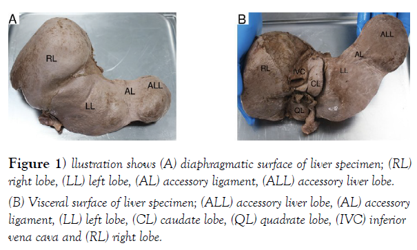
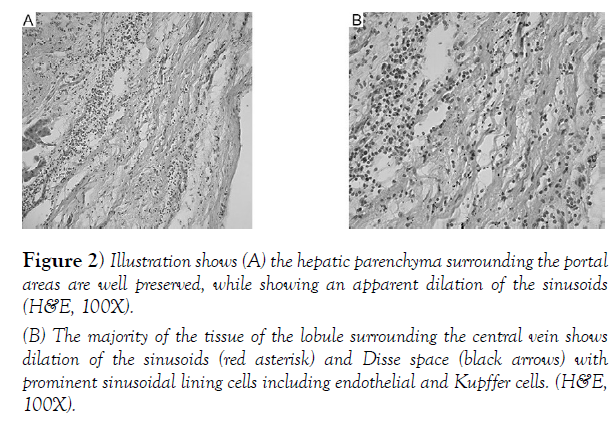
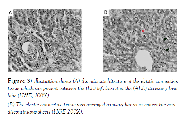
The dissected cadaver showed a left variant (Figure 1A). The variation was present from the left lobe separated by a well-defined fissure. The ALL displayed a ligamentous attachment, accessory ligament (AL) with a flexible conformation alongside the lateral edge of the left lobe. The atypical left sided ALL weighted 1,134 g and measured 8.5 cm in length by 2.3 cm in width. The quadrate lobe, caudate lobe, and fissure by the ligamentum teres were located on the visceral part of the specimen (Figure 1B). Ligamentum teres were found embedded in the substance of the liver on its interior surface. The position and the measurements of the gallbladder were found anatomically normal. The falciform ligament was attached at its average size. The color, consistency, and texture of the atypical left variant ALL or elongated segment were found to be similar to normal anatomy liver parenchyma. Branches of the hepatic artery, portal vein, and a small bile duct were observed in the ALL. Thus, the extrahepatic lobe received the vascularity from the left hepatic artery and left branch of the portal vein. Furthermore, this variation displayed a structural difference from both continuous and ectopic ALL, which had not joined to the liver via a connecting stalk, nor is it separated. For the histological examination of the accessory liver, hepatic tissue from the ALL were used to evaluate the structural organization. The tissue was fixed in 10% neutral buffered formaldehyde, serially sectioned and stained with a hematoxylin–eosin (H&E) stain to confirm the histological sample as an ALL. The histological analysis of the ALL indicated a normal liver lobular architecture composed of polyhedral hepatocytes and sinusoids which are interspersed with central venules and portal tracts. Portal tract contained a portal vein, a branch of the hepatic artery and a bile duct surrounded by loose stromal connective tissue (Figure 2A). The nucleus was centrally located, round and contained one or more nucleoli. The abundant cytoplasm was eosinophilic and contained fine basophilic granules representing rough endoplasmic reticulum. The glycogen components allowed a fine, reticulated, and foamy appearance to the cytoplasm. The darker staining of hepatic parenchyma surrounding the portal areas are well preserved. The hepatic sinusoids separate cords of hepatocytes and are lined by sinusoidal lining cells supported by reticulin fibers. Subsequently, the hepatocytes radiated outwards from a central vein while around the central veins showed sinusoidal dilatation (Figure 2B). Sinusoidal dilatation is a common finding. From a morphological perspective, sinusoidal dilatation is present focally in a biopsy, which is unlikely to be a significant finding. Blood flow studies are necessary to determine the presence of or rule out an obstructive lesion. The structural organization of the AL revealed connective tissue suggesting the appearance of elastic fibers and collagen bundles continued with the capsule of the liver (Figure 3A). The elastic fibers are thinner than collagen fibers and are interspersed among collagen fibers (Figure 3B). Additionally, elastic fibers are responsible for conferring specific properties on the tissue.
Figure 1) llustration shows (A) diaphragmatic surface of liver specimen; (RL) right lobe, (LL) left lobe, (AL) accessory ligament, (ALL) accessory liver lobe.
(B) Visceral surface of liver specimen; (ALL) accessory liver lobe, (AL) accessory ligament, (LL) left lobe, (CL) caudate lobe, (QL) quadrate lobe, (IVC) inferior vena cava and (RL) right lobe.
Figure 2) Illustration shows (A) the hepatic parenchyma surrounding the portal areas are well preserved, while showing an apparent dilation of the sinusoids (H&E, 100X).
(B) The majority of the tissue of the lobule surrounding the central vein shows dilation of the sinusoids (red asterisk) and Disse space (black arrows) with prominent sinusoidal lining cells including endothelial and Kupffer cells. (H&E, 100X).
Furthermore, fibrous tissue was absent, indicating that the elongated parts were not the appendix of the liver. There were no malignant tumor cells. Therefore, the structural organization where the classical hexagonal lobule arrangement of the atypical variant ALL confirmed the presence of tissues suggestive of typical liver architecture. The organs of the abdomen and surrounding structures in the cadaver were anatomically normal.
Discussion
The Accessory liver lobes are typically asymptomatic and are frequently an incidental finding during laparotomy, laparoscopy, or autopsy [9–11]. In the literature, four main types of ALLs are mentioned: (1) an ectopic ALL which attaches to the gallbladder or the intra abdominal ligaments; (2) a microscopic ectopic ALL found in the gallbladder wall; (3) a large continuous ALL attached to the main liver by a stalk, and (4) a small accessory liver lobe (10–30 g in weight) which is attached to the main liver [12 -15]. Instead, this variant is joined by a fibrous ligamentous attachment on the left side.
The ALL in the subject appears to belong to class 4, but the ligamentous attachment seems to differ from previously described sessile or pedunculated attachments. A laparoscopic observational study revealed the incidence of liver anomalies to be 19.3% [11]. In the same study, the accessory liver lobe and ectopic liver incidence was 0.7% [11]. In another laparoscopic series, it was 0.56% [10]. The occurrence of the ALL may suggested to be associated with mesothelial inclusion cysts in an omphalocele [16] and biliary atresia [17]. The dilation of the hepatic sinusoids between the hepatic cords is a common finding observed in patients with obstruction of blood flow at the level of the vena cava or the heart will lead to diffuse changes in the liver parenchyma [18].
Previously, a case of asymptomatic accessory liver lobe attached to the gastrohepatic ligament was reported, which demonstrated an abnormally positioned liver divided into big accessory hepatic lobes (30 g), small accessory hepatic lobes (30 g), ectopic lobes with no hepatic connection and microscopic accessory lobes in the gallbladder wall [19].
Medical literature also describes common normal variants in liver morphology including a horizontal elongation of the lateral segment of the left hepatic lobe, which can extend into the left upper abdominal quadrant and eventually wrap around the splenic contour. Incidentally, these tend to be more usual in females [20]. In this report, the atypical ALL was found on the left lobe in an elderly female cadaver. At the left end of the adult left lobe, a fibrous band (fibrous appendix of the liver) may also appear as an atrophied remnant of the more extensive part of the left lobe found in children, but this was not observed in this specimen.
Given recent advances in imaging methods, medical literature recommends increasing the number of studies assessing variation in vivo, a technique that aided previous quantitative studies seeking to establish ALL prevalence in other populations [10-17]. Knowledge of the incidence of accessory liver lobes is of clinical importance during the diagnosis and treatment of liver tumors and intrathoracic mass [15-17,19]. However, to reach a definitive diagnosis, histopathological examination is necessary to demonstrate the normal hepatic parenchyma tissue [2]. Frequently ALL are anatomical variations without any clinical consequences other than as a source of confusion with tumors or the exceptional torsion of a pedunculated form. Knowledge of the incidence of accessory liver lobes is of clinical importance during the diagnosis and treatment of liver tumors and intrathoracic mass [15-17,19]. Surgeons generally do not realize how to identify accessory or ectopic liver lobes before surgery or autopsy using imaging studies, possibly due to the absence of symptoms, and the small size of structures. In case of complications or after diagnosis of pedunculated liver tumors, laparoscopy is well indicated for the removal of these lobes or tumors [21]. Subsequently, for liver transplantation, anatomical variants concerning liver lobes can be accommodates without donor complications or complex reconstructions [22]. Surgeons generally do not realize how to identify accessory or ectopic liver lobes before surgery or autopsy using imaging studies, possibly due to the absence of symptoms, and the small size of structures. The extrahepatic mass can be detected by hepatobiliary iminodiacetic acid scan, ultrasonography, and if possible dynamic dual- or triple-phase helical computerized tomography. A percutaneous imaging-guided biopsy may also be the best option to reveal the normal liver parenchyma [23].
Author Contributions
J.Q.R, R.A., Writing - original draft, conceived and designed the study, conducted research, provided research materials, collected data, and wrote the initial draft of the article. All authors have critically reviewed and approved the final draft and are responsible for the content and similarity index of the manuscript.
I.S., Formal analysis, analyzed and interpreted data and wrote final draft of article and provided logistic support. All authors have critically reviewed and approved the final draft and are responsible for the content and similarity index of the manuscript.
Acknowledgements
We acknowledge Dr. Astrid Zayas (UCC-SoM) for providing reagents, assistance for histochemistry and critical feedback. We thank Dr. Maryví González-Solá (Texas Women’s University), Dr. Catalina Villamil (UCC – School of Chiropractic), Dr. Thomas Schikorski (UCC – School of Medicine), Dr. Sofía Jiménez-Dietsch (UCC – School of Medicine) and Dr. Saqib Mansoor (Bakhtawar Ameen Medical and Dental College) for providing critical feedback. In addition, we would like to thank to the Graduate Program in Biomedical Sciences for providing the funding for this article. Most importantly, we would like to recognize and be grateful for the people who donated their bodies and their families for making this study possible.
REFERENCES
- Drake RL, Vogl WM, Gray H, et al. In Gray's anatomy for students. Elsevier 4th Edition: 2020;328–30.
- Wang C. Accessory lobes of the liver: A report of 3 cases and review of the literature. Intractable & Rare Diseases Research. 2012;86-1.
- Glenisson M, Salloum C, Lim C, et al. Accessory liver lobes: Anatomical description and clinical implications. Journal of Visceral Surgery. 2014;151(6):451-55.
- Savopoulos C, Kakaletsis N, Kaiafa G, et al. Riedel's lobe of the liver: a case report. Medicine. 2015;94(3):430.
- Fogh J, Tromholt N, Jørgensen F. Persistent impairment of liver function caused by a pendulated accessory liver lobe. Eur J Nucl Med. 1989;15: 326-327.
- Jambhekar K, Pandey T, Kaushik C, et al. Intermittent torsion of accessory hepatic lobe: An unusual cause of recurrent right upper quadrant pain. Indian J. Radiol. 2010;20(2):135–137.
- Pujari BD, Deodhare SG. Symptomatic accessory lobe of liver with a review of the literature. Postgraduate medical journal.1976;52(606):234–36.
- Tancredi A, Cuttitta A, de Martino D, et al. Ectopic hepatic tissue misdiagnosed as a tumor of lung. Updates In Surgery. 2010;62(2):121-23.
- Baruah P, Choudhury PR. Tongue-like elongation of the left lobe of liver. OA Case Reports. 2013;2(17):161.
- Caygill CP, Gatenby PA. Ectopic liver and hepatocarcinogenesis. Eur J Gastroenterol Hepatol. 2004;16(8):727–29.
- Serdar Han, Lutfi Soylu. Accessory Liver Lobe in the Left Thoracic Cavity. Ann Thorac Surg. 2009;87(6):1933-34.
- Al-Anwar S, Askari A, Alvi A. Right Iliac fossa pain due to torsion and ischemia of accessory liver lobe. J. Surg. Case Rep. 2019;5.
- Nayak SB, Kumar N, Sirasanagandla SR,et al. A mini accessory liver lobe in the fissure for ligamentum teres and its clinical significance: a case report. J. clin. diagn. 2013;7(11):2573–2574.
- Ravindra Kumar Boddeti, Subhadra Devi Velichety, Usha Rani U. Accessory left lobe of the liver. Int J Intg Med Sci. 2014;1(1):17-19.
- Caygill CP, Gatenby PA. Ectopic liver and hepatocarcinogenesis. Eur J Gastroenterol Hepatol. 2004;16(8):727–29.
- Rougemont AL, Sartelet H, Oligny LL, et al. Accessory liver lobe with mesothelial inclusion cysts in an omphalocele: a new malformative association. Pediatr Dev Pathol. 2007;10:224–228
- Pereira RM, Simo ̃es E, Silva AC, et al. Successful management of concomitant omphalocele, accessory hepatic lobe, and biliary atresia in a 44-day-old boy. J Pediatr Surg. 2005;40:21–E 24
- Nakhleh RE. Pathology and differential diagnosis of hepatic venous outflow impairment. Clin Liver Dis. 2017;10:49–52.
- Massaro M, Valencia MP, Guzman M, et al. Accessory hepatic lobe mimicking an intra-abdominal tumor. J Comput Assist Tomogr. 2007; 31:572–573
- Haaga JR, Dogra VS, Forsting M, et al. CT and MRI of the whole body in gastrointestinal imaging. Philadelphia: Mosby Elsevier; 2009;5(2)1458.
- Glenisson M, Salloum C, Lim C, et al. Accessory liver lobes: Anatomical description and clinical implications. J. Visc. Surg. 2014;151(6):451–55.
- Marcos A, Ham JM, Fisher RA, et al. Surgical Management of Anatomical Variations of the Right Lobe in Living Donor Liver Transplantation. Ann Surg. 2000;231:824–31.
- Hierro CR, Rueda FV, Cruz VV, et al. Focal nodular hyperplasia on accessory lobe of the liver: Preoperative diagnosis and management. J Pediatr Surg. 2013;48:251–54.


