A Didelphys Uterus with twins - A very rare reproductive system anatomical variation: Case Report Tanzania
2 Department of Surgery, Faculty of Medicine, Saint Francis University College of Health and Allied Sciences, P.O. BOX 175, Ifakara, Tanzania
3 Department of Obstetrics and Gynecology, Faculty of Medicine, Saint Francis University College of Health and Allied Sciences, P.O. BOX 175, Ifakara, Tanzania
Received: 06-Oct-2023, Manuscript No. ijav-23-6794; Editor assigned: 09-Oct-2023, Pre QC No. ijav-23-6794; Accepted Date: Oct 30, 2023; Reviewed: 23-Oct-2023 QC No. ijav-23-6794; Revised: 28-Oct-2023, Manuscript No. ijav-23-6794; Published: 31-Oct-2023, DOI: 10.37532/1308-4038.16(10).310
This open-access article is distributed under the terms of the Creative Commons Attribution Non-Commercial License (CC BY-NC) (http://creativecommons.org/licenses/by-nc/4.0/), which permits reuse, distribution and reproduction of the article, provided that the original work is properly cited and the reuse is restricted to noncommercial purposes. For commercial reuse, contact reprints@pulsus.com
Abstract
Uterine Didelphys (UD) is one of the rare congenital condition which may be associated with significant complications during pregnant and during labor. It rarely affects infertility and generally is asymptomatic. 32 years female reported below was incidental diagnosed during caesarian section and was found to have twins in separate UD. Serial sonography simplify the diagnosis but with limitation in primary health centers in low and middle income countries.
Keywords
Mullerian duct anomalies; Infertility; Sonography; Twin
List of abbreviation
ANC Ante Natal Clinic
AN visit Ante Natal Visit
APGAR Appearance, Pulse, Grimace, Activity, and Respiration
CVD Cervical Vertex Delivery
CS Caesarian Section GA Gestational Age
MD Mullerian duct anomalies
UD Uterus Didelphys
INTRODUCTION
Uterus Didelphys (UD) is a condition of uterine fusion defect characterized by failure to fuse resulting the individual to have two hemi uterus and cervices [1 ]. It constitutes approximately 5% of the Mullerian duct anomalies (MD) [1 , 2]. It is a lateral fusion defect of the MD [3, 4]. Incomplete fusion of Müller’s ducts occurs between 12 and 16 weeks of fetal life [5]. UD is associated with miscarriage, premature labor, and premature rupture of the membranes, obstructed labor and malpresentations [3 , 6]. It is a rare uterine anomaly occurring in 0.1%-0.5% of healthy fertile population [1 ]. However, most of these congenital anomalies are undiagnosed or unrecognized [1 , 4]. Most of pregnant mother with UDs are singleton whereby twin deliveries occur 1:1,000,000 [6]. The mother with UD is usually diagnosed during ANC visit which may be uneasy in some areas especially in Low and middle income areas due to limited availability of ultrasound. Herein, we report a 32 years female case that was detected to have UD during CS. The aim of this case report is to increase a building capacity among health workers at both tertiary and primary health centers such that all risks of complex deliveries should be excluded and put into considerations to prevent the possibility of unfavorable outcomes
CASE REPORT
A 32 years female gravid 4, GA at 36/40 Para 3 by cervical vertex deliveries (CVD) living 3, who came at health center presenting with pelvic pain. She denied any history of dyspareunia, or abortion. She denied also medical condition before and during recent pregnancy but rather she was given antimalaria prophylaxis as Tanzania lutein for malaria in pregnancy prevention.
The review of other systems was found to be uneventful. On physical exam, the patient was found to have normal vital signs, while the gravid uterus was extended 39 cm and fetal movements were normal with good activity, no any other pregnant related physical anomalies were detected such as conjunctiva paleness or lower extremity edema. The patient was admitted in labor ward with diagnosis of G4P3L3 at GA
36/40 at term with labor pain. The patography was initiated to monitor labor progression. At admission cervical dilatation was 4cm with minimal uterine contraction, while fetal head descend and fetal lie were not indicated on the patography, four hours later there were no change. However, after 8 hours the mother reported as reduced abdominal pain and fetal movements. Abdominal assessment noted absence of contraction, by the use of a fetoscope, there was increased fetal heart rate (166min). The patient was planned for emergence caesarian section with diagnosis of poor progress of labor and fetal distress. The patient was prepared for emergence caesarian.
DISCUSSION
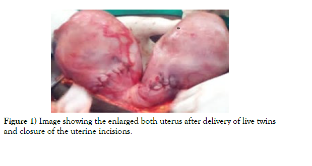
Congenital uterine malformation results from defects in lateral fusion [1 ,3 ,5] UD is a rare case of uterine malformation associated with two complete uterus [1 ,3 ,7 ] The pregnant mother with UD is on high risk for unfavorable pregnancy outcomes such as preterm labor, fetal malpresentations, intrauterine fetal death, uterine rupture and even perinatal mortality [1 -3 ,5,6]. UD may not be suspected before although some women with UD experience dyspareunia as a result of a vaginal septum [5] the condition which was uneventful to our patient. During child birth, women with UD have successful full-term pregnancies [1 , 7 ]. However, they still belong to a high-risked group since may have breech presentation in both multiple pregnancy and single tone pregnant. In the later, non-pregnant uterus may block the pelvic inlet causing obstructed labor [3 ] while in the former there may be difficult of fetal descend simultaneously in distinct uterus as it happened in our case. Usually it is unilateral pregnancy with few cases of twins in UD [6] including our case. In contrast to our case, the patient with UD may be detected during examination through presence of vaginal septum which normally is present in complete uterine separation [1 , 5]. Very few cases are reported to have missing the vaginal septum [3] including our case, failure to detect it probably could be due to the lack of knowledge about the vaginal anatomy among the health workers who were examining the patient in previous and late pregnancies. Patients with uterus didelphys belong to a high risk group and deserve meticulous prenatal care [2]. Therefore, serial sonograms are necessary to evaluate fetal well-being and growth, and hence prediction of safe delivery. In our case it was difficult to do sonography during AN visit due to limitation of availability of ultrasound machines and sonographers in health centers at our health care protocols. This limit the probability of early diagnosis of hidden maternal risk factor of this kind. The UDs are not a direct cause of infertility but may be associated with high risks for labor complications [1 -3 , 5, 6] and hence during ANC visit could be important to do all investigation to exclude any probability of maternal congenital anomaly which can be fatal to both baby and the mother especially during delivery (Figure 1).
CONCLUSION
The scant clinical signs of uterine didelphys in limited availability of sonographic imaging in remote rural health centers limit early diagnosis of this rare obstetric condition. From this case shows that, UD is rarely associated with infertility, but also safe and normal deliveries do occur. However the clinicians should be aware on the rare cases since may cause fatal complications during any mode of deliveries. Whenever possible, diagnostic imaging should be done to all pregnant women as it is recommended to be a diagnostic tool.
ETHICAL APPROVAL
Institutional and international research regulations were observed during interacting with the patient and the preparation of the manuscript.
CONSENT
A written informed consent was obtained from the patient for the case details to be published, and it has been kept by the corresponding author.
CONFLICTS OF INTEREST
The authors declare that there are no conflicts of interest regarding the publication of this article.
ACKNOWLEDGEMENT
We thank medical in charge at Rupilo health center who gave details of the patient clinical information.
REFERENCES
- Ravikanth R. Case Report Bicornuate Uterus with Pregnancy. 2017; 2–51.
- Cassar OA, Dalli SM. Pregnancy in a bicornuate uterus with contraceptive coil in situ - a case report. 2017; 7: 52–249.
- Paknejad O, Bryant D, Peterkin C, Wilcox W. Uterine Didelphys in a Pregnant Mother. 2018; 30–1423.
- Agarwal I, Tayade S, Sharma S. A Case Report of a Normal Pregnancy in a Bicornuate Uterus through In Vitro Fertilization Case Presentation. 2022; 14(9).
- Borzyszkowska D, Golara A, Tuczy N, Kozłowski M, Cwiertnia A, et al. The Impact of Uterus Didelphys on Fertility and Pregnancy. 2022.
- K RM, Saheb W, Jarjour I, El-tal R, Meita Z, et al. Delayed-interval-delivery of twins in didelphys uterus complicated with chorioamnionitis : a case report and a brief review of literature. J Matern Neonatal Med. 2020; 1–5.
- Ross C, El-hassan H, Lakasing L. Uterus Didelphys : Two pregnancies two term breech caesarean deliveries. 2018; 9–2017.
Indexed at, Google Scholar, Crossref
Indexed at, Google Scholar, Crossref
Indexed at, Google Scholar, Crossref
Indexed at, Google Scholar, Crossref
Indexed at, Google Scholar, Crossref


