A duplex kidney with dromedary hump showing altered hilar anatomy
Sreekanth Tallapaneni* and Mahesh Vemarapu
Department of Anatomy, Shadan Institute of Medical Sciences Teaching Hospital & Research Centre, Hyderabad, Andhra Pradesh, India.
- *Corresponding Author:
- Sreekanth Tallapaneni, MD
Assistant Professor, Department of Anatomy, Shadan Institute of Medical Sciences, Teaching Hospital & Research Centre, Hyderabad, Andhra Pradesh, India.
Tel: +91 40 23345429
E-mail: anatomysreekanth18@yahoo.com
Date of Received: July 17th, 2010
Date of Accepted: December 19th, 2010
Published Online: February 22nd, 2011
© IJAV. 2011; 4: 34–36.
[ft_below_content] =>Keywords
double pelvis,lobulations,segmental branches
Introduction
The kidneys are paired reddish brown retroperitoneal organs lying in the paravertebral gutters of posterior abdominal wall. Multiple lobulations are seen throughout the fetal life [1]. Most of them disappear during the first year of birth but varying degrees of lobulations may persist in the adult life It is not unusual to see a focal bulge in the mid lateral contour of the kidney, refered to as dromedary hump. It is a normal variation occurring due to the downward pressure of spleen on the left side [2]. The renal vascular pedicle classically described as single artery, larger vein lying anterior to the artery entering via the hilum. Behind them lies the renal pelvis, which is the collecting system of the kidney. The renal artery typically divides into a more constant posterior division and anterior division. The anterior division shows apical, upper, middle, and lower anterior segmental branches. All the segmental arteries are end arteries supplying their respective segments. A duplex kidney is one that has two separate pelvicalyceal systems. It has an upper pole and lower pole the ureters may join at any point. If they join at level of uretero-pelvic junction the configuration is termed bifid system. If the urters join more distally or proximal to the bladder called bifid ureters [2].
The kidney and ureter develop in the mesoderm on the dorsal wall of the coelomic cavity. The distal part of the duct of the transient pronephros receives tubules of mesonephros to develop and termed the mesonephric duct. A diverticulum appears at lower end of mesonephric duct, which develops into the metanephric duct or ureteric bud. On the the top of the ureteric bud a cap of tissue differentiates to form the metanephros which develops into the definitive kidney [3]. The ureteric bud arises from the mesonephric duct around fifth week of intrauterine life [4]. The branching and growth of the ureteric bud is stimulated by the glial derived neurotropic factor (GDNF) and scatter factor (hepatocyte growth factor) which are produced by the transcription factor WT1 expressed by the mesenchyme. The tyrosine kinase receptors RET, for GDNF & MET, for HGF, are synthesized by epithelium of ureteric buds establishing signaling pathways between the two tissues [5]. The ureteric bud forms the ureter which dialates at the upper end to form the major calyces and in turn divides to form the minor calyces and collecting tubules [6].
Case Report
The altered hilar anatomy with double pelvis was observed in the left duplex kidney of a 55-year-old female cadaver during the routine dissection hours for first year M.B.B.S. students at the Department of Anatomy, Shadan Institute of Medical Sciences, Teaching Hospital & Research Centre, Hyderabad, N.T.R. University, India.
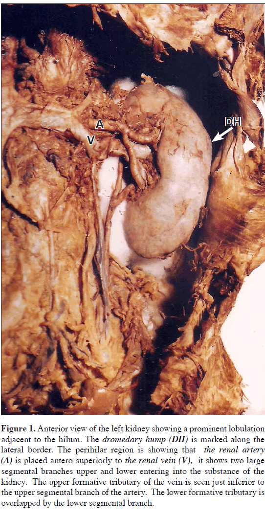
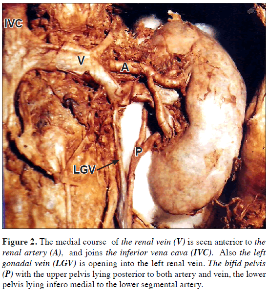
The specimen was a lobulated duplex left kidney, which showed prominent lobulation on the anterior surface just adjacent to the hilar region, also the mid-lateral portion of the convex lateral border showed a small focal bulge – the dromedary hump (Figure 1). The perihilar region showed that the renal vasculature was reversed in its antero-posterior disposition (Figure 1). The renal artery was the 1st structure encountered with two anterior segmental branches, the upper branch and lower branch (Figure 1). The renal vein which was seen behind the artery was formed by two tributaries as superior and inferior. They were arising from the hilum and running towards the inferior vena cava. A left gonadal vein was seen joining the left renal vein (Figure 2). The superior AVPIVCLGVformative tributary is seen clearly, lying just inferior to the upper segmental arterial branch. The inferior formative tributary is seen overlapped by the lower segmental anterior branch of renal artery. When viewed medially, after merging with each other the renal vein was seen larger and was lying anterior to the renal artery and overlapping it. The double pelvis with its upper component is seen lying posterior to both artery and vein. The lower pelvis is seen infero-medial to the lower segmental artery (Figure 2). Both pelvices were uniting to form the ureter, medial and just below the hilum the ureter extended downwards and opened in to the bladder as usual.
Figure 1: Anterior view of the left kidney showing a prominent lobulation adjacent to the hilum. The dromedary hump (DH) is marked along the lateral border. The perihilar region is showing that the renal artery (A) is placed antero-superiorly to the renal vein (V), it shows two large segmental branches upper and lower entering into the substance of the kidney. The upper formative tributary of the vein is seen just inferior to the upper segmental branch of the artery. The lower formative tributary is overlapped by the lower segmental branch.
Figure 2: The medial course of the renal vein (V) is seen anterior to the renal artery (A), and joins the inferior vena cava (IVC). Also the left gonadal vein (LGV) is opening into the left renal vein. The bifid pelvis (P) with the upper pelvis lying posterior to both artery and vein, the lower pelvis lying infero medial to the lower segmental artery.
Discussion
In the present case, two lobulations were seen one on the anterior surface adjacent to the hilum extending laterally, and the other was a small focal bulge on the lateral border –the dromedary hump– which is a normal variation. The persistent fetal lobulations must be distinguished from pathological scarring by the site of irregularity in the renal surface. In fetal lobulation the divisions lie between calyces where as in the cortical scarring the loss of cortex overlies a calyx. Also the lobulations cause foldings of the column of cortex demonstrable in enhanced CT images. The dimercapto succinic acid scintigram (DMSA) is also used to investigate the renal scarring [3]. A duplex ureter is a common variation of kidneys, which is more common in females than males having prevalence of about 2%. Of subjects with duplication anomaly, 20% have bilateral abnormalities. In the present case, it was an isolated double pelvis without any other associated anomaly. Mostly such anomalies go unnoticed. An unilateral isolated bifid ureter on right side in a female with two limbs of ureter had their respective pelves coming out as separate entities from the hilus of the right kidney is being reported by Das S [4]. Double pelvis is usually unilateral and it is the cause of premature division of the ureteric bud near its termination [5]. The bifid ureter is reported to be more common on the right side by Rege [7]. In the present case, it was seen on the left side. Nevertheless complications including frequent urinary tract infection and the occurrence of calculi were reported by Giannokopoulos [8] and ureteric stenosis by Busslinger [9]. Bifid pelvis/bifid ureters are usually detected as incidental findings at autopsy or if symptomatic detected during routine investigation such as intravenous pyelography (IVP),ultrasound, computed tomography, magnetic resonance. In the present case two segmental arteries (upper and lower) were seen in the perihilar region. Each renal artery has about segmental branches that is apical upper middle and lower segmental branches from the anterior division. The antero-posterior disposition of the vein artery and pelvis was seen. The perihilar branching of the main renal artery is highly inconsistent and antero-posterior disposition also can be reversed. The awareness of all possible variations is essential for urologists when considering implantation of stent in the renal arteries [10] and radiologists interpreting renal angiograms.
References
- Williams PL, Bannister LH, Berry MM, Collins P, Dyson M, Dussek JE, Ferguson MWJ. Gray’s Anatomy. 38th Ed., Edinburgh, Churchill Livingstone. 1995; 1815–1836.
- Walsh PC, Retik AB, Vaughan ED Jr., Wein AJ, Kavoussi LR, Novick AC, Partin AW, Peters CA. Campbell’s Urology. 8th Ed., 2002; Vol.1; 20–21, 25–28; Vol.3; 2007.
- Butler P, Mitchell AWM, Ellis H. Applied Radiological Anatomy. 2007; 259–267.
- Das S, Dhar P, Mehra RD. Unilateral isolated bifid ureter – a case report. J Anat Soc India. 2001; 50: 43–44.
- Saddler TW. Langman’s Medical Embryology. 11th Ed., Lippincott; Williams & Wilkins. 2009; 239.
- Snell RS. Clinical Anatomy. 7th Ed., Lippincott Williams & Wilkins. 2003; 286–289.
- Rege VM, Deshmukh SS, Borwankar SS, Gandhi RK. Blind ending bifid ureter (a case report). J Postgrad Med. 1986; 32: 233–235.
- Giannakopoulos X, Chambilomatis P, Thirothoulakis M, Seferiadis G. The blind-ending bifid ureter. Int Urol Nephrol. 1994; 26: 161–165.
- Busslinger MI, Kaiser G. Surgical significance of the duplex kidney with bifid ureter. Eur J Pediatr Surg. 1992; 2: 150–151.
- Garcier JM, De Fraissinette B, Filaire M, Gayard P, Therre T, Ravel A, Boyer L. Origin and initial course of the renal arteries: a radiological study. Surg Radiol Anat. 2001; 23: 51–55.
Sreekanth Tallapaneni* and Mahesh Vemarapu
Department of Anatomy, Shadan Institute of Medical Sciences Teaching Hospital & Research Centre, Hyderabad, Andhra Pradesh, India.
- *Corresponding Author:
- Sreekanth Tallapaneni, MD
Assistant Professor, Department of Anatomy, Shadan Institute of Medical Sciences, Teaching Hospital & Research Centre, Hyderabad, Andhra Pradesh, India.
Tel: +91 40 23345429
E-mail: anatomysreekanth18@yahoo.com
Date of Received: July 17th, 2010
Date of Accepted: December 19th, 2010
Published Online: February 22nd, 2011
© IJAV. 2011; 4: 34–36.
Abstract
A comprehensive knowledge of the wide range of variations of renal vasculature and renal pelvis is mandatory to the anatomists for a better understanding of the embryology. It remains as the key issue in determining the technical feasibility of various endourologic procedures and innumerable intervention techniques besides kidney retrievals for transplantation. In the present case the duplex kidney showed lobulations on the anterior surface just adjacent to the hilar region. The midlateral portion of the convex lateral border of the kidney showed a small focal bulge –dromedary hump. At the hilum reversed anterio-posterior disposition of renal vasculature with anteriorly placed renal artery which bifurcated into two upper and lower anterior segmental branches. The renal vein formed by large tributaries arising from the hilum running towards the inferior vena cava. The renal pelvis was most posteriorly placed which showed a double pelvis. The upper pelvis was seen arising behind the renal vein and the lower pelvis arising inferomedial to the lower anterior segmental artery. Both the pelvises were seen uniting medial to the lower part of hilum and continued as a single ureter which opened into the bladder. The thorough knowledge of these anatomical variations is necessary to avoid iatrogenic injuries and enable the surgeon and radiologists approach unusual situations with confidence rather than surprise.
-Keywords
double pelvis,lobulations,segmental branches
Introduction
The kidneys are paired reddish brown retroperitoneal organs lying in the paravertebral gutters of posterior abdominal wall. Multiple lobulations are seen throughout the fetal life [1]. Most of them disappear during the first year of birth but varying degrees of lobulations may persist in the adult life It is not unusual to see a focal bulge in the mid lateral contour of the kidney, refered to as dromedary hump. It is a normal variation occurring due to the downward pressure of spleen on the left side [2]. The renal vascular pedicle classically described as single artery, larger vein lying anterior to the artery entering via the hilum. Behind them lies the renal pelvis, which is the collecting system of the kidney. The renal artery typically divides into a more constant posterior division and anterior division. The anterior division shows apical, upper, middle, and lower anterior segmental branches. All the segmental arteries are end arteries supplying their respective segments. A duplex kidney is one that has two separate pelvicalyceal systems. It has an upper pole and lower pole the ureters may join at any point. If they join at level of uretero-pelvic junction the configuration is termed bifid system. If the urters join more distally or proximal to the bladder called bifid ureters [2].
The kidney and ureter develop in the mesoderm on the dorsal wall of the coelomic cavity. The distal part of the duct of the transient pronephros receives tubules of mesonephros to develop and termed the mesonephric duct. A diverticulum appears at lower end of mesonephric duct, which develops into the metanephric duct or ureteric bud. On the the top of the ureteric bud a cap of tissue differentiates to form the metanephros which develops into the definitive kidney [3]. The ureteric bud arises from the mesonephric duct around fifth week of intrauterine life [4]. The branching and growth of the ureteric bud is stimulated by the glial derived neurotropic factor (GDNF) and scatter factor (hepatocyte growth factor) which are produced by the transcription factor WT1 expressed by the mesenchyme. The tyrosine kinase receptors RET, for GDNF & MET, for HGF, are synthesized by epithelium of ureteric buds establishing signaling pathways between the two tissues [5]. The ureteric bud forms the ureter which dialates at the upper end to form the major calyces and in turn divides to form the minor calyces and collecting tubules [6].
Case Report
The altered hilar anatomy with double pelvis was observed in the left duplex kidney of a 55-year-old female cadaver during the routine dissection hours for first year M.B.B.S. students at the Department of Anatomy, Shadan Institute of Medical Sciences, Teaching Hospital & Research Centre, Hyderabad, N.T.R. University, India.
The specimen was a lobulated duplex left kidney, which showed prominent lobulation on the anterior surface just adjacent to the hilar region, also the mid-lateral portion of the convex lateral border showed a small focal bulge – the dromedary hump (Figure 1). The perihilar region showed that the renal vasculature was reversed in its antero-posterior disposition (Figure 1). The renal artery was the 1st structure encountered with two anterior segmental branches, the upper branch and lower branch (Figure 1). The renal vein which was seen behind the artery was formed by two tributaries as superior and inferior. They were arising from the hilum and running towards the inferior vena cava. A left gonadal vein was seen joining the left renal vein (Figure 2). The superior AVPIVCLGVformative tributary is seen clearly, lying just inferior to the upper segmental arterial branch. The inferior formative tributary is seen overlapped by the lower segmental anterior branch of renal artery. When viewed medially, after merging with each other the renal vein was seen larger and was lying anterior to the renal artery and overlapping it. The double pelvis with its upper component is seen lying posterior to both artery and vein. The lower pelvis is seen infero-medial to the lower segmental artery (Figure 2). Both pelvices were uniting to form the ureter, medial and just below the hilum the ureter extended downwards and opened in to the bladder as usual.
Figure 1: Anterior view of the left kidney showing a prominent lobulation adjacent to the hilum. The dromedary hump (DH) is marked along the lateral border. The perihilar region is showing that the renal artery (A) is placed antero-superiorly to the renal vein (V), it shows two large segmental branches upper and lower entering into the substance of the kidney. The upper formative tributary of the vein is seen just inferior to the upper segmental branch of the artery. The lower formative tributary is overlapped by the lower segmental branch.
Figure 2: The medial course of the renal vein (V) is seen anterior to the renal artery (A), and joins the inferior vena cava (IVC). Also the left gonadal vein (LGV) is opening into the left renal vein. The bifid pelvis (P) with the upper pelvis lying posterior to both artery and vein, the lower pelvis lying infero medial to the lower segmental artery.
Discussion
In the present case, two lobulations were seen one on the anterior surface adjacent to the hilum extending laterally, and the other was a small focal bulge on the lateral border –the dromedary hump– which is a normal variation. The persistent fetal lobulations must be distinguished from pathological scarring by the site of irregularity in the renal surface. In fetal lobulation the divisions lie between calyces where as in the cortical scarring the loss of cortex overlies a calyx. Also the lobulations cause foldings of the column of cortex demonstrable in enhanced CT images. The dimercapto succinic acid scintigram (DMSA) is also used to investigate the renal scarring [3]. A duplex ureter is a common variation of kidneys, which is more common in females than males having prevalence of about 2%. Of subjects with duplication anomaly, 20% have bilateral abnormalities. In the present case, it was an isolated double pelvis without any other associated anomaly. Mostly such anomalies go unnoticed. An unilateral isolated bifid ureter on right side in a female with two limbs of ureter had their respective pelves coming out as separate entities from the hilus of the right kidney is being reported by Das S [4]. Double pelvis is usually unilateral and it is the cause of premature division of the ureteric bud near its termination [5]. The bifid ureter is reported to be more common on the right side by Rege [7]. In the present case, it was seen on the left side. Nevertheless complications including frequent urinary tract infection and the occurrence of calculi were reported by Giannokopoulos [8] and ureteric stenosis by Busslinger [9]. Bifid pelvis/bifid ureters are usually detected as incidental findings at autopsy or if symptomatic detected during routine investigation such as intravenous pyelography (IVP),ultrasound, computed tomography, magnetic resonance. In the present case two segmental arteries (upper and lower) were seen in the perihilar region. Each renal artery has about segmental branches that is apical upper middle and lower segmental branches from the anterior division. The antero-posterior disposition of the vein artery and pelvis was seen. The perihilar branching of the main renal artery is highly inconsistent and antero-posterior disposition also can be reversed. The awareness of all possible variations is essential for urologists when considering implantation of stent in the renal arteries [10] and radiologists interpreting renal angiograms.
References
- Williams PL, Bannister LH, Berry MM, Collins P, Dyson M, Dussek JE, Ferguson MWJ. Gray’s Anatomy. 38th Ed., Edinburgh, Churchill Livingstone. 1995; 1815–1836.
- Walsh PC, Retik AB, Vaughan ED Jr., Wein AJ, Kavoussi LR, Novick AC, Partin AW, Peters CA. Campbell’s Urology. 8th Ed., 2002; Vol.1; 20–21, 25–28; Vol.3; 2007.
- Butler P, Mitchell AWM, Ellis H. Applied Radiological Anatomy. 2007; 259–267.
- Das S, Dhar P, Mehra RD. Unilateral isolated bifid ureter – a case report. J Anat Soc India. 2001; 50: 43–44.
- Saddler TW. Langman’s Medical Embryology. 11th Ed., Lippincott; Williams & Wilkins. 2009; 239.
- Snell RS. Clinical Anatomy. 7th Ed., Lippincott Williams & Wilkins. 2003; 286–289.
- Rege VM, Deshmukh SS, Borwankar SS, Gandhi RK. Blind ending bifid ureter (a case report). J Postgrad Med. 1986; 32: 233–235.
- Giannakopoulos X, Chambilomatis P, Thirothoulakis M, Seferiadis G. The blind-ending bifid ureter. Int Urol Nephrol. 1994; 26: 161–165.
- Busslinger MI, Kaiser G. Surgical significance of the duplex kidney with bifid ureter. Eur J Pediatr Surg. 1992; 2: 150–151.
- Garcier JM, De Fraissinette B, Filaire M, Gayard P, Therre T, Ravel A, Boyer L. Origin and initial course of the renal arteries: a radiological study. Surg Radiol Anat. 2001; 23: 51–55.


