A duplicate obturator foramen – a report of rare variation
Nitin Radhakishan Mudiraj* and Prakash Laxman Jahagirdar
Department of Anatomy, Bharati Vidyapeeth Deemed University, Medical College and Hospital, Sangli, Maharashtra, India
- *Corresponding Author:
- Nitin Radhakishan Mudiraj
Associate Professor, Department of Anatomy, Bharati Vidyapeeth Deemed University, Medical College and Hospital, Sangli, Maharashtra, India
Tel: +91 942 1858613
E-mail: dr.nitinmudiraj09@gmail.com
Date of Received: December 3rd, 2010
Date of Accepted: August 5th, 2011
Published Online: September 14th, 2011
© Int J Anat Var (IJAV). 2011; 4: 155–156.
[ft_below_content] =>Keywords
obturator foramen, obturator membrane, iliac foramen, transobturator sling, pelvic osteotomy
Introduction
The obturator foramen is a large opening in the hip bone. It is bounded superiorly by the obturator surface of the pubic body and the obturator groove; inferiorly by the ischial and inferior pubic rami; anteriorly by the superior and inferior pubic rami; and posteriorly by the ischial ramus.
Materials and Methods
The obturator foramen is almost closed by obturator membrane which is attached to its margins, except above near obturator groove, where communication remains between the pelvis and thigh (obturator canal); this free edge is attached to an anterior obturator tubercle at the anterior end of the inferior border of the superior pubic ramus and a posterior obturator tubercle on the anterior border of acetabular notch. The contents of the obturator canal include nerve to the obturator externus and obturator artery, (superomedially), obturator vein (inferomedially), and anterior and posterior divisions of the obturator nerve (superolaterally), which lay within the obturator groove [1].
Knowledge and awareness of variations in the foramina associated with the obturator foramen may be of immense clinical importance to surgeons and radiologists.
Case Report
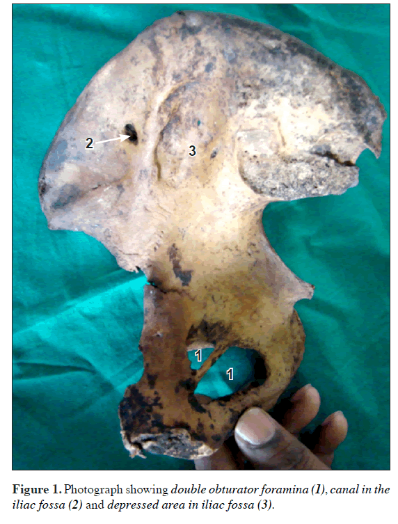
We found the variant of obturator foramen in a young right hip bone. Large and oval obturator foramen and everted conjoint ischiopubic rami confirmed the male sex. Its anatomical features were studied in detail by examination of the lateral and pelvic surfaces and appropriate morphometric measurements were taken. Obturator canal was closed by a plate of bone inferiorly to form a complete opening. The plate of bone surrounding the obturator canal could be traced to the superior pubic ramus. The maximum transverse and vertical dimensions of the opening measured 1.6 cm and 1.7 cm, respectively. In the same innominate bone, canal was seen in the iliac fossa 6.0 cm from anterior superior iliac spine and 2.0 cm from iliac crest. In addition to that, there was large depressed area close to the medial border of ilium.
Discussion
Conversion of obturator groove into bony foramen has been reported [2]. Karantanas et al. found a case of a double obturator foramen, which had been detected in x-ray film [3]. A triplicate obturator foramina was also reported by Das et al. [4]. The presence of a plate of bone in the obturator foramen may lead to compression of the nerves and blood vessels with neurological and vascular effects. The literature available on morphology of the obturator foramen and associated openings is less. The obturator foramen has been studied in detail to find out the potential risks to the dorsal nerve of the penis and to the obturator canal when different slings are used [5]. Researchers have also studied the obturator region to analyze the relationships of the trans-obturator sling and anatomical structures within the obturator region [6]. The obturator region has also been used for cystocele repair by a synthetic vaginal mesh, which is secured anteriorly through the obturator foramen [7]. This region is also a route for the management of short-pedicled undescended testicle [8].
Pelvic osteotomy also requires prior anatomical knowledge of the obturator region. In the light of the above facts, the importance of the obturator foramen cannot be overlooked in clinical practice.
Such a rare variation may be due to ossification of upper margin of obturator membrane. Second author in his experience of more than four decades in anatomy has found this type of variant for the first time. The exact cause for this ossification is not clearly understood but possibly could be due to unusual obturator vessels.
The canal seen in the iliac fossa could be for communication of an external pelvic vein with an internal pelvic vein [2].
Detailed knowledge of regional anatomy is required when exploring new techniques. Thus the use and popularity of a new technique, tension-free vaginal tape has lead to significant vascular and bowel injuries that may have been avoided with improved familiarity with the anatomy of obturator region.
References
- Williams PL, Bannister LH, Berry MM, Collins P, Dyson M, Dussek JE, Ferguson MWJ. Gray’s Anatomy. 38th Ed., Edinburgh,Churchill Livingstone. 1995; 668–669.
- Bergman RA, Thompson SA, Afifi AK, Saadeh FA. Compendium of Human Anatomic Variation. Text; Catalog, Atlas and World Literature. Baltimore, Urban and Schwarzenberg. 1988; 207.
- Karantanas A, Velesiotou K, Sakellariou E. Double obturator foramen. AJR Am J Roentgenol. 2002; 178: 245.
- Das S, Suri R, Kapur V. A triplicate obturator foramen. Folia Morphol (Warsz). 2006; 65: 164–166.
- Achtari C, McKenzie BJ, Hiscock R, Rosamilia A, Schierlitz L, Briggs CA, Dwyer PL. Anatomical study of the obturator foramen and dorsal nerve of the clitoris and their relationship to minimally invasive slings. Int Urogynecol J Pelvic Floor Dysfunct. 2006; 17: 330–334.
- Whiteside JL, Walters MD. Anatomy of the obturator region: relations to a trans-obturator sling. Int Urogynecol J Pelvic Floor Dysfunct. 2004; 15: 223–226.
- Yan A, Anne M, Karine A, Vanessa F, Christophe P,Anne T, Patrick M. Cystocele repair by a synthetic vaginal mesh secured anteriorly through the obturator foramen. Eur J Obstet Gynecol Reprod Biol. 2004; 115: 90–94.
- Shafik A. Obturator foramen approach. II. A new surgical approach for management of the short-pedicled undescended testicle. Am J Surg. 1982; 144: 381–384.
Nitin Radhakishan Mudiraj* and Prakash Laxman Jahagirdar
Department of Anatomy, Bharati Vidyapeeth Deemed University, Medical College and Hospital, Sangli, Maharashtra, India
- *Corresponding Author:
- Nitin Radhakishan Mudiraj
Associate Professor, Department of Anatomy, Bharati Vidyapeeth Deemed University, Medical College and Hospital, Sangli, Maharashtra, India
Tel: +91 942 1858613
E-mail: dr.nitinmudiraj09@gmail.com
Date of Received: December 3rd, 2010
Date of Accepted: August 5th, 2011
Published Online: September 14th, 2011
© Int J Anat Var (IJAV). 2011; 4: 155–156.
Abstract
In present study, occurrence of double obturator foramen and canal in the iliac fossa of right hip bone has been found. Knowledge of such variant may be clinically important for radiologists interpreting radiograms and surgeons performing operative procedures in the hip region.
-Keywords
obturator foramen, obturator membrane, iliac foramen, transobturator sling, pelvic osteotomy
Introduction
The obturator foramen is a large opening in the hip bone. It is bounded superiorly by the obturator surface of the pubic body and the obturator groove; inferiorly by the ischial and inferior pubic rami; anteriorly by the superior and inferior pubic rami; and posteriorly by the ischial ramus.
Materials and Methods
The obturator foramen is almost closed by obturator membrane which is attached to its margins, except above near obturator groove, where communication remains between the pelvis and thigh (obturator canal); this free edge is attached to an anterior obturator tubercle at the anterior end of the inferior border of the superior pubic ramus and a posterior obturator tubercle on the anterior border of acetabular notch. The contents of the obturator canal include nerve to the obturator externus and obturator artery, (superomedially), obturator vein (inferomedially), and anterior and posterior divisions of the obturator nerve (superolaterally), which lay within the obturator groove [1].
Knowledge and awareness of variations in the foramina associated with the obturator foramen may be of immense clinical importance to surgeons and radiologists.
Case Report
We found the variant of obturator foramen in a young right hip bone. Large and oval obturator foramen and everted conjoint ischiopubic rami confirmed the male sex. Its anatomical features were studied in detail by examination of the lateral and pelvic surfaces and appropriate morphometric measurements were taken. Obturator canal was closed by a plate of bone inferiorly to form a complete opening. The plate of bone surrounding the obturator canal could be traced to the superior pubic ramus. The maximum transverse and vertical dimensions of the opening measured 1.6 cm and 1.7 cm, respectively. In the same innominate bone, canal was seen in the iliac fossa 6.0 cm from anterior superior iliac spine and 2.0 cm from iliac crest. In addition to that, there was large depressed area close to the medial border of ilium.
Discussion
Conversion of obturator groove into bony foramen has been reported [2]. Karantanas et al. found a case of a double obturator foramen, which had been detected in x-ray film [3]. A triplicate obturator foramina was also reported by Das et al. [4]. The presence of a plate of bone in the obturator foramen may lead to compression of the nerves and blood vessels with neurological and vascular effects. The literature available on morphology of the obturator foramen and associated openings is less. The obturator foramen has been studied in detail to find out the potential risks to the dorsal nerve of the penis and to the obturator canal when different slings are used [5]. Researchers have also studied the obturator region to analyze the relationships of the trans-obturator sling and anatomical structures within the obturator region [6]. The obturator region has also been used for cystocele repair by a synthetic vaginal mesh, which is secured anteriorly through the obturator foramen [7]. This region is also a route for the management of short-pedicled undescended testicle [8].
Pelvic osteotomy also requires prior anatomical knowledge of the obturator region. In the light of the above facts, the importance of the obturator foramen cannot be overlooked in clinical practice.
Such a rare variation may be due to ossification of upper margin of obturator membrane. Second author in his experience of more than four decades in anatomy has found this type of variant for the first time. The exact cause for this ossification is not clearly understood but possibly could be due to unusual obturator vessels.
The canal seen in the iliac fossa could be for communication of an external pelvic vein with an internal pelvic vein [2].
Detailed knowledge of regional anatomy is required when exploring new techniques. Thus the use and popularity of a new technique, tension-free vaginal tape has lead to significant vascular and bowel injuries that may have been avoided with improved familiarity with the anatomy of obturator region.
References
- Williams PL, Bannister LH, Berry MM, Collins P, Dyson M, Dussek JE, Ferguson MWJ. Gray’s Anatomy. 38th Ed., Edinburgh,Churchill Livingstone. 1995; 668–669.
- Bergman RA, Thompson SA, Afifi AK, Saadeh FA. Compendium of Human Anatomic Variation. Text; Catalog, Atlas and World Literature. Baltimore, Urban and Schwarzenberg. 1988; 207.
- Karantanas A, Velesiotou K, Sakellariou E. Double obturator foramen. AJR Am J Roentgenol. 2002; 178: 245.
- Das S, Suri R, Kapur V. A triplicate obturator foramen. Folia Morphol (Warsz). 2006; 65: 164–166.
- Achtari C, McKenzie BJ, Hiscock R, Rosamilia A, Schierlitz L, Briggs CA, Dwyer PL. Anatomical study of the obturator foramen and dorsal nerve of the clitoris and their relationship to minimally invasive slings. Int Urogynecol J Pelvic Floor Dysfunct. 2006; 17: 330–334.
- Whiteside JL, Walters MD. Anatomy of the obturator region: relations to a trans-obturator sling. Int Urogynecol J Pelvic Floor Dysfunct. 2004; 15: 223–226.
- Yan A, Anne M, Karine A, Vanessa F, Christophe P,Anne T, Patrick M. Cystocele repair by a synthetic vaginal mesh secured anteriorly through the obturator foramen. Eur J Obstet Gynecol Reprod Biol. 2004; 115: 90–94.
- Shafik A. Obturator foramen approach. II. A new surgical approach for management of the short-pedicled undescended testicle. Am J Surg. 1982; 144: 381–384.


