A foot bearing load of multiple variations
Dharwal Kumud*, Mahajan Anupama
Department of Anatomy, Shri Guru Ram Das Institute of Medical Sciences and Research, Amritsar (PB), India.
- *Corresponding Author:
- Dr. Dharwal Kumud
Dharwal Clinic, Cheel Mandi Near Ramgarhia School, Amritsar (PB) 143001, India.
Tel: +91 9872737679
E-mail: kdharwal@gmail.com
Date of Received: February 21st, 2012
Date of Accepted: June 10th, 2012
Published Online: February 14th, 2013
© Int J Anat Var (IJAV). 2013; 6: 41–44.
[ft_below_content] =>Keywords
sole, muscle, multiple, variations
Introduction
Muscle variations are commonly encountered. Basically data regarding accessory musculature has been based on serendipitous findings at surgery or during cadaveric dissections. Flexor hallucis brevis (FHB), a muscle of the 3rd muscular layer of the sole of the foot showed a 3rd head. These accessory slips may be useful as replacement tendons in pathology of the flexor hallucis longus (FHL) and flexor digitorum longus (FDL) tendons, which are commonly damaged due to overuse and these slips also affect the results of FHL tendon transfer [1]. There is usually a communication between the FHL and FDL muscles [2]. The multiple communications, present in this specimen, indicate a more interaction and influence of the two muscles on each other [3]. The missing adductor longus transverse head is quite frequent and can be a missing part of contrahentes [4]. The presence of the abductor of fifth metatarsal (abductor ossis metatarsi quinti muscle) and the opponens digiti minimi muscle can strengthen the parent muscles and can be used as replacement flaps in tendon injuries [1]. The contrahentes and other additional slips also have an anthropological importance [4,5].
Case Report
During routine cadaveric dissection of the sole of the foot as a part of the undergraduate teaching, five variations were encountered in this particular left foot of a 59-year-old male.
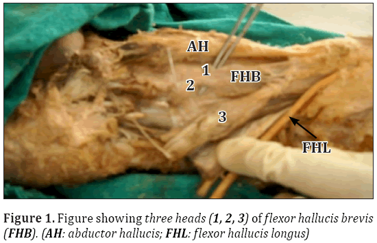
• FHB, apart from its usual 2 heads of origin from the fascia of tibialis posterior and cuboid, had a third head in the specimen (Figure 1). It was an aponeurotic head, arising from medial tubercle of the calcaneal tuberosity deep to the origin of abductor hallucis muscle; 6.4 mm broad, 17.1 mm long; then formed a thick flashy belly with bipennate arrangement and by far the thickest and the major >50%, contributor of the muscle mass. This head after turning fleshy spanned 9.3 cm, then joined almost midway the muscle belly arising from other 2 heads, about 4.7 cm from the insertion. Further on it became a flat tendon near the metatarsal heads but continued separately enclosing the tendon of FHL from its posterolateral aspect (lateral portion is sometimes named as the first Interosseous plantaris). The muscle continued towards the lateral side of the proximal phalanx to be inserted on lateral side of its base.
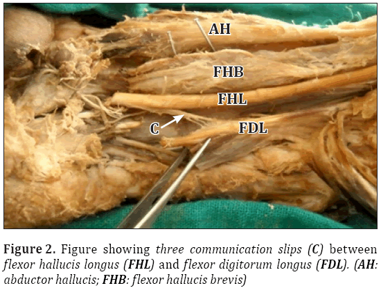
• Usually there may or may not be a single communication between the FHL and FDL muscles at the knot of Henry. In this specimen (Figure 2) there were three communicating slips between these two muscles.
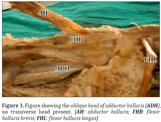
• The adductor hallucis (AH) muscle (Figure 3) had quite bulky oblique head of origin but there was no transverse head of its origin in this specimen.
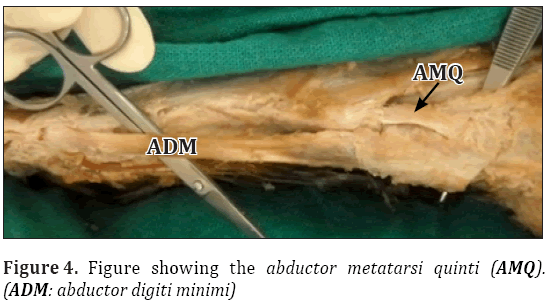
• The abductor ossis metatarsi quinti or the abductor of fifth metatarsal (Figure 4): Normally abductor digiti minimi is inserted on the lateral side of base of the proximal phalanx of fifth ray but sometimes it has variable additional insertions which are known as abductor ossis metatarsi quinti. As is seen in this specimen about 50% of the bulk of abductor digiti minimi is inserted on the tuberosity of the 5th metatarsal.
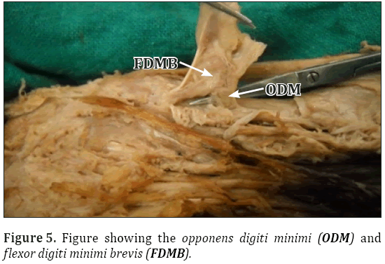
• The opponens digiti minimi (Figure 5): Usually flexor digiti minimi brevis is inserted on the lateral side of base of the proximal phalanx. But in this specimen an additional insertion was on the shaft of the fifth metatarsal known as opponens digiti minimi.
Discussion
A large number of accessory muscles have been described in the anatomy, surgery and radiology literature. Accessory muscles are anatomic variants representing additional distinct muscles that are encountered along with the usual complement of muscles [6].
However, in vast majority of cases these accessory muscles are asymptomatic but quite often these may be the cause of painful conditions like tarsal tunnel syndrome [7] by obstruction, hallux rigidus or hallux valgus by simple mechanical traction, may result in compression neuropathies [6] or rigid hind foot [8,9].
The third head of FHB (Figure 1), this variant is quoted in standard text books of anatomy [2] but no reference about its prevalence could be retrieved. This strong slip very well can create a problem to the individual and an additional pull may be a causative factor in producing the hallux valgus. This muscle along with FHL and FDL are important muscles used in ballet dancing and in actions requiring a forced and prolonged plantar flexion [6] as in association with some dance forms; in athletic discipline like long jump, triple jump, football etc. Pathology of the FHL and FDL tendons is commonly related to overuse, direct trauma and, less commonly to the inflammatory disease like tenosynovitis leading to partial/complete tears. These injuries are of paramount importance because loss of person’s livelihood is at stake.
The tendon ruptures often require a flap replacement. In that case these additional slips can be useful as replacement tendons to the FHL [6]. If by MRI, the presence of this slip is established, then this easily dispensable slip becomes the first choice to be used as a replacement flap so that other important muscles can be spared. These variants are to be kept in mind and properly evaluated beforehand by MRI while doing FHL tendon transfer [1].
Normally a tendinous slip of the lateral portion of the FHL tendon joins with the FDL tendon in the sole of the foot at the knot of Henry. This communication at times may be missing 29% [10]. But in this specimen (Figure 2) there were three slips of communication between these two muscles. Thereby, indicating a more interaction and influence of the two muscles on each other [3]. This slip provides a tethering mechanism that prevents excessive retraction of a proximal FHL tendon fragment after rupture [6]. In majority of cases of repair of tibialis posterior dysfunction result in retention of function of both, the hallux and the lesser digits, due to this strong communication [3].
Adductor hallucis muscle belongs to third layer of muscles [2,11] and shows a bulky oblique head of origin but no transverse head (Figure 3). The role of this muscle as adductor of hallux has become quite limited in human foot. Absence of transverse head of AH is quoted as 6% [5]. In the presence of other long and short flexors, it seems that the muscle, especially its transverse head has a tendency of evolutionary regression [11]. They have more of an anthropological value as they belong to contrahentes an atavistic group of muscles [12], which are present as a muscular sheet in embryo, most of which disappear and only parts of it persist in human foot [13]. In humans, contrahens 1 has survived and is the adductor hallucis [14], but the other contrahentes may persist as rare variations [13].
The abductor of fifth metatarsal, named as abductor ossis metatarsi quinti is an additional insertion of abductor digiti minimi brevis (ADMB) on the tuberosity of fifth metatarsal [2] or to middle or anterior part of fifth metatarsal and its prevalence is found to be 40% [15]. In the present specimen (Figure 4), a separate slip of the ADMB was inserted on the tuberosity of the fifth metatarsal. In the literature a few more variations regarding the ADMB are quoted as being inserted on middle phalanx [16] or having 2 bellies [17] or 3 bellies [18].
The flexor digiti minimi brevis (FDMB) muscle inserts on lateral sides of base of proximal phalanx of fifth ray but as seen in present specimen (Figure 5) has an accessory slip inserted on shaft of fifth metatarsal known as a separate entity, the opponens digiti minimi [2,19]. To the best of our knowledge not much could be found about this variant, except one report of insertion of FDMB on both medial and lateral sides of base of proximal phalanx of fifth ray [20]. These slips provide an additional control of these muscles on metatarsals, helping in tight griping of lateral edge of the foot on uneven ground and additive components in maintenance of lateral longitudinal and transverse arches of foot [10], an important requisite of bipedal gait. These accessory muscles do provide additional replacement flaps for flexor injury repairs.
The vast majority of, these accessory muscles are asymptomatic and tend to represent incidental findings at surgery or imaging. However, accessory muscles may result in symptoms in some cases. An accessory muscle can manifest as a painless/painful soft tissue mass or fullness in the ankle or may not be detected as obvious mass instead appears as isointense shadow with respect to muscle on all imaging sequences. If we know about their existence then only we can look for them in predictable locations [21].
Furthermore, accessory muscles may result in compression neuropathies. Careful evaluation of fibro-osseous tunnels for an accessory muscle may help identify such a muscle as a causative factor, which can easily be overlooked unless these slips of muscles are specifically sought for during the review process [8].
Conclusion
The knowledge of these variations in foot muscle architecture is of utmost importance to orthopedist, radiologists and podiatrists in analysis of foot function, biomechanical modeling of the foot and prosthesis designing. In painful and disabling conditions of the foot during differential diagnosis the presence of accessory muscles of foot should be kept in mind as causative factors, which can easily be overlooked unless these are specifically not sought for during the review process.
These accessory slips in the sole can strengthen the parent muscle and can be used as replacement flaps in tendon ruptures. Any additional muscle slips can be tormentors but can be a blessing in disguise as in tendon ruptures in forced plantar flexion in ballet dancers and athletes to be used as replacement flaps and thus spare other important muscles. They are a part of an atavistic group of muscle the ‘contrahentes’ in primates and so have an anthropological importance.
Acknowledgement
We are grateful to our teachers and colleagues.
References
- Pichler W, Tesch NP, Grechenig W, Tanzer K, Grasslober M. Anatomical variations of the flexor hallucis longus muscle and the consequences for tendon transfer. A cadaver study. SurgRadiol Anat. 2005; 27: 227–231.
- Standring S, ed. Gray’s Anatomy. 40th Ed., Churchill Livingstone, Elsevier. 2008; 1450–1455.
- O’Sullivan E, Carare-Nnadi R, Greenslade J, Bowyer G. Clinical significance of variations in the interconnections between flexor digitorum longus and flexor hallucis longus in the region of the knot of Henry. Clin Anat. 2005;18:121–125.
- Hirsch BE, Vekkos LE. Anomalous contrahentes muscles in human feet. Anat Anz.1984;155:123–129.
- Cralley JC, Schuberth JM. Transverse head of adductor hallucis. Anat Anz.1979; 46: 400–409.
- Smith SE, Flye CW. Flexor Hallucis Longus Tendon Injury Imaging. http://emedicine.medscape.com/article/386171-overview (accessed May 2011).
- Rosenberg ZS, Beltran J, Bencardino JT. MR imaging of the ankle and foot. Radiographics. 2000; 20:153–179.
- Sookur PA, Naraghi AM, Bleakney RR, Jalan R, Chan O, White LM. Accessory muscles: anatomy, symptoms and radiologic evaluation. Radiographics. 2008; 28: 481–499.
- Carroll JF. MRI Web Clinic – November 2008. Accessory Muscles of the Ankle. http://www.radsource.us/clinic/0811 (accessed January 2012).
- Fernandes R, Aguiar R, Trudell D, Resnick D. Tendons in the plantar aspect of the foot: MR imaging and anatomic correlation in cadavers. Skeletal Radiol. 2007; 36: 115–122.
- Arakawa T, Sekiya S, Kumaki K, Terashima T. Intramuscular nerve distribution pattern of the oblique and transverse heads of the adductor hallucis muscles in the human foot. Anat Sci Int. 2006; 81: 187–196.
- Stark HH, Otter TA, Boyes JH, Rickard TA. “Atavistic contrahentes digitorum” and associated muscle abnormalities of the hand: a cause of symptoms. Report of three cases. J Bone Joint Surg Am. 1979; 61: 286–289.
- Yamamoto C, Murakami T, Ohtsuka A. Homology of the adductor pollicis and contrahentes muscles: a study of monkey hands. Acta Med Okayama.1988; 42: 215–226.
- Tubbs RS, Salter EG, Oakes WJ. Contrahentes digitorum muscle. Clin Anat. 2005; 18: 606–608.
- Bergmann RA, Afifi AK, Miyauchi R. Illustrated encylopedia of human anatomic variation: A.Abductor Digiti Minimi (pedis). http://www.anatomyatlases.org/AnatomicVariants/MuscularSystem/Text/A/03Abductor.shtml (accessed July 2010).
- Asomugha AL, Chukwuanukwu TO, Nwajagu GI, Ukoha U. An accessory flexor of the fifth toe. Niger J Clin Pract. 2005; 8: 130 132.
- Nayyar A, Mehta V, Gupta V, Suri RK, Rath G. Clinico-anatomical considerations of unilateral bipartite abductor digiti minimi muscle of the foot: a case report. Anatomy. 2010; 4: 72–75.
- Kopuz C, Tetik S, Ozbenli S. A rare anomaly of the abductor digiti minimi muscle of the foot. Cells Tissues Organs. 1999; 164: 174–176.
- Schuenke M, Schulte E, Schumacher U, Ross ML, Lamperti ED, eds. Thieme Atlas of Anatomy, International Edition. Georg Thieme Verlag. 2006; 438–460.
- Rana KK, Das S. Anomalous attachment of the flexor digiti minimi muscle of the foot: an anatomical study with clinical implications. Eur J Anat. 2006; 10: 153–155.
- Link SC, Erickson SJ, Timins ME. MR imaging of the ankle and foot: normal structures and anatomic variants that may simulate disease. AJR Am J Roentgenol. 1993; 16: 607–612.
Dharwal Kumud*, Mahajan Anupama
Department of Anatomy, Shri Guru Ram Das Institute of Medical Sciences and Research, Amritsar (PB), India.
- *Corresponding Author:
- Dr. Dharwal Kumud
Dharwal Clinic, Cheel Mandi Near Ramgarhia School, Amritsar (PB) 143001, India.
Tel: +91 9872737679
E-mail: kdharwal@gmail.com
Date of Received: February 21st, 2012
Date of Accepted: June 10th, 2012
Published Online: February 14th, 2013
© Int J Anat Var (IJAV). 2013; 6: 41–44.
Abstract
During routine cadaveric dissection of a sole of foot, multiple variations were encountered. The flexor hallucis brevis muscle had a third head, arising from the medial tubercle of the calcaneal tuberosity. The flexor hallucis longus and the flexor digitorum longus tendons had multiple communications between them. The adductor hallucis muscle had a large oblique head and missing transverse head. The abductor of fifth metatarsal was present. The opponens digiti minimi muscle was present. These additional muscle slips may pose some problems to an individual, but can be godsend awards to be used as reconstruction flaps, in flexor injuries thereby sparing the use of other important long flexors. The knowledge of these variations in foot muscle architecture is of utmost importance in analysis of foot function, biomechanical modeling of the foot and prosthesis designing and to the orthopedist, radiologists and podiatrists. These additional slips also have an anthropological importance.
-Keywords
sole, muscle, multiple, variations
Introduction
Muscle variations are commonly encountered. Basically data regarding accessory musculature has been based on serendipitous findings at surgery or during cadaveric dissections. Flexor hallucis brevis (FHB), a muscle of the 3rd muscular layer of the sole of the foot showed a 3rd head. These accessory slips may be useful as replacement tendons in pathology of the flexor hallucis longus (FHL) and flexor digitorum longus (FDL) tendons, which are commonly damaged due to overuse and these slips also affect the results of FHL tendon transfer [1]. There is usually a communication between the FHL and FDL muscles [2]. The multiple communications, present in this specimen, indicate a more interaction and influence of the two muscles on each other [3]. The missing adductor longus transverse head is quite frequent and can be a missing part of contrahentes [4]. The presence of the abductor of fifth metatarsal (abductor ossis metatarsi quinti muscle) and the opponens digiti minimi muscle can strengthen the parent muscles and can be used as replacement flaps in tendon injuries [1]. The contrahentes and other additional slips also have an anthropological importance [4,5].
Case Report
During routine cadaveric dissection of the sole of the foot as a part of the undergraduate teaching, five variations were encountered in this particular left foot of a 59-year-old male.
• FHB, apart from its usual 2 heads of origin from the fascia of tibialis posterior and cuboid, had a third head in the specimen (Figure 1). It was an aponeurotic head, arising from medial tubercle of the calcaneal tuberosity deep to the origin of abductor hallucis muscle; 6.4 mm broad, 17.1 mm long; then formed a thick flashy belly with bipennate arrangement and by far the thickest and the major >50%, contributor of the muscle mass. This head after turning fleshy spanned 9.3 cm, then joined almost midway the muscle belly arising from other 2 heads, about 4.7 cm from the insertion. Further on it became a flat tendon near the metatarsal heads but continued separately enclosing the tendon of FHL from its posterolateral aspect (lateral portion is sometimes named as the first Interosseous plantaris). The muscle continued towards the lateral side of the proximal phalanx to be inserted on lateral side of its base.
• Usually there may or may not be a single communication between the FHL and FDL muscles at the knot of Henry. In this specimen (Figure 2) there were three communicating slips between these two muscles.
• The adductor hallucis (AH) muscle (Figure 3) had quite bulky oblique head of origin but there was no transverse head of its origin in this specimen.
• The abductor ossis metatarsi quinti or the abductor of fifth metatarsal (Figure 4): Normally abductor digiti minimi is inserted on the lateral side of base of the proximal phalanx of fifth ray but sometimes it has variable additional insertions which are known as abductor ossis metatarsi quinti. As is seen in this specimen about 50% of the bulk of abductor digiti minimi is inserted on the tuberosity of the 5th metatarsal.
• The opponens digiti minimi (Figure 5): Usually flexor digiti minimi brevis is inserted on the lateral side of base of the proximal phalanx. But in this specimen an additional insertion was on the shaft of the fifth metatarsal known as opponens digiti minimi.
Discussion
A large number of accessory muscles have been described in the anatomy, surgery and radiology literature. Accessory muscles are anatomic variants representing additional distinct muscles that are encountered along with the usual complement of muscles [6].
However, in vast majority of cases these accessory muscles are asymptomatic but quite often these may be the cause of painful conditions like tarsal tunnel syndrome [7] by obstruction, hallux rigidus or hallux valgus by simple mechanical traction, may result in compression neuropathies [6] or rigid hind foot [8,9].
The third head of FHB (Figure 1), this variant is quoted in standard text books of anatomy [2] but no reference about its prevalence could be retrieved. This strong slip very well can create a problem to the individual and an additional pull may be a causative factor in producing the hallux valgus. This muscle along with FHL and FDL are important muscles used in ballet dancing and in actions requiring a forced and prolonged plantar flexion [6] as in association with some dance forms; in athletic discipline like long jump, triple jump, football etc. Pathology of the FHL and FDL tendons is commonly related to overuse, direct trauma and, less commonly to the inflammatory disease like tenosynovitis leading to partial/complete tears. These injuries are of paramount importance because loss of person’s livelihood is at stake.
The tendon ruptures often require a flap replacement. In that case these additional slips can be useful as replacement tendons to the FHL [6]. If by MRI, the presence of this slip is established, then this easily dispensable slip becomes the first choice to be used as a replacement flap so that other important muscles can be spared. These variants are to be kept in mind and properly evaluated beforehand by MRI while doing FHL tendon transfer [1].
Normally a tendinous slip of the lateral portion of the FHL tendon joins with the FDL tendon in the sole of the foot at the knot of Henry. This communication at times may be missing 29% [10]. But in this specimen (Figure 2) there were three slips of communication between these two muscles. Thereby, indicating a more interaction and influence of the two muscles on each other [3]. This slip provides a tethering mechanism that prevents excessive retraction of a proximal FHL tendon fragment after rupture [6]. In majority of cases of repair of tibialis posterior dysfunction result in retention of function of both, the hallux and the lesser digits, due to this strong communication [3].
Adductor hallucis muscle belongs to third layer of muscles [2,11] and shows a bulky oblique head of origin but no transverse head (Figure 3). The role of this muscle as adductor of hallux has become quite limited in human foot. Absence of transverse head of AH is quoted as 6% [5]. In the presence of other long and short flexors, it seems that the muscle, especially its transverse head has a tendency of evolutionary regression [11]. They have more of an anthropological value as they belong to contrahentes an atavistic group of muscles [12], which are present as a muscular sheet in embryo, most of which disappear and only parts of it persist in human foot [13]. In humans, contrahens 1 has survived and is the adductor hallucis [14], but the other contrahentes may persist as rare variations [13].
The abductor of fifth metatarsal, named as abductor ossis metatarsi quinti is an additional insertion of abductor digiti minimi brevis (ADMB) on the tuberosity of fifth metatarsal [2] or to middle or anterior part of fifth metatarsal and its prevalence is found to be 40% [15]. In the present specimen (Figure 4), a separate slip of the ADMB was inserted on the tuberosity of the fifth metatarsal. In the literature a few more variations regarding the ADMB are quoted as being inserted on middle phalanx [16] or having 2 bellies [17] or 3 bellies [18].
The flexor digiti minimi brevis (FDMB) muscle inserts on lateral sides of base of proximal phalanx of fifth ray but as seen in present specimen (Figure 5) has an accessory slip inserted on shaft of fifth metatarsal known as a separate entity, the opponens digiti minimi [2,19]. To the best of our knowledge not much could be found about this variant, except one report of insertion of FDMB on both medial and lateral sides of base of proximal phalanx of fifth ray [20]. These slips provide an additional control of these muscles on metatarsals, helping in tight griping of lateral edge of the foot on uneven ground and additive components in maintenance of lateral longitudinal and transverse arches of foot [10], an important requisite of bipedal gait. These accessory muscles do provide additional replacement flaps for flexor injury repairs.
The vast majority of, these accessory muscles are asymptomatic and tend to represent incidental findings at surgery or imaging. However, accessory muscles may result in symptoms in some cases. An accessory muscle can manifest as a painless/painful soft tissue mass or fullness in the ankle or may not be detected as obvious mass instead appears as isointense shadow with respect to muscle on all imaging sequences. If we know about their existence then only we can look for them in predictable locations [21].
Furthermore, accessory muscles may result in compression neuropathies. Careful evaluation of fibro-osseous tunnels for an accessory muscle may help identify such a muscle as a causative factor, which can easily be overlooked unless these slips of muscles are specifically sought for during the review process [8].
Conclusion
The knowledge of these variations in foot muscle architecture is of utmost importance to orthopedist, radiologists and podiatrists in analysis of foot function, biomechanical modeling of the foot and prosthesis designing. In painful and disabling conditions of the foot during differential diagnosis the presence of accessory muscles of foot should be kept in mind as causative factors, which can easily be overlooked unless these are specifically not sought for during the review process.
These accessory slips in the sole can strengthen the parent muscle and can be used as replacement flaps in tendon ruptures. Any additional muscle slips can be tormentors but can be a blessing in disguise as in tendon ruptures in forced plantar flexion in ballet dancers and athletes to be used as replacement flaps and thus spare other important muscles. They are a part of an atavistic group of muscle the ‘contrahentes’ in primates and so have an anthropological importance.
Acknowledgement
We are grateful to our teachers and colleagues.
References
- Pichler W, Tesch NP, Grechenig W, Tanzer K, Grasslober M. Anatomical variations of the flexor hallucis longus muscle and the consequences for tendon transfer. A cadaver study. SurgRadiol Anat. 2005; 27: 227–231.
- Standring S, ed. Gray’s Anatomy. 40th Ed., Churchill Livingstone, Elsevier. 2008; 1450–1455.
- O’Sullivan E, Carare-Nnadi R, Greenslade J, Bowyer G. Clinical significance of variations in the interconnections between flexor digitorum longus and flexor hallucis longus in the region of the knot of Henry. Clin Anat. 2005;18:121–125.
- Hirsch BE, Vekkos LE. Anomalous contrahentes muscles in human feet. Anat Anz.1984;155:123–129.
- Cralley JC, Schuberth JM. Transverse head of adductor hallucis. Anat Anz.1979; 46: 400–409.
- Smith SE, Flye CW. Flexor Hallucis Longus Tendon Injury Imaging. http://emedicine.medscape.com/article/386171-overview (accessed May 2011).
- Rosenberg ZS, Beltran J, Bencardino JT. MR imaging of the ankle and foot. Radiographics. 2000; 20:153–179.
- Sookur PA, Naraghi AM, Bleakney RR, Jalan R, Chan O, White LM. Accessory muscles: anatomy, symptoms and radiologic evaluation. Radiographics. 2008; 28: 481–499.
- Carroll JF. MRI Web Clinic – November 2008. Accessory Muscles of the Ankle. http://www.radsource.us/clinic/0811 (accessed January 2012).
- Fernandes R, Aguiar R, Trudell D, Resnick D. Tendons in the plantar aspect of the foot: MR imaging and anatomic correlation in cadavers. Skeletal Radiol. 2007; 36: 115–122.
- Arakawa T, Sekiya S, Kumaki K, Terashima T. Intramuscular nerve distribution pattern of the oblique and transverse heads of the adductor hallucis muscles in the human foot. Anat Sci Int. 2006; 81: 187–196.
- Stark HH, Otter TA, Boyes JH, Rickard TA. “Atavistic contrahentes digitorum” and associated muscle abnormalities of the hand: a cause of symptoms. Report of three cases. J Bone Joint Surg Am. 1979; 61: 286–289.
- Yamamoto C, Murakami T, Ohtsuka A. Homology of the adductor pollicis and contrahentes muscles: a study of monkey hands. Acta Med Okayama.1988; 42: 215–226.
- Tubbs RS, Salter EG, Oakes WJ. Contrahentes digitorum muscle. Clin Anat. 2005; 18: 606–608.
- Bergmann RA, Afifi AK, Miyauchi R. Illustrated encylopedia of human anatomic variation: A.Abductor Digiti Minimi (pedis). http://www.anatomyatlases.org/AnatomicVariants/MuscularSystem/Text/A/03Abductor.shtml (accessed July 2010).
- Asomugha AL, Chukwuanukwu TO, Nwajagu GI, Ukoha U. An accessory flexor of the fifth toe. Niger J Clin Pract. 2005; 8: 130 132.
- Nayyar A, Mehta V, Gupta V, Suri RK, Rath G. Clinico-anatomical considerations of unilateral bipartite abductor digiti minimi muscle of the foot: a case report. Anatomy. 2010; 4: 72–75.
- Kopuz C, Tetik S, Ozbenli S. A rare anomaly of the abductor digiti minimi muscle of the foot. Cells Tissues Organs. 1999; 164: 174–176.
- Schuenke M, Schulte E, Schumacher U, Ross ML, Lamperti ED, eds. Thieme Atlas of Anatomy, International Edition. Georg Thieme Verlag. 2006; 438–460.
- Rana KK, Das S. Anomalous attachment of the flexor digiti minimi muscle of the foot: an anatomical study with clinical implications. Eur J Anat. 2006; 10: 153–155.
- Link SC, Erickson SJ, Timins ME. MR imaging of the ankle and foot: normal structures and anatomic variants that may simulate disease. AJR Am J Roentgenol. 1993; 16: 607–612.


