A highly mobile intra-atrial big mass: Image focus
Received: 16-Aug-2017 Accepted Date: Aug 18, 2017; Published: 22-Aug-2017
Citation: Laachach H, Ofkir M, Berrajaa M, et al. A highly mobile intra-atrial big mass: Image focus. Clin Cardiol J 2017;1(1):14.
This open-access article is distributed under the terms of the Creative Commons Attribution Non-Commercial License (CC BY-NC) (http://creativecommons.org/licenses/by-nc/4.0/), which permits reuse, distribution and reproduction of the article, provided that the original work is properly cited and the reuse is restricted to noncommercial purposes. For commercial reuse, contact reprints@pulsus.com
Abstract
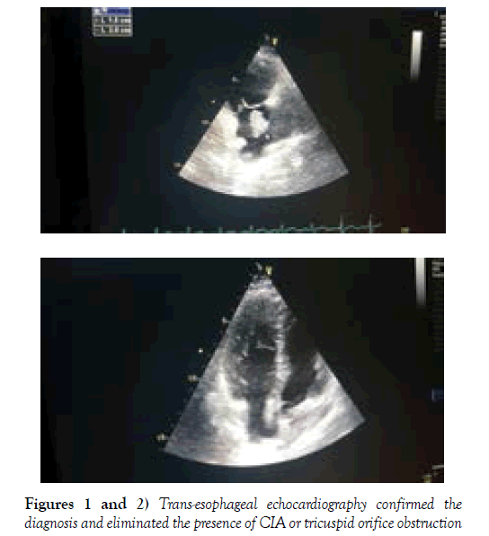
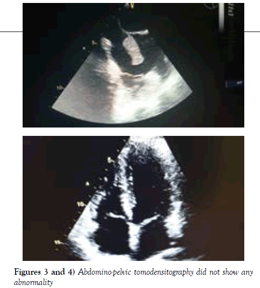
A 28-year-old woman, with no pathological history, admitted for acute dyspnea. The hemodynamic state was unusual with a respiratory rate of 26 cycles per minute, saturation at 92% in ambient air and a temperature at 38 degrees Celsius. There were no abnormalities in the X-ray of the chest. Thoracic angio-CT revealed distal bilateral pulmonary embolism. Transthoracic echocardiography has showed a pediculated right intra-atrial mass, highly mobile and finely attached to the inter-articular septum; with fragments at the end of the vena cava (Figures 1 and 2). Trans-esophageal echocardiography confirmed the diagnosis and eliminated the presence of CIA or tricuspid orifice obstruction (Figure 3). Given the imminent risk of detachment of the mass and its migration, the patient was transferred to cardiac surgery after the anticoagulant treatment; she benefited from extraction of a thrombus and aspiration of several thrombi. The short and medium-term outcome was good (Figure 4). Abdomino-pelvic tomodensitography did not show any abnormality. On the other hand, no sign pointing towards a disease behcet, whereas the anti-phospholipid antibody assay is negative and the thrombophilia assessment have not been performed.
Clinical Image
A 28-year-old woman, with no pathological history, admitted for acute dyspnea. The hemodynamic state was unusual with a respiratory rate of 26 cycles per minute, saturation at 92% in ambient air and a temperature at 38 degrees Celsius. There were no abnormalities in the X-ray of the chest. Thoracic angio-CT revealed distal bilateral pulmonary embolism. Transthoracic echocardiography has showed a pediculated right intra-atrial mass, highly mobile and finely attached to the inter-articular septum; with fragments at the end of the vena cava (Figures 1 and 2). Trans-esophageal echocardiography confirmed the diagnosis and eliminated the presence of CIA or tricuspid orifice obstruction (Figure 3). Given the imminent risk of detachment of the mass and its migration, the patient was transferred to cardiac surgery after the anticoagulant treatment; she benefited from extraction of a thrombus and aspiration of several thrombi. The short and medium-term outcome was good (Figure 4). Abdomino-pelvic tomodensitography did not show any abnormality. On the other hand, no sign pointing towards a disease behcet, whereas the anti-phospholipid antibody assay is negative and the thrombophilia assessment have not been performed.
Figures 1 and 2: Trans-esophageal echocardiography confirmed the diagnosis and eliminated the presence of CIA or tricuspid orifice obstruction


