A histological, histochemical and ultrastructural characterization of uterine vessels at early stages of pregnancy
Received: 01-Nov-2018 Accepted Date: Nov 28, 2018; Published: 30-Nov-2018
Citation: Fatma El-Zahraa AM, Abd-Elhafez EA. A Histological, histochemical and ultrastructural characterization of uterine vessels at early stages of pregnancy. J Histol Histopathol Res 2018;2(2): 41-7.
This open-access article is distributed under the terms of the Creative Commons Attribution Non-Commercial License (CC BY-NC) (http://creativecommons.org/licenses/by-nc/4.0/), which permits reuse, distribution and reproduction of the article, provided that the original work is properly cited and the reuse is restricted to noncommercial purposes. For commercial reuse, contact reprints@pulsus.com
Abstract
The integrity of uterine circulation is essential for normal pregnancy outcome. The present study describes for the first time the histology, histochemistry, and ultrastructure of the different types of vessels in the uterus at critical early stages of pregnancy. Arteries were congested and showed PAS-positive granules. veins characterized by its wide lumen and thick adventitia rich in collagen and elastic fibers and showed PAS-positive reaction in their wall. Lymphatics showed a positive reaction with PAS-AB combination. Many arteriols observed in the uterine wall and showed PAS-positive granules and venules appeared congested in the endometrium. Spiral artery and arterial sinuses also demonstrated. Anastomosis between arterioles and venules was recorded. Blood vessels of a special structure were demonstrated presenting a vascular wall of variable thickness, unevenly distributed smooth muscle in the wall, smooth muscle fibers arranged in a different direction. Spirally oriented arterioles and venules were also demonstrated. Sclerotic changes observed in the wall of some vessels. Glomus cells observed in the wall of many arteries, arterioles, spirally oriented arterioles, and arteriovenous anastomosis. filopodia-like protrusions and caveolae were the most characteristic feature of the endothelium.
Keywords
Blood vessels; Lymphatics; Glomus cells; Telocyte; Filopodia; Early pregnancy
The integrity of uterine circulation is essential for normal pregnancy outcome. The clinical relevance of maternal uterine vascular adaptation during pregnancy is underscored by the fact that its aberrance is associated with several common gestational pathologies, including intrauterine growth restriction, gestational diabetes, and preeclampsia [1]. Preeclampsia is a major cause of maternal and neonatal mortality and morbidity [2].
It is known that the vascularization of the uterus is of primary importance in the pregnancy success [3], and vascular disorders may play a role in pathological pregnancies [4]. Furthermore, normal uterine vascular development may have critical effects on the growth and development of the fetus and insufficient uterine vascular establishment is associated with increased risk for cardiovascular morbidity in adult life [5].
The lymphatic circulation plays an important role in regulating content of interstitial fluid and adaptive immunity [6] and lymphatic vessels could also play a role in maternal and fetal immunity [7]. The uterine lymphatics during pregnancy have been scantily studied.
Rabbit has a duplex uterus, with the main uteroovarian arteries and veins running parallel to, but well outside of, the uterine wall within the mesometrium. The vessels of the mesometrium are perfused by arterial blood coming from either the uterine or the ovarian end. Secondary vessels analogous to the arcuate arteries in humans may form redundant loops with the main artery, and tertiary radial arteries connect the arcuate loops with the uterine wall. These radial arteries divided into premyometrial arteries. The premyometrial arteries enter the uterine wall supplies the myometrium [1]. A better understanding of the basic morphological structure of the blood vessels of the uterus of the rabbit may guide the clinicians to the design of strategies to improve their reproductive efficiency and help in solve pregnancy problems which related to the blood vessels. Yet, the available literature lacks any detailed information concerning the uterine vessels during early stages of pregnancy.
The aim of the present investigation was to study the different types of vessels in the uterus at critical early stages of pregnancy. The current study focused on arteries, veins, lymphatics, arterioles, venules, blood vessels of special structure.
Material and Methods
Sample collection
The study was approved by the Ethics Committee of Assiut University, Egypt. The material included in this work was originated from the female genitalia of 6 healthy New Zealand rabbits at 0 and 7days of pregnancy. The uterus was taken immediately after slaughtering.
Histological preparation
The collected materials were dissected as soon as possible and immediately fixed in Bouin’s fluid for 22 hours. The fixed materials were dehydrated in an ascending series of ethanol, cleared in methyl benzoate and then embedded in paraffin wax. Transverse paraffin sections at 1-7 μm in thickness were cut and stained with Harris haematoxylin and eosin for general histological examination, Crossmon’s Trichrome for identification of collagenous and muscle fibers and Wigert’s resorcin fuchsine for identification of elastic fibers. For carbohydrate histochemistery, sections were stained with Periodic Acid Schiff (PAS) technique. For demonstration of neutral mucopolysaccharides, combined alcian blue-PAS technique were used [8].
Semithin sections and transmission electron microscopic preparations
Other small specimens of the uterus were fixed in a mixture of 2.5% paraformaldehyde and 2.5% glutaraldehyde in 0.1M Na-cacodylate buffer, pH 7.3 for 4 hours at 4 Cº. They were washed in the same buffer used and then post-fixed in 1% osmic acid in 0.1M Na-cacodylate buffer for further 2 hours at room temperature. The samples were then dehydrated in ethanol and embedded in Araldite-Epon mixture. Semithin sections (1μm in thickness) were cut and stained with Toluidine blue and examined under a light microscope. Thin sections, obtained by a Reichert ultramicrotome, were stained with uranyl acetate and lead citrate [9] and examined with a Philips EM 400.
Results
0 day of pregnancy
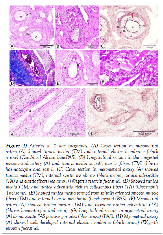
Mesometrial arteries were congested and demonstrated in varies directions cross and longitudinally oriented arteries (Figure 1A and 1B). The wall of these arteries was formed mainly of 3 - 5 layers of smooth muscle fibers tunica media, intermingled by elastic fibers. The tunica intima consisted of a thin layer of flat endothelial cells separated from the coating muscular media by a well-developed highly tortuous internal elastic membrane. The adventitia contained abundant elastic fibers and rich in collagenous fibers (Figure 1A- 1D). In addition, there were arteries recorded in the mesometrium with spirally oriented smooth muscle fibers in the tunica media (Figure 1E).
Figure 1: Arteries at 0 day pregnancy. (A) Cross section in mesometrial artery (A) showed tunica media (TM) and internal elastic membrane (black arrow) (Combined Alcian blue-PAS). (B) Longitudinal section in the congested mesometrial artery (A) and tunica media smooth muscle fibers (TM) (Harris haematoxylin and eosin). (C) Cross section in mesometrial artery (A) showed tunica media (TM), internal elastic membrane (black arrow), tunica adventitia (TA) and elastic fibers (red arrow) (Wigert’s resorcin fuchsine). (D) Showed tunica media (TM) and tunica adventitia rich in collagenous fibers (TA) (Crossmon’s Trichrome). (E) Showed tunica media formed from spirally oriented smooth muscle fibers (TM) and internal elastic membrane (black arrow) (PAS). (F) Myometrial artery (A) showed tunica media (TM) and vascular tunica adventitia (TA) (Harris haematoxylin and eosin). (G) Longitudinal section in myometrial artery (A) demonstrate PAS-positive granules (blue arrow) (PAS). (H) Myometrial artery (A) showed well developed internal elastic membrane (black arrow) (Wigert’s resorcin fuchsine).
Myometrial arteries possessed a wide lumen and surrounded by a 2-4 layer of smooth muscle fiber. In addition, the adventitia showed several blood vessels. These arteries demonstrate PAS-positive granules in their wall and well developed internal elastic membrane (Figure 1F-1H).
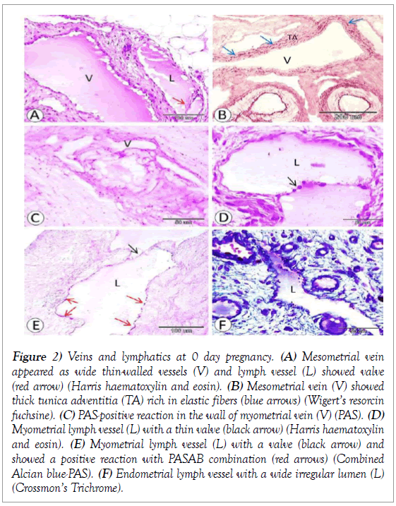
Mesometrial veins were numerous wide thin-walled vessels of different size. The tunica intima was surrounded by a thin tunica media composing of a 1-2 layer of smooth muscle fibers encircled by a relatively thicker adventitia rich in collagen and elastic fibers (Figure 2A and 2B). At the myometrial region, venae showed PAS-positive reaction in their wall (Figure 2C).
Figure 2: Veins and lymphatics at 0 day pregnancy. (A) Mesometrial vein appeared as wide thin-walled vessels (V) and lymph vessel (L) showed valve (red arrow) (Harris haematoxylin and eosin). (B) Mesometrial vein (V) showed thick tunica adventitia (TA) rich in elastic fibers (blue arrows) (Wigert’s resorcin fuchsine). (C) PAS-positive reaction in the wall of myometrial vein (V) (PAS). (D) Myometrial lymph vessel (L) with a thin valve (black arrow) (Harris haematoxylin and eosin). (E) Myometrial lymph vessel (L) with a valve (black arrow) and showed a positive reaction with PASAB combination (red arrows) (Combined Alcian blue-PAS). (F) Endometrial lymph vessel with a wide irregular lumen (L) (Crossmon’s Trichrome).
Several thin- walled wide-lumened lymphatics were recorded within the mesometrium. Their wall is composed of an intimal tunic surrounded by a thick fibrous layer consisting of collagenous, elastic fibers and connective tissue cells. Some of these vessels demonstrate valves (Figure 2A).
Myometrial lymphatics were composed of an intimal tunic surrounded by a fibrous layer of collagenous and elastic fibers. The valves of these vessels were mainly thin and consisted of a single cellular layer. The wall of myometrial lymph vessels showed a positive reaction with PAS-AB combination (Figure 2D and 2E). The lymphatic vessel in the endometrium appeared as wide irregular thin walled vessels (Figure 2F).
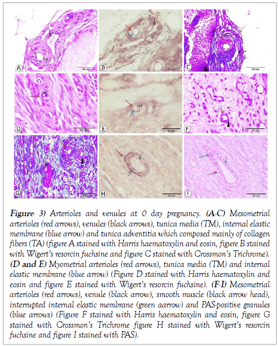
Several arterioles were demonstrated within the mesometrium. These blood vessels were composed of 1-2 smooth muscle fiber-thick tunica media encircling a thin tunica intima with interrupted internal elastic membrane and surrounded by tunica adventitia which composed mainly of collagen fibers (Figure 3A-3C). In addition, arterioles of the myometrium were composed of 1-2 smooth muscle fiber-thick tunica media surrounded a thin intimal tunic with interrupted internal elastic membrane and was surrounded by fibrous adventitial tunic which mainly consisted of collagen fibers (Figure 3D and 3E).
Figure 3: Arterioles and venules at 0 day pregnancy. (A-C) Mesometrial arterioles (red arrows), venules (black arrows), tunica media (TM), internal elastic membrane (blue arrow) and tunica adventitia which composed mainly of collagen fibers (TA) (figure A stained with Harris haematoxylin and eosin, figure B stained with Wigert’s resorcin fuchsine and figure C stained with Crossmon’s Trichrome). (D and E) Myometrial arterioles (red arrows), tunica media (TM) and internal elastic membrane (blue arrow) (Figure D stained with Harris haematoxylin and eosin and figure E stained with Wigert’s resorcin fuchsine). (F-I) Mesometrial arterioles (red arrows), venule (black arrow), smooth muscle (black arrow head), interrupted internal elastic membrane (green aarrow) and PAS-positive granules (blue arrows) (Figure F stained with Harris haematoxylin and eosin, figure G stained with Crossmon’s Trichrome figure H stained with Wigert’s resorcin fuchsine and figure I stained with PAS).
Many arterioles were observed within the endometrium with 1-2 smooth muscle fiber-thick tunica media encircling a thin tunica intima with interrupted internal elastic membrane and surrounded by tunica adventitia which contained mainly elastic and collagenous fibers. In addition, few PASpositive granules observed on the wall (Figure 3F-3I).
Venules wall consists of an intimal tunic surrounded by one cell-thick tunica media of circularly arranged smooth muscle fibers coated by thick adventitial tunic consisting of collagenous fibers and connective tissue cellular elements. However, venules of endometrium appeared as dilated thin wall vessels engorged with blood (Figure 3A and 3G).
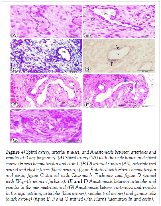
Spiral artery also demonstrated and characterized by the wide lumen and spiral course. Its wall consists of tunica intima of endothelial cells surrounded by a thin layer of smooth muscle fibers and thin adventitia (Figure 4A). In addition, arterial sinuses demonstrated and characterized by wide irregular lumen, one layer of smooth muscle tunica media and unevenly disreputed elastic fibers (Figure 4B-4D).
Figure 4: Spiral artery, arterial sinuses, and Anastomosis between arterioles and venules at 0 day pregnancy. (A) Spiral artery (SA) with the wide lumen and spiral course (Harris haematoxylin and eosin). (B-D) arterial sinuses (AS), arteriole (red arrow) and elastic fibers (black arrows) (figure B stained with Harris haematoxylin and eosin, figure C stained with Crossmon’s Trichrome and figure D stained with Wigert’s resorcin fuchsine). (E and F) Anastomosis between arterioles and venules in the mesometrium and (G) Anastomosis between arterioles and venules in the myometrium, arterioles (blue arrows), venules (red arrows) and glomus cells (black arrows) (figure E, F and G stained with Harris haematoxylin and eosin).
Anastomosis between arterioles and venules was recorded in mesometrium and showed an abrupt change from the arteriole to a venule. Another arteriovenous anastomosis in the mesentery with several glomus cells and its lumen is closed (Figure 4E and 4F). Anastomosis between arterioles and venules was also recorded in the myometrium and (Figure 4G).
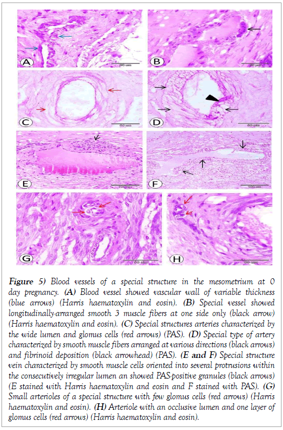
Blood vessels of a special structure were demonstrated within the mesometrium presenting a vascular wall of variable thickness. The thicker portions of the wall showed a tunica media of 2-3 layers of smooth muscle fibers. However, the thin portions of the wall showed tunica media consisting of an interrupted layer of smooth muscle fibers (Figure 5A). In addition, another vessel showed 2-3 layers of longitudinally-arranged smooth muscle fibers at one side only (Figure 5B).
Figure 5: Blood vessels of a special structure in the mesometrium at 0 day pregnancy. (A) Blood vessel showed vascular wall of variable thickness (blue arrows) (Harris haematoxylin and eosin). (B) Special vessel showed longitudinally-arranged smooth 3 muscle fibers at one side only (black arrow) (Harris haematoxylin and eosin). (C) Special structures arteries characterized by the wide lumen and glomus cells (red arrows) (PAS). (D) Special type of artery characterized by smooth muscle fibers arranged at various directions (black arrows) and fibrinoid deposition (black arrowhead) (PAS). (E and F) Special structure vein characterized by smooth muscle cells oriented into several protrusions within the consecutively irregular lumen an showed PAS-positive granules (black arrows) (E stained with Harris haematoxylin and eosin and F stained with PAS). (G) Small arterioles of a special structure with few glomus cells (red arrows) (Harris haematoxylin and eosin). (H) Arteriole with an occlusive lumen and one layer of glomus cells (red arrows) (Harris haematoxylin and eosin).
Various special structures arteries observed within the mesometrium. Characterized by the wide lumen and 2-3 layer of glomus cells (Figure 5C). Another special type of artery characterized by smooth muscle fibers arranged at various directions and fibrinoid deposition (Figure 5D).
Various special structures veins observed within the mesometrium and characterized by a wide irregular lumen. The flattened endothelial cell lining was surrounded by 2-3 cell thick muscular media composing of circularly arranged smooth muscle fibers. These muscle cells were oriented into several protrusions, within the consecutively irregular lumen, bearing as muscular pads. The relatively thick adventitia mainly consists of collagenous and elastic elements. In addition, several islets of longitudinally-arranged smooth muscle fibers surrounded these vessels and characterized by PAS-positive granules (Figure 5E and 5F).
Small arterioles of a special structure were also observed and lined with 2-3 endothelial cells and surrounded by smooth muscle fibers of tunica media with few glomus cells (Figure 5G). Another arteriole with an occlusive lumen and one layer of glomus cells also demonstrated (Figure 5H).
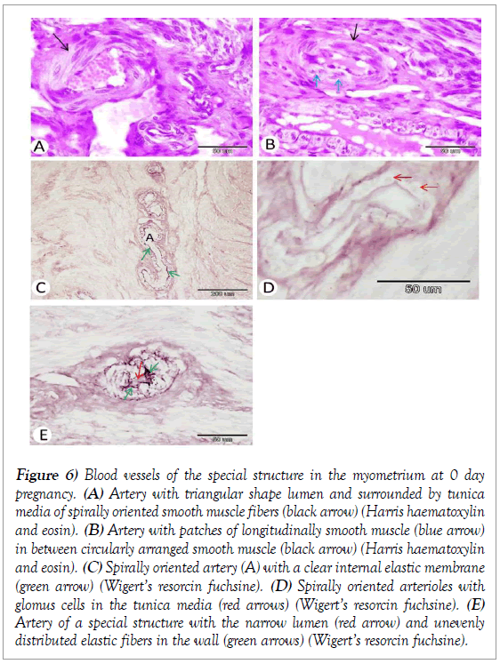
Blood vessels of the special structure were demonstrated also within the myometrium. Our observation revealed that there were arteries with triangular shape lumen and surrounded by tunica media of spirally oriented smooth muscle fibers (Figure 6A). In addition, some arteries demonstrate 2-4 layer of circularly arranged smooth muscle fibers and patches of longitudinally smooth muscle fibers in between (Figure 6B).
Figure 6: Blood vessels of the special structure in the myometrium at 0 day pregnancy. (A) Artery with triangular shape lumen and surrounded by tunica media of spirally oriented smooth muscle fibers (black arrow) (Harris haematoxylin and eosin). (B) Artery with patches of longitudinally smooth muscle (blue arrow) in between circularly arranged smooth muscle (black arrow) (Harris haematoxylin and eosin). (C) Spirally oriented artery (A) with a clear internal elastic membrane (green arrow) (Wigert’s resorcin fuchsine). (D) Spirally oriented arterioles with glomus cells in the tunica media (red arrows) (Wigert’s resorcin fuchsine). (E) Artery of a special structure with the narrow lumen (red arrow) and unevenly distributed elastic fibers in the wall (green arrows) (Wigert’s resorcin fuchsine).
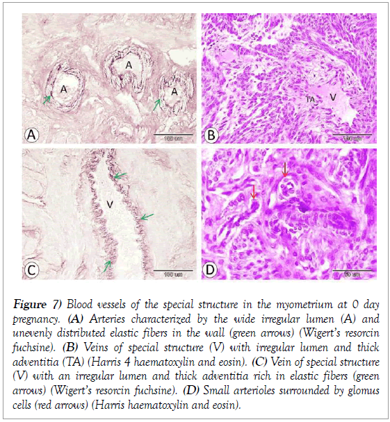
Spirally oriented arteries were present within the myometrium with a wide lumen, clear internal elastic membrane and elastic fibers distributed at a various layer of the vessels wall (Figure 6C). Another spirally oriented arterioles observed and characterized by clear glomus cells in the tunica media (Figure 6D). In addition, there was another special type of arteries which characterized by the narrow lumen, thick wall and unevenly distributed elastic fibers in the wall (Figure 6E). Although, there was another type of arteries characterized by the wide irregular lumen and unevenly distributed elastic fibers in the wall (Figure 7A).
Figure 7: Blood vessels of the special structure in the myometrium at 0 day pregnancy. (A) Arteries characterized by the wide irregular lumen (A) and unevenly distributed elastic fibers in the wall (green arrows) (Wigert’s resorcin fuchsine). (B) Veins of special structure (V) with irregular lumen and thick adventitia (TA) (Harris 4 haematoxylin and eosin). (C) Vein of special structure (V) with an irregular lumen and thick adventitia rich in elastic fibers (green arrows) (Wigert’s resorcin fuchsine). (D) Small arterioles surrounded by glomus cells (red arrows) (Harris haematoxylin and eosin).
Veins of special structure could also be observed within myometrium bearing an irregular lumen and relatively thick fibrillar adventitia mainly consists of collagenous and elastic elements (Figure 7B and 7C). Small arterioles of a special structure were lined with 2-3 endothelial cells and surrounded by glomus cells (Figure 7D).
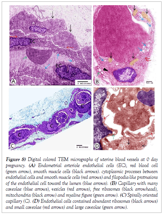
Transmission electron microscopic observations of uterine blood vessels at 0day pregnancy showed that there were endometrial arterioles with endothelial cells surrounded by two smooth muscle layers. Endothelial cells and the inner smooth muscle cells gave cytoplasmic processes to each other and numerous filopodia-like protrusions of the endothelial cell toward the lumen (Figure 8A). Capillary with overlapping endothelial cell ends also observed and characterized by many caveolae of different size distributed singly or in small groups, numerous small vesicles, many free ribosomes, some mitochondria, and myaline figure (Figure 8B). In addition, spirally oriented capillary demonstrated and characterized by abundant ribosomes and rich in caveolae which small in size and arranged in rows or large in size and distributed singly (Figure 8C and 8D)
Figure 8: Digital colored TEM micrographs of uterine blood vessels at 0 day pregnancy. (A) Endometrial arteriole endothelial cells (EC), red blood cell (green arrow), smooth muscle cells (black arrows). cytoplasmic processes between endothelial cells and smooth muscle cells (red arrows) and filopodia-like protrusions of the endothelial cell toward the lumen (blue arrows). (B) Capillary with many caveolae (blue arrows), vesicles (red arrows), free ribosomes (black arrowhead), mitochondria (black arrow) and myaline figure (green arrow). (C) Spirally oriented capillary (C). (D) Endothelial cells contained abundant ribosomes (black arrows) and small caveolae (red arrows) and large caveolae (green arrow).
7 days of pregnancy
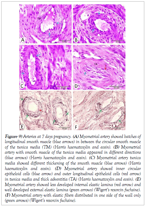
The most prominent characteristic features of this stage of pregnancy were the observation of numerous vessels of special morphological character. Some arteries in the myometrium contained batches of longitudinal smooth muscle in between the circular smooth muscle of the tunica media (Figure 9A). In some arteria, smooth muscle of the tunica media appeared in different directions (Figure 9B). Other arterial tunica media showed different thickening of the smooth muscle from 1 to 3 layer (Figure 9C). In addition, there was arteria showed inner circular and outer longitudinal epitheloid cells in tunica media and thick adventitia (Figure 9D).
Figure 9: Arteries at 7 days pregnancy. (A) Myometrial artery showed batches of longitudinal smooth muscle (blue arrows) in between the circular smooth muscle of the tunica media (TM) (Harris haematoxylin and eosin). (B) Myometrial artery with smooth muscle of the tunica media appeared in different directions (blue arrows) (Harris haematoxylin and eosin). (C) Myometrial artery tunica media showed different thickening of the smooth muscle (blue arrows) (Harris haematoxylin and eosin). (D) Myometrial artery showed inner circular epitheloid cells (blue arrow) and outer longitudinal epitheloid cells (red arrow) in tunica media and thick adventitia (TA) (Harris haematoxylin and eosin). (E) Myometrial artery showed less developed internal elastic lamina (red arrow) and well developed external elastic lamina (green arrows) (Wigert’s resorcin fuchsine). (F) Myometrial artery with elastic fibers distributed in one side of the wall only (green arrows) (Wigert’s resorcin fuchsine).
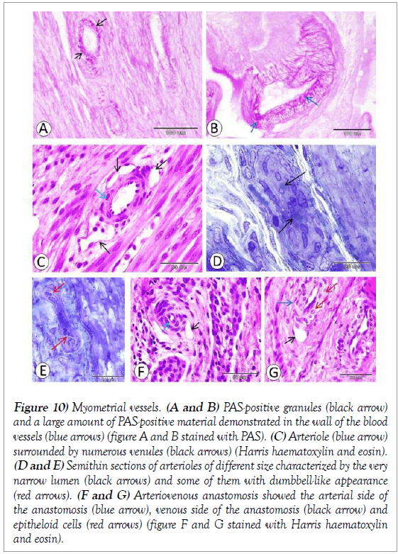
Our observation also revealed that Internal elastic lamina of some arteries appeared less developed but the external elastic lamina clearly well developed (Figure 9E) and in other arteries the elastic fibers distributed in one side of the wall only (Figure 9F). PAS-positive granules and PAS-positive material demonstrated in the wall of the blood vessels in the large amount (Figure 10A and 10B). Moreover, our observation also revealed that some arterioles surrounded by numerous venules (Figure 10C). Other arterioles of different size characterized by a very narrow lumen and some of them with dumbbelllike appearance (Figure 10D and 10E).
Figure 10: Myometrial vessels. (A and B) PAS-positive granules (black arrow) and a large amount of PAS-positive material demonstrated in the wall of the blood vessels (blue arrows) (figure A and B stained with PAS). (C) Arteriole (blue arrow) surrounded by numerous venules (black arrows) (Harris haematoxylin and eosin). (D and E) Semithin sections of arterioles of different size characterized by the very narrow lumen (black arrows) and some of them with dumbbell-like appearance (red arrows). (F and G) Arteriovenous anastomosis showed the arterial side of the anastomosis (blue arrow), venous side of the anastomosis (black arrow) and epitheloid cells (red arrows) (figure F and G stained with Harris haematoxylin and eosin).
At this stage of pregnancy Arteriovenous anastomosis also observed with thick fibrous adventitia. The arterial side of the anastomosis may show narrow lumen and thick media (Figure 10F). However, another artery tunica media formed from epitheloid cells (Figure 10G).
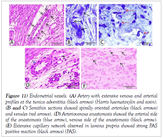
Endometrial arteries showing special character one of them showed extensive venous or arterial profiles at the tunica adventitia of many arteries (Figure 11A). Spirally oriented arterioles and venules also observed at the endometrium (Figure 11B and 11C). In addition, arteriovenous anastomosis observe at the endometrium (Figure 11D). Extensive capillary network observed in lamina propria and take strong PAS positive reaction (Figure 11E).
Figure 11: Endometrial vessels. (A) Artery with extensive venous and arterial profiles at the tunica adventitia (black arrows) (Harris haematoxylin and eosin). (B and C) Semithin sections showed spirally oriented arterioles (black arrows) and venules (red arrows). (D) Arteriovenous anastomosis showed the arterial side of the anastomosis (blue arrow), venous side of the anastomosis (black arrow). (E) Extensive capillary network observed in lamina propria showed strong PAS positive reaction (black arrows) (PAS).
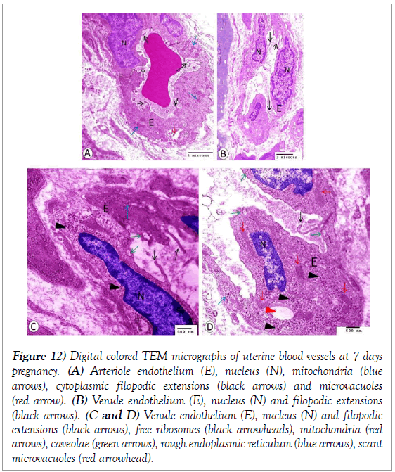
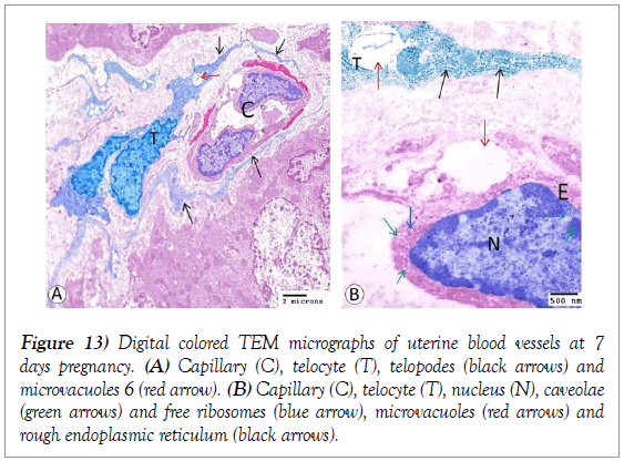
At this stage of pregnancy transmission electron microscopic observations of uterine blood vessels showed that endothelial cells of the arteriole appeared swell to a plump with abundant mitochondria, numerous cytoplasmic filopodic extensions toward the lumen and few microvacuoles. The nuclei of these endothelial cells change to large nuclei (Figure 12A). However, the endothelium of the venule characterized by the presence of abundant free ribosomes, many mitochondria, some caveolae, few rough endoplasmic reticulum, scant microvacuoles and several filopodic extensions toward the lumen (Figure 12B-12D). Capillary also observed and encircled by telocyte (Figure 13A). Endothelium of the capillary contained caveolae and free ribosomes and large microvacuoles resemble the microvacuoles which present in the nearby telocyte (Figure 13B).
Figure 12: Digital colored TEM micrographs of uterine blood vessels at 7 days pregnancy. (A) Arteriole endothelium (E), nucleus (N), mitochondria (blue arrows), cytoplasmic filopodic extensions (black arrows) and microvacuoles (red arrow). (B) Venule endothelium (E), nucleus (N) and filopodic extensions (black arrows). (C and D) Venule endothelium (E), nucleus (N) and filopodic extensions (black arrows), free ribosomes (black arrowheads), mitochondria (red arrows), caveolae (green arrows), rough endoplasmic reticulum (blue arrows), scant microvacuoles (red arrowhead).
Figure 13: Digital colored TEM micrographs of uterine blood vessels at 7 days pregnancy. (A) Capillary (C), telocyte (T), telopodes (black arrows) and microvacuoles 6 (red arrow). (B) Capillary (C), telocyte (T), nucleus (N), caveolae (green arrows) and free ribosomes (blue arrow), microvacuoles (red arrows) and rough endoplasmic reticulum (black arrows).
Discussion
Rabbit is considered one of the widely used laboratory animals. Rabbits have been used extensively for basic research in drug and bacteria, toxicology, healing, tissue and organ culture, mycology, skin sensitivity, immunology, ophthalmology, oncology and reproductive biology [10]. The rabbit is believed to be a helpful model for comparative biology in humans, concerning sperm capacitation and the general reactivity of the female genital tract during the reproductive cycle [11].
In this study, we established for the first time a detailed investigation of the arterial, venous and lymphatic mapping of the uterus at early stages of pregnancy in the rabbit. The viability of this vessels were very important for the female reproduction and fetus maintenance. It was recognized in the present study that arteries were congested and demonstrated in varies directions in the mesometrial region.
Rabbit has uteroovarian arteries running within the mesometrium and giving secondary and tertiary vessels (radial arteries). These radial arteries give the premyometrial radial arteries enter the uterine wall supplies the myometrium. The uterus is drained by a venous system that parallels the arterial tree, with closely apposed arteries and veins [1].
The present study demonstrated that arteries of mesometrial and myometrial regions were characterized by well-developed elastic lamina which helps the vessel to bear changes in blood pressure during pregnancy.
Large increases in uteroplacental blood flow during gestation are essential for normal foetal growth and survival and occur in every mammalian species studied, including human beings [12]. During pregnancy, the passive (fully dilated) diameter of the human uterine artery is approximately doubled [13], with similar changes reported in rodents, sheep, pigs and guinea pigs [14,15].
The myometrial arterial supply that course between the outer and middle thirds of the myometrium and this zone is referred to as the ‘vascular zone’. Because of their semicircular course, these arteries are referred to as ‘arcuate’ arteries [16]. Our observation revealed that myometrial arteries characterized by its wide lumen, thin muscular coat, and vascularized adventitia.
Arteries and veins are arranged in close apposition in a number of species [17]. Our observation revealed that mesometrial and myometrial veins characterized by its thin wall encircled by a thick adventitia rich in collagen and elastic fibers.
The uterus is an immunologically unique organ in that it must be able to protect itself effectively from invading pathogens, but at the same time is faced with periodic exposures to foreign cells and tissues, namely allogeneic spermatozoa and the fetal-placental unit. Even though the uterus seems to be an effective site of immunization for some antigens [18]. In rats, mice, and similar small mammals, there is only one main lymphatic plexus lying between the circular and longitudinal muscles. In addition, Endometrium possessed few or no lymphatics [19].
Our observation revealed that several thin- walled wide-lumened lymphatics were recorded within the mesometrium. Their wall is composed of an intimal tunic surrounded by a thick fibrous layer consisting of collagenous, elastic fibers and connective tissue cells. Some of these vessels demonstrate valves. The rat showed similar observation [20].
Myometrial lymphatics were composed of an intimal tunic surrounded by a fibrous layer of collagenous and elastic fibers. The valves of these vessels were mainly thin and consisted of a single cellular layer. The wall of myometrial lymph vessels showed a positive reaction with PAS-AB combination. The lymphatic vessel in the endometrium appeared as wide irregular thin walled vessels similar observation obtained in the rat [21].
Rabbit arterial sinuses as arterial device affecting the pulse pressure and linear velocity of maternal blood flow and regulate optimal condition for nutritional exchange [22]. The present study revealed that Several arterioles, venules observed at the different layer of the uterus. in addition, spiral artery also observed with a wide lumen and spiral course. As well as, arterial sinuses characterized by wide irregular lumen, a single layer of smooth muscle and unevenly distributed elastic fibers.
Arteriovenous anastomosis demonstrated at a various layer of the uterus. Some of this vessels showed several glomus cells. Arteriovenous anastomoses are one of the key adaptations for thermoregulation [23]. The glomus cell observed at this study in the wall of arteries, arterioles, arteriovenous anastomosis, and spirally oriented arterioles. the glomus cells can be considered as a highly reactive special type of cell, which cause a reduction in the diameter of the lumen by their ability to swell or even complete obliteration of the vessel lumen [24-26].
The histological and histochemical investigation of the uterine vasculature in the rabbit revealed several blood vessels of special regulatory devices which indicate a unique function. These vessels were observed at different uterine parts.
Our observation revealed that there was fibrinoid deposition at some arteries similar observation reported in women [27,28]. In addition, Fibrinoid deposition and elastic fibers contents of vessels will increase at 7 days pregnancy of pregnancy. gestation sclerosis discused as a type of sclerosis developed due to circulatory demands increased [29]. However, gestational sclerosis considered as physiological processes as they are not uniform in nature, intensity and distribution [26].
The present study demonstrated some spirally oriented arterioles with extreme thickening of their wall by glomus cells in addition to arterioles with a narrow lumen. This constriction of arterioles increases peripheral resistance to blood flow giving an important role in the regulation of systemic blood pressure. Similar observations were recorded in camel ovaries [26] and camel skin [30].
In our study, Various vessels of special structure detected at different parts of the uterus. The vessels of the special structure are supposed to possess an important regulatory function for the peripheral circulation. They exert an active function on the blood flow and pressure regulation. This function is attained either through the contraction of the smooth muscle fibers or by the presence of glomus cells which cause a reduction in the diameter of the lumen by their ability to swell [25].
Ultrastructure study revealed that numerous filopodia-like protrusions at the arteriole endothelium toward the lumen. The formation of filopodial structures described under inflammatory conditions and in response to the inflammatory mediators [31]. Mild but significant inflammatory activity is involved in the development of normal pregnancy, which might have important physiological roles [32,33]. The maternal inflammatory response is supposed to be modulated to allow the establishment and maintenance of a viable pregnancy.
Abundant caveolae of different sizes and different arrangement were observed in the endothelial cells of the capillary. The permeability of endothelial cells is usually described in electron microscope studies as related to the presence of caveolae [34]. With the advancement of pregnancy, endothelial cells of some vessels appeared swell to a plump and Similar observation obtained at early stages of human pregnancy [35].
Our data revealed that telocyte encircled the capillary with similar vacuoles in both. The telocyte discussed as play a role in juxta and paracrine signaling [36]. In addition, telocyte expressed estrogen and progesterone receptors and act as a hormonal sensor [37,38]. However, it always needs to be noted the exceptional cases out of such rules, and cutaneous horns at the gross level should prompt consideration of biopsy for the definite diagnosis. Our case #10 also seems to support such speculation.
REFERENCES
- Osol G, Mandala M. Maternal uterine vascular remodeling during pregnancy. Physiology. 2009;24(1):58-71.
- Antonios TF, Nama V, Wang D, et al. Microvascular remodeling in preeclampsia: quantifying capillary rarefaction accurately and independently predicts preeclampsia. American journal of hypertension. 2013;26(9):1162-9.
- Klauber N, Rohan RM, Flynn E, et al. Critical components of the female reproductive pathway are suppressed by the angiogenesis inhibitor AGM-1470. Nature medicine. 1997;3(4):443.
- Roberts JM. Endothelial dysfunction in preeclampsia. Semin Reprod Endocrinol. 1998;16:5-15.
- Barker DJ. Fetal growth and adult disease. BJOG: An International Journal of Obstetrics & Gynaecology. 1992;99(4):275-6.
- Oliver G. Lymphatic vasculature development. Nature Reviews Immunology. 2004;4(1):35.
- Red-Horse K. Lymphatic vessels in the uterine wall. Placenta. 2008;29 (Suppl A): S55-9.
- Bancroft JD, Gamble M. Theory and practice of histological and histochemical Techniques, 2002.
- Reynolds ES. The use of lead citrate at high pH as an electronopaque stain in electron microscopy. J Cell Biol 1963;17:208212.
- Hafez ESE. Reproduction and Breeding Techniques for Laboratory Animals. Lea and Febiger. Philadelphia, 1970.
- Barberini F, Correr S, De Santis F, et al. The Mucosa of the Rabbit Vagina a Morphodynamic study by transmission and Scanning Electron Microscopy. Prog. Clin. Biol. Res. 1992;256:15-420.
- Mandala M, Osol G. Physiological remodelling of the maternal uterine circulation during pregnancy. Basic & clinical pharmacology & toxicology. 2012;110(1):12-8.
- Palmer S K, Zamudio S, Coffin C, et al. Quantitative estimation of human uterine artery blood flow and pelvic blood flow redistribution in pregnancy. Obstetrics and gynecology. 1992;80(6):1000-6.
- Annibale DJ, Rosenfeld, CR, Stull, JT, et al. Protein content and myosin light chain phosphorylation in uterine arteries during pregnancy. American Journal of Physiology-Cell Physiology. 1990;259(3):C484-9.
- van der Heijden OW, Essers YP, Spaanderman ME, et al. Uterine artery remodeling in pseudopregnancy is comparable to that in early pregnancy. Biology of reproduction. 2005;73(6):1289-93.
- Farrer-Brown G, Beilby JOW, Tarbit MH. The blood supply of the uterus. BJOG: An International Journal of Obstetrics & Gynaecology. 1970;77(8):673-81.
- Celia G, Osol G. Uterine venous permeability in the rat is altered in response to pregnancy, vascular endothelial growth factor, and venous constriction. Endothelium 2005;12(1-2):81-8.
- Beer AE, Billingham RE. Implantation, transplantation, and epithelial-mesenchymal relationships in the rat uterus. Journal of Experimental Medicine. 1970;132(4):721-36.
- Hoggan G, Hoggan FE. On the comparative anatomy of the lymphatics of the uterus. Journal of anatomy and physiology 1881;16(Pt 1),50.
- Head JR, Lande IJ. Uterine lymphatics: passage of ink and lymphoid cells from the rat’s uterine wall and lumen. Biology of reproduction. 1983;28(4):941-55.
- Lauweryns JM, Cornillie FJ. Topography and ultrastructure of the uterine lymphatics in the rat. European Journal of Obstetrics & Gynecology and Reproductive Biology. 1984;18(5-6):309-27.
- Hayashi K, Kusakabe KT, Khan H, et al. Characteristic patterns of maternal and fetal arterial construction in the rabbit placenta. Medical molecular morphology 2014;47(2):76-82.
- Tarasoff FJ, Fisher HD. Anatomy of the hind flippers of two species of seals with reference to thermoregulation. Canadian Journal of Zoology 1970;48(4):821-9.
- Hussein MM, Hassan AHS. Special Histological Features of the Angioarchitecture of the Ovaries in the Egyptian Buffaloes (Bubalus bubalis). Journal of Advanced Microscopy Research 2018;13(3):298-305.
- Mohamden DM. Angioarchitecture of the lungs of the camel (camelus dromedarius). M.V.Sc. Egypt: Assuit University, 2009.
- Mokhtar DM, Abd‐Elhafez EA. Morphological Studies on the Peripheral Circulation of the Ovary in One-Humped Camel (Camelus dromedarius). Anatomia, Histologia, Embryologia 2016;45(4):319-28.
- Kam EP, Gardner L, Loke YW, et al. The role of trophoblast in the physiological change in decidual spiral arteries. Human Reproduction 1999;14(8):2131-8.
- Pijnenborg R, Dixon G, Robertson WB, et al. Trophoblastic invasion of human decidua from 8 to 18 weeks of pregnancy. Placenta 1980;1(1):3-19.
- Dahme E. Morphological changes in the vessel wall in spontaneous animal arteriosclerosis. J. Atheroscler. Res. 1962;2,153-160.
- Fath-Elbab MR, Abou-Elhamd AS. Special cutaneous vascular elements in one-humped camel (Camelus dromedarius). Journal of Advanced Veterinary and Animal Research 2016;3(2):106-11.
- De Smet F, Segura I, De Bock K, et al. Mechanisms of vessel branching: filopodia on endothelial tip cells lead the way. Arteriosclerosis, thrombosis, and vascular biology 2009;29(5):639-49.
- Guilbert L, Robertson SA, Wegmann TG. The trophoblast as an integral component of a macrophage-cytokine network. Immunology and cell biology 1993;71(1):49.
- Palm M., Axelsson O, Wernroth L, et al. Involvement of inflammation in normal pregnancy. Acta obstetricia et gynecologica Scandinavica 2013;92(5):601-5.
- Peek M, Landgren BM, Johannisson E. The endometrial capillaries during the normal menstrual cycle: a morphometric study. Human Reproduction 1992;7(7):906-11.
- Pijnenborg R, Bland JM, Robertson WA, et al. Uteroplacental arterial changes related to interstitial trophoblast migration in early human pregnancy. Placenta 1983;4(4):397-413
- Popescu LM, Hinescu ME, Ionescu N, et al. Interstitial cells of Cajal in pancreas. Journal of cellular and molecular medicine 2005;9(1):169-90.
- Hutchings G, Williams O, Cretoiu D, et al. Myometrial interstitial cells and the coordination of myometrial contractility. Journal of cellular and molecular medicine 2009;13(10):4268-82.
- Cretoiu SM, Simionescu DCA, Popescu LM () Telocytes in human fallopian tube and uterus express estrogen and progesterone receptors. In Sex Steroids. Intech., 2012


