A horseshoe kidney with a unique constellation of renal vasculature: A case report
Received: 19-Dec-2018 Accepted Date: Dec 26, 2018; Published: 07-Jan-2019, DOI: 10.37532/1308-4038.19.12.002
Citation: Zhang G, Schmidt RR. A horseshoe kidney with a unique constellation of renal vasculature: A case report. Int J Anat Var. Mar 2019;12(1): 002-004.
This open-access article is distributed under the terms of the Creative Commons Attribution Non-Commercial License (CC BY-NC) (http://creativecommons.org/licenses/by-nc/4.0/), which permits reuse, distribution and reproduction of the article, provided that the original work is properly cited and the reuse is restricted to noncommercial purposes. For commercial reuse, contact reprints@pulsus.com
Abstract
Horseshoe kidney with variable vascular supply is of interest to clinicians and anatomists due to its immense surgical significance. A horseshoe kidney encountered during cadaveric dissection is reported here. The left kidney is vertical and the right kidney is slightly horizontal. The isthmus is narrow and 1.5 cm inferior to the inferior mesenteric artery. The hilum of the right kidney opens anteriorly and the left opens anteromedially. The ureters lie on the anterior surfaces of both kidneys. In addition to renal arteries arising from the aorta described in previous reports, a unique renal artery (Renal Artery 3) was found supplying the right kidney. This RA3 originates from the aorta at L3 vertebral level and takes a long and tortuous course to reach the hilum of the right kidney. Such arterial variation is important for surgical procedures involving the abdominal aorta.
Keywords
Horseshoe kidney; Renal arteries; Anatomic variation
Introduction
Horseshoe kidney (HSK) is a common congenital renal defect found in 0.25% of the general population [1]. In most instances of HSK, both kidneys fuse at their lower poles with the isthmus bridging the two renal masses. HSKs usually demonstrate three anatomical anomalies: ectopic position, malrotation, and variable vasculature [1]. Although some patients with HSK are asymptomatic, HSK is associated with abnormalities such as secondary renal pathology, ureteropelvic junction obstruction, vesicoureteral reflux, and certain renal tumors [2].
The arterial supply of HSK is widely variable. It is reported that 80% of HSK is associated with abnormalities in arterial supply [3]. In a study of 90 HSKs, 387 renal arteries were identified [4]. The accessory renal arteries may originate directly from the aorta at various locations, from the common iliac artery and inferior mesenteric artery. Management of the aberrant renal vasculature poses significant risk to surgical procedures involving the abdominal aorta in patients with HSK [5]. Sacrificing or ligation of any of these renal arteries may lead to renal infarction and as result surgeons make a concerted effort to preserve the renal vasculature [6]. Here we report a case of HSK with a unique and rare renal vasculature encountered during medical students’ cadaveric dissection.
Case Report
The horseshoe kidney described in the present report was found in an 83- year old male during routine cadaver dissection at Sidney Kimmel Medical College, Philadelphia. This was the first HSK case encountered among 532 donations [0.19%] in the dissection laboratory at SKMC during 2008-2018. The donated body at SKMC was embalmed with Maryland State Anatomical Solution Concentrate and had the following composition: Formaldehyde (2.0-3.0%), methanol (24.0-28.0%), Phenol (22.0-26.0%) and Glycerine (38.0-42.0%). The concentrate was diluted 1:2 with water and another 16- 32 oz of glycerine and 4.0 oz of Saturol/liter were added for the embalming procedure.
Differential malrotation of both kidneys
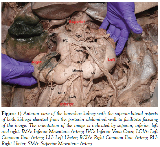
This was an asymmetric horseshoe kidney with a narrow isthmus that deviated slightly to the left. The isthmus connected the inferior poles of the two kidneys (Figure 1). It was situated anterior to the abdominal aorta at the third lumbar vertebral level (L3), about 1.5 cm inferior to the main trunk of the inferior mesenteric artery. The latter lay anterior to the isthmus and generated a shallow impression on the surface of the isthmus.
Figure 1) Anterior view of the horseshoe kidney with the superior-lateral aspects of both kidneys elevated from the posterior abdominal wall to facilitate focusing of the image. The orientation of the image is indicated by superior, inferior, left and right. IMA: Inferior Mesenteric Artery; IVC: Inferior Vena Cava; LCIA: Left Common Iliac Artery; LU: Left Ureter; RCIA: Right Common Iliac Artery; RU: Right Ureter; SMA: Superior Mesenteric Artery.
The left kidney was larger than the right. It measured 13 cm in length (from superior to inferior pole), 7 cm in width, and 5 cm in thickness. The right kidney measured 12.5 cm in length, 6.5 cm in width and 4.5 cm in thickness. Both kidneys exhibited malrotation. However, the malrotation was not symmetrical. The left kidney was largely vertical, while the right kidney was horizontal. The hilum of the left kidney faced anteriorly and medially, whereas the hilum of right kidney was directed completely anteriorly. The right ureter descended along the anterior surface of the kidney and created a deep indentation delineating the kidney into a 2/3 superior portion (positioned laterally) and a 1/3 inferior portion. The inferior pole was located in a medial position, anterior to the inferior vena cava. Owing the rather vertical orientation of the left kidney, the left ureter left the hilum in an anterior/medial position and imparted a shallow impression on the inferior pole of the left kidney.
Arterial supply and venous drainage
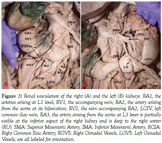
Two renal arteries (RA1 Left and RA1 Right) arose from either side of the aorta at L1 vertebral level, immediately inferior to the superior mesenteric artery (Figures 2A and 2B). These arteries travelled posterosuperior to the accompany renal veins (RV1 Left and RV1 Right) and reached the hilum of each kidney. Both of these renal arteries measured 5 mm in diameter. Both accompanying renal veins drained to the inferior vena cava at approximately the L1 vertebral level.
Figure 2) Renal vasculature of the right (A) and the left (B) kidneys. RA1, the arteries arising at L1 level; RV1, the accompanying vein; RA2, the artery arising from the aorta at its bifurcation; RV2, the vein accompanying RA2; LCIV, left common iliac vein; RA3, the artery arising from the aorta at L3 lever is partially visible at the inferior aspect of the right kidney and is deep to the right ureter (RU). SMA: Superior Mesenteric Artery; IMA: Inferior Mesenteric Artery; RCIA: Right Common Iliac Artery; RGVS: Right Gonadal Vessels; LGVS: Left Gonadal Vessels; are all labeled for orientation.
Inferiorly, an artery (RA2) arose from the anterior aspect of the aorta at its bifurcation. This artery had a diameter of 5 mm. It sent three branches to the inferior pole of the right kidney and the isthmus. Its larger terminal branch entered the inferior pole of the left kidney. The vein accompanying this artery (RV2) drained to the left common iliac vein. This vein also drained the small vein accompanying the artery (RA3) described below. The median sacral artery branched from posterior aspect of the aorta at its bifurcation and followed a typical course descending down to pelvis on the anterior surface of the sacrum.
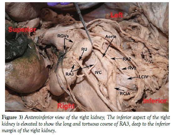
Another artery, renal artery 3 (RA3), was unique to this case and different from the renal arteries described in previous reports. RA3 measured about 3 mm in diameter, arose from the right and posterior aspect of the aorta at the L3 vertebral level, posterior to the inferior pole of the right kidney. It took a long and tortuous course deep to the inferior pole of the right kidney, coursed in the fibrous adipose tissue around the inferior margin of the right kidney and deep to the right ureter. It entered the hilum from an inferior/ anterior aspect of the kidney and lateral to the right ureter (Figure 3). The vein accompanying RA3 drained to RV2, the companying vein of RA2.
Renal calyces and trajectory of the ureters
The hilum of each kidney was covered by the renal capsule. The renal sinus of the right kidney was broad and opened anteriorly and superiorly. Four major renal calyces and some minor calyces were visible in the renal sinus and external to renal tissue. The renal pelvis was anterior to the calyces. The right ureter left the hilum, descended medial to the psoas major and lateral to the right common iliac artery (Figure 2A), and reached the urinary bladder at the normal entering point.
The left kidney occupied a vertical position with its hilum directed anteromedially. Three major calyces were seen external of the renal tissue, but the minor calyces were located deep into the renal tissue. The left ureter took a normal course, descending into the pelvis and entering the urinary bladder.
Discussion
Owing the immense surgical significance, numerous reports have attempted to map the various arterial supplies of HSK. Graves proposed a classification system consisting of six basic patterns for the arterial supply of HSK in 1969 [7]. Subsequently, many authors have categorized the renal arterial supply of HSK according to Graves’ classification system. However, more recent investigations of HSK cases demonstrate the renal vasculature associated with HSK exhibits even more extensive variations beyond these six basic patterns [8].
The arterial supply of HSK reported here is different from any of the six basic patterns described previously by Graves. The RA3 supplying the right kidney is embedded in fibrous adipose tissue behind the kidney and follows a long and tortuous course, before reaching the hilum of the kidney. Management of a renal artery such as this in a surgical procedure can be challenging due to its hidden position. Unexpected disruption of such arteries may cause serious complications for the patient. Therefore, pre-procedural imaging is essential for any surgery associated with HSK. While ultrasound is sufficient for diagnosis of horseshoe kidney, detection of accessory vasculature and surrounding structures are best achieved via CT and MRI. Meanwhile, future data collection is essential for mapping the extensive and complex patterns of renal arterial supply in HSK.
Horseshoe kidneys usually fail to migrate superiorly to their normal position in the retroperitoneal renal fossa. It is often regarded that the U-shaped kidney is arrested in its ascent by the root of the inferior mesenteric artery. The case reported here does not support this theory since the isthmus is located about 1.5 cm inferior to the root of the IMA. Rather, the artery arising from the abdominal aorta at its bifurcation (RA2) supports the hypothesis that persistent embryonic arteries may also inhibit the ascent of the horseshoe kidney [9].
The HSK reported here is asymmetrically fused and its narrow isthmus deviates to the left. Asymmetrical or laterally fused horsesshoe kidneys may result from differential displacement of embryonic renal masses [10]. It is also observed that the right kidney shows non-rotation and the left kidney incomplete rotation. The different degrees of malrotation of the kidneys in the present case suggest differential rotation processes of the two kidneys. Due to the malrotation, especially seen in the right kidney, the junction between the ureter and the renal pelvis forms a sharp angle which may lead to urinary drainage problems and stasis.
Acknowledgements
The authors wish to thank all who donated their bodies for the training of the future healthcare professionals. The authors also acknowledge Dr. Bruce Fenderson from the Department of Pathology, Anatomy & Cell Biology at Sidney Kimmel Medical College for editing.
REFERENCES
- Natsis K, Piagkou M, Skotsimara A, et al. Horseshoe kidney: a review of anatomy and pathology. Surg Radiologic Anat. 2014;36:517-26.
- Castro JE, Green NA. Complications of horseshoe kidney. Urology. 1975;6:344-7.
- Connelly TL, McKinnon W, Smith RB III, et al. Abdominal aortic surgery and horseshoe kidney: report of six cases and a review. Arch Surg. 1980;115:1459-63.
- Glodny B, Peterson J, Hofmann KJ, et al. Kidney fusion anomalies revisited: clinical and radiological analysis of 209 cases of crossed fused ectopia and horseshoe kidney. BJU Int. 2009;103:224-35.
- Lotina SL, Davidovic LB, Kostic DM, et al. Surgery of abdominal aorta with horseshoe kidney. Srp Arh Celok Lek. 1997;125:36-44.
- Sharma K, Babrowski T, Milner R. A novel chimney approach for management of horseshoe kidney during EVAR. EJVES Short Rep. 2016;33: 16-9.
- Graves FT. The arterial anatomy of the congenitally abnormal kidney. Br J Surg. 1969;56:533-41.
- Tijerina GO, Uresti J, Urrutia VE, et al. Anatomical study of the horseshoe kidney. Int J Morphol. 2009;27:491-4.
- Yoshinaga K, Kodama K, Tanii I, et al. Morphological study of a horseshoe kidney with special reference to the vascular system. Anat Sci Int. 2002;77:134-9.
- Cook WA, Stephens FD. Fused kidneys: morphologic study and theory of embryogenesis. Birth Defects Orig Artic Ser. 1976;13:327-40.


