A muscle variant in the region of the popliteal fossa associated with the semitendinosus muscle
Received: 23-Oct-2018 Accepted Date: Oct 31, 2018; Published: 13-Nov-2018
Citation: Elliott GE. A muscle variant in the region of the popliteal fossa associated with the semitendinosus muscle. Int J Anat Var. Dec 2018;11(4):127-128.
This open-access article is distributed under the terms of the Creative Commons Attribution Non-Commercial License (CC BY-NC) (http://creativecommons.org/licenses/by-nc/4.0/), which permits reuse, distribution and reproduction of the article, provided that the original work is properly cited and the reuse is restricted to noncommercial purposes. For commercial reuse, contact reprints@pulsus.com
Abstract
Posterior thigh pain is frequently caused by straining the posterior thigh muscles. Because of these clinical implications, it is important for surgeons to have a comprehensive knowledge of muscle variations. During a routine dissection class at Ross University School of Medicine, a 57-year-old male cadaver presented with a unilateral muscle variation of the semitendinosus.
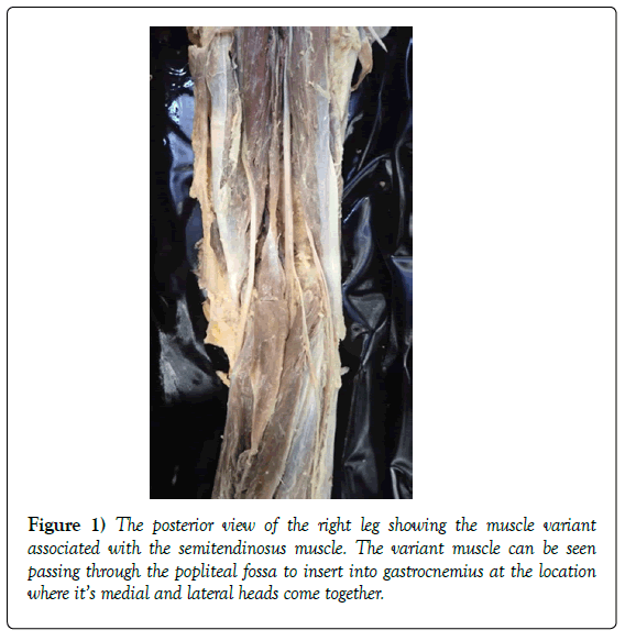
In this case, an accessory muscle appears to originate from the belly of the semitendinosus muscle and passes down, through the popliteal fossa, to insert between the medial and lateral heads of the gastrocnemius muscle. The variant muscle may have compressed the popliteal vein and part of the tibial nerve during its pathway with potential implications for muscle function in the leg and foot. To the best of the author’s knowledge, such a rare anatomical variation has not yet been reported in the literature before.
Keywords
Gastrocnemius; Muscle; Popliteal; Semitendinosus; Variant
Introduction
Posterior thigh pain is often associated with straining of the hamstring muscles [1]. Because of the association of these muscles with pain, it is important to be aware of anatomical variants of the muscles that may cause unusual or new symptoms [1-3]. The hamstrings consist of the semitendinosus, semimembranosus and biceps femoris muscles. All three of these muscles are involved in extension of the hip joint and flexion of the knee joint [4,5].
The biceps femoris has two heads, one arises from the femur, and one from the ischial tuberosity, while the semitendinosus and semimembranosus muscles are simple muscles that originate on the ischial tuberosity and insert onto the medial aspect of the tibia [4,5]. In this instance, an extra muscle appears to arise from the belly of the semitendinosus, passing superficially through the popliteal fossa, before inserting between the two heads of the gastrocnemius muscle and has not been documented before in the literature.
Case Report
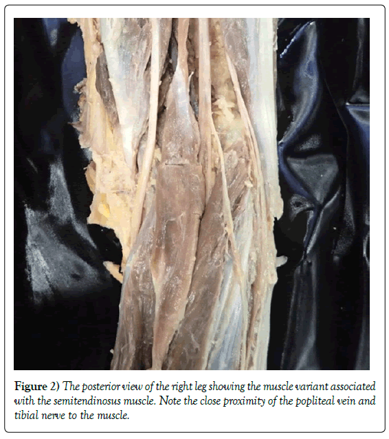
During a routine dissection of the posterior thigh and popliteal fossa at Ross University School of Medicine, a muscle variant was discovered in a 57-year-old male cadaver. This unilateral muscle variant, located in the right leg, appears to be an additional belly of the semitendinosus muscle (Figures 1 and 2). The variant muscle arises from the belly of the semitendinosus muscle, via a small tendinous slip, before passing superficially through the center of the popliteal fossa (Figure 1).
In the leg, this variant muscle passes between the medial and lateral heads of the gastrocnemius, inserting via a small tendon at the location where these two heads join. The muscle is located superficially in the popliteal fossa, passing superior to the popliteal vein and artery. Close proximity of this muscle to the popliteal vein may have caused a compression effect on the vein (Figure 2). In this particular case, compression of the tibial nerve may have also occurred due to the close relationship of the muscle with the nerve in relation to the gastrocnemius muscle (Figure 2). Along its length, the muscle measured 12 inches and at its widest, 1 inch, in the middle of the popliteal fossa. No medical history for the patient indicates prior knowledge of the existence of this muscle variant.
Discussion
Interestingly, a number of case studies have documented similar muscle variants in the thigh and popliteal fossa with different origins and insertions [6-10]. These muscle variations in the hamstring region have potential clinical implications because of their close proximity to the neurovasculature of the popliteal fossa. For example, Barry and Bothroyd [8] identified the tensor fasciae suralis muscle that originates from two fascial slips of the fascia lata and fascia on deep and superficial surfaces of the biceps femoris, respectively. The authors noted that the muscle passed down into the popliteal fossa deep to the fascia before inserting into the deep surface of the fascia cruris [8]. Parsons [9] also noted a small muscle in the fascia lata of the popliteal fossa, with fibers that ran in a transverse orientation. Kumar and Bhagwat [6] reported a Y-shaped muscle that originated by two small slips from the semitendinosus and long head of biceps femoris, before inserting into gastrocnemius [6]. They suggested the muscle was a variant of the tensor fasciae suralis muscle or that it could be a variant of the third head of the gastrocnemius [6]. Finally, Somayaji et al. [7] also described the muscle formation as previously documented by Kumar and Bhagwat [6]. Somayaji, et al. [7] also highlighted the rare nature of this muscle, commenting that it must have a very low incidence rate because it was only present in one of 300 individuals they had dissected over multiple years at the institute.
The authors [7] speculated on the muscle’s clinical significance, suggesting that it may compress the popliteal vein. Kim and colleagues [10] also identify this muscle as previously described [6,7] but observe that it originated from the biceps femoris only and inserted into the medial head of gastrocnemius [10]. The muscle variant described in this case study is similar to that described by Kumar and Bhagwat [6] and Somayaji, et al. [7], with the notable exception that it has no association with the biceps femoris muscle.
In this case, the muscle does run in close proximity to the popliteal vein so may result in entrapment of the vein and compression of the tibial nerve. Entrapment of the popliteal vein can result in pain in the posterior compartment of the leg, deep vein thrombosis, and swelling [11] Compression of the tibial nerve in the popliteal fossa can be diagnosed by pain in the muscles of the posterior compartment in the leg, as well as a positive Tinel sign [12]. Typically, tibial nerve compression neuropathy is rare, occurs distal/ within the tarsal tunnel, and may be the result of intrinsic entrapment [13,14]. Intrinsic entrapment can be caused by various factors including popliteal cysts, and variant muscles [15]. As a result, surgeons need to be aware that such muscles may be the cause of impingement findings of the tibial nerve and MRI would be the most effective diagnostic imaging tool for these purposes.
Acknowledgement
The author would like to thank those who donate their bodies to medical education for their selfless act of kindness.
REFERENCES
- Fraser PR, Wood AR, Rosales AA. Anatomical variation of the semitendinosus muscle origin. Int J Anat Var. 2013;6:225-7.
- Chakravarthi KK. Unusual unilateral multiple muscular variants of back of thigh. Ann Med Health Sci Res. 2013;3:S1-S2.
- Prasad AM, Nayak S, Deepthinath R, et al. Clinically important variations in the lower limb: A case report. Eur J Anat. 2005;9:167-9.
- Hollinshead WH. Anatomy for surgeons: The back and limbs. Philadelphia: Lipincott Williams and Wilkins. 1982;199-201.
- Gray’s Anatomy: The anatomical basis of clinical practice, wrist and hand. 2006;pp:926-31.
- Kumar GR, Bhagwat SS. An anomalous muscle in the region of the popliteal fossa: A case report. J Anat Soc. 2006;55:65-8.
- Somayaji SN, Vincent R, Bairy KL. An anomalous muscle in the region of the popliteal fossa: Case report. J Anat. 1998;192:307-8.
- Barry D, Bothroyd JS. Tensor fasciae suralis. J Anat. 1924;58:382-90.
- Parsons FG. Note on an abnormal muscle in the popliteal space. J Anat. 1920;54:170-8.
- Kim DI, Kim HJ, Shin C, et al. An abnormal muscle in the superficial region of the popliteal fossa. Anatomical Science International. 2009;84: 61-3.
- Gerkin TM, Beebe HG, Williams DM, et al. Popliteal vein entrapment presenting as deep vein thrombosis and chronic venous insufficiency. 1993; 18:760-6.
- Shookster L, Falke GI, Ducic I, et al. Fibromyalgia and Tinel’s sign in the foot. J Am Podiatr Med Assoc. 2004;9:400-3.
- Tani JC. Fibrous band compression of the tibial nerve branch of the sciatic nerve. Am J Orthop. 1995; 24: 910-2.
- Reichner DR, Evans GRD. Tibial and common peroneal nerve compression in the popliteal fossa: A case report and literature review. The Internet J Plastic Surg. 2004;2:1.
- Sansone V, Sosio C, da Gama Malcher M, et al. Two cases of tibial nerve compression caused by uncommon popliteal cysts. Arthroscopy. 2002;18:E8.


