A rare anatomical variation of the course of the ulnar nerve
2 Department of Nursing, Hamamatsu University School of Medicine, Hamamatsu, Japan
3 Department of Anatomy & Neuroscience, Hamamatsu University School of Medicine, Hamamatsu, Japan
Received: 12-Jan-2018 Accepted Date: Feb 13, 2018; Published: 05-Mar-2018, DOI: 10.37532/1308-4038.18.11.35
Citation: Sato T, Hirano Y, Sato N, et al. A rare anatomical variation of the course of the ulnar nerve. Int J Anat Var. 2018;11(1):035-036.
This open-access article is distributed under the terms of the Creative Commons Attribution Non-Commercial License (CC BY-NC) (http://creativecommons.org/licenses/by-nc/4.0/), which permits reuse, distribution and reproduction of the article, provided that the original work is properly cited and the reuse is restricted to noncommercial purposes. For commercial reuse, contact reprints@pulsus.com
Abstract
A rare case of anatomical variation of the course of the ulnar nerve is presented. The variation consisted of the ulnar nerve piercing, and being tethered to, the fascia connecting the medial intermuscular septum and medial condyle of the right humerus. In addition, the nerve was also twisted as it pierced the fascia, at a distance of 65 mm proximal to the medial epicondyle. Such an anatomical variation of the nerve could potentially cause strain and mechanical damage to the nerve. An awareness of the possibility of such a variation may be of importance in the evaluation of entrapment neuropathy, because presence of such a rare variation could obscure the precise cause of the entrapment neuropathy.
Keywords
Ulnar nerve; Anatomical variation; Entrapment neuropathy; Clinical anatomy; Twisting of nerve
Introduction
The ulnar nerve is the terminal branch of the medial cord of the brachial plexus and descends along the upper arm to the distal third of the humerus, where it runs along the medial intermuscular septum. Thereafter, the nerve courses through the ulnar groove behind the medial epicondyle of the humerus and runs under the aponeurosis between the two head of the flexor carpi ulnaris. Distal to the elbow, the ulnar nerve travels on the medial side of the elbow, giving off several branches innervating flexor carpi ulnaris and flexor digitorum profundus muscles, then coursing to the hand to supply motor and sensory fibers to enable functioning of the hand.
The ulnar nerve is known to susceptible to entrapment on the medial side of the elbow. There are four sites where the ulnar nerve can be trapped: the arcade of Struthers, the ulnar groove, cubital tunnel, and the exit between the two heads of flexor carpi ulnaris. In particular, cubital tunnel is a wellknown location for entrapment, being the second most common site for nerve entrapment in the upper extremity after the carpal tunnel [1]. Although many studies have reported anatomical variations of the ulnar nerve [2-5], in most cases, they pertain to anatomical branch or abnormal communication of the ulnar nerve with other nerves in the arm or hand [2-4]. Reports of anatomical variations of the course of the ulnar nerve are rare [5].
Herein, we present a cadaver case with anatomical variation of the course of the ulnar nerve above the medial epicondyle.
Case Report
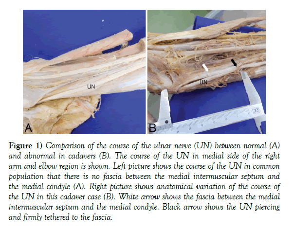
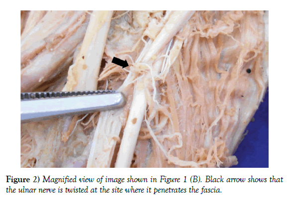
During routine educational dissection at XXXX University School of Medicine in 2015, brachial plexus of 93-year-old Japanese female was dissected and exposed. This cadaver had no surgical incisions on the skin or any fractures of the bones of the arm. After the ulnar nerve was identified in the right upper arm, it was traced distally. When the course of the ulnar nerve between the medial head of the triceps brachii and brachialis muscles was followed, we observed the fascia connecting between the medial intermuscular septum and the medial condyle of the right humerus. The ulnar nerve pierced and was firmly tethered to the fascia (Figures 1 and 2) and had little room to move within the fascia.
Figure 1) Comparison of the course of the ulnar nerve (UN) between normal (A) and abnormal in cadavers (B). The course of the UN in medial side of the right arm and elbow region is shown. Left picture shows the course of the UN in common population that there is no fascia between the medial intermuscular septum and the medial condyle (A). Right picture shows anatomical variation of the course of the UN in this cadaver case (B). White arrow shows the fascia between the medial intermuscular septum and the medial condyle. Black arrow shows the UN piercing and firmly tethered to the fascia.
Furthermore, the ulnar nerve was twisted at the site where it pierced fascia connecting between the medial intermuscular septum and the medial condyle of the humerus. The site of penetration of the fascia at which the nerve was twisted was 65 mm proximal to the medial epicondyle of the right humerus (Figure 1). Besides this twisting of the nerve at the medial intermuscular septum, no other anatomical variation of the nerve was observed.
Discussion
Previous studies have reported anatomical variations of the ulnar nerve, consisting of abnormal branches of the nerve or abnormal communications between the ulnar nerve/its branches and other nerves of the hand. To the best of our knowledge, few studies have reported an abnormal course of the ulnar nerve, which is uncommon. Previous study reported that the ulnar nerve was positioned anteriorly to the medial epicondyle of the humerus instead of posteriorly to the medial epicondyle of the humerus [5].
We observed a fibrous band from the medial intermuscular septum to the medial epicondyle of the right humerus, which is different from the arcade of Struthers, connecting the medial head of the triceps brachii and the medial intermuscular septum. Furthermore, the ulnar nerve was twisted where it crossed the fibrous band, at a site 65 mm proximal to the medial epicondyle. Karatas et al. [6] reported observing a fibrous band from the medial intermuscular septum to the medial epicondyle of the humerus, crossing over the ulnar nerve in several cadavers. However, they did not find any abnormal course of the ulnar nerve, which we found in our cadaver case reported herein. Thus, the anatomical variation of the course of the ulnar nerve found in our cadaver and reported has not been reported previously.
Entrapment of the ulnar nerve is a common cause of nerve injury in the arm, which is clinically important as it can cause pain at the elbow, atrophy, weakness of the ulnar side of the hand, decreased grip strength and decreased activities of daily living and work. The common sites of ulnar nerve entrapment are the arcade of Struthers, the ulnar groove, cubital tunnel. Although exact etiologies of entrapment are not clear, it could be caused by external compression or traction during elbow flexion [7]. Wright et al. [8] revealed that elbow flexion is responsible for increasing the nerve tension (strain) by 29%, is deleterious to the nerve because it is associated with increase of the intraneural pressure with decreased cross-sectional area [9], and also a decrease in blood flow in stretched nerve [10]. We considered that anatomical variation herein could predispose to nerve entrapment even with a normal range of motion of the elbow, because the ulnar nerve was tethered at the site of penetration of the fascia and also twisted in this region, resulting in a limited range of excursion and pathologic stretch of the nerve.
Conclusion
In conclusion, we believe that the rare anatomical variation of the course of the ulnar nerve reported here could confound diagnosis of entrapment neuropathies. An awareness of the possibility of this anatomical variation may provide clues to a precise diagnosis of the possible cause of ulnar nerve entrapment in patients presenting with ulnar nerve symptoms in which the clinician finds it difficult to identify the exact cause. This case report may be helpful, in particular, after the frequency (true occurrence) of this anatomical variation is determined by watching for it in more cadavers in the future.
REFERENCES
- Bergfield TG, Aulicino PL. Variation of the deep motor branch of the ulnar nerve at the wrist. J Hand Surg Am. 1998;13:368-9.
- Chow JCY, Papachristos AA, Ojeda A. An aberrant anatomic variation along the course of the ulnar nerve above the elbow with coexistent cubital tunnel syndrome. Clin Anat. 2006;19:661-4.
- Gelberman RH, Yamaguchi K, Hollstien SB, et al. Changes in interstitial pressure and cross-sectional area of the cubital tunnel and of the ulnar nerve with flexion of the elbow. An experimental study in human cadavera. J Bone Joint Surg Am. 1998;80:492-501.
- Karatas A, Apaydin N, Uz A, et al. Regional anatomic structures of the elbow that may potentially compress the ulnar nerve. J Shoulder Elbow Surg. 2009;18:627-63.
- McCarthy RE, Nalebuff EA. Anomalous volar branch of the dorsal cutaneous ulnar nerve: a case report. J Hand Surg Am. 1980;5:19-20.
- Mondelli M, Giannini F, Ballerini M, et al. Incidence of ulnar neuropathy at the elbow in the province of Siena (Italy). J Neurol Sci. 2005;234:5-10.
- Ogata K, Naito M. Blood flow of peripheral nerve effects of dissection, stretching and compression. J Hand Surg Br. 1986;11:10-4.
- Omejec G, Podnar S. Precise localization of ulnar neuropathy at the elbow. Clin Neurophysiol. 2015;126: 2390-6.
- Satteson ES, Li Z. Anteriorly positioned ulnar nerve at the elbow: a rare anatomical event: case report. J Hand Surg Am. 2015;40:984-6.
- Wright TW, Glowczewskie F Jr, Cowin D, et al. Ulnar nerve excursion and strain at the elbow and wrist associated with upper extremity motion. J Hand Surg Am. 2001;26:655-62.


