A rare bilateral variant branching pattern of the axillary artery
2 Associate Professor, Department of Anatomy, American University of Antigua College of Medicine, Antigua
3 Student, American University of Antigua College of Medicine, Antigua
Received: 14-Aug-2018 Accepted Date: Aug 23, 2018; Published: 07-Sep-2018
Citation: Joshua SA, D’ Costa S, Johal R, et al. A rare bilateral variant branching pattern of the axillary artery. Int J Anat Var. Sep 2018;11(3):101-102.
This open-access article is distributed under the terms of the Creative Commons Attribution Non-Commercial License (CC BY-NC) (http://creativecommons.org/licenses/by-nc/4.0/), which permits reuse, distribution and reproduction of the article, provided that the original work is properly cited and the reuse is restricted to noncommercial purposes. For commercial reuse, contact reprints@pulsus.com
Abstract
Arterial injuries in upper extremities can occur in athletes performing forceful, repetitive, high velocity arm movements or during cannulation. Variations in the course and branching pattern of axillary artery would always be predisposition factor for such vascular injuries. We present an unusual bilateral variation in the branching pattern of the axillary artery which was observed in a 69-year-old female cadaver. A common trunk was observed arising from the second part of axillary artery which divided into lateral thoracic artery, thoracodorsal artery, circumflex scapular artery, posterior circumflex humeral artery, anterior circumflex humeral and profunda brachii artery. Knowledge of such variations is of utmost importance in preoperative planning for surgical procedures involving the axilla or axillary artery.
Keywords
Axillary artery; Common trunk; Anatomical variation; Vascular injuries
Introduction
The course of axillary artery in the region of axilla and its relation to brachial plexus and axillary lymph nodes has always been a concern for vascular surgeons. Operating on injuries or aneurysm related to axillary artery always poses greater challenge to surgeons, especially if there are variations in the branching pattern of this artery. The axillary artery being the continuation of the subclavian artery from the lateral margin of the first rib to the lower margin of the teres major muscle is the sole supply to the upper limb (1). Studies by McCarthy et al. and Durham et al. have shown that failure to recognize the vascular variations would prove fatal during surgeries or injuries, especially as seen in athletes who are involved in sporting activities with more usage of upper limbs in games like tennis, badminton and volley ball or during axillary artery cannulation (2,3).
Case Report
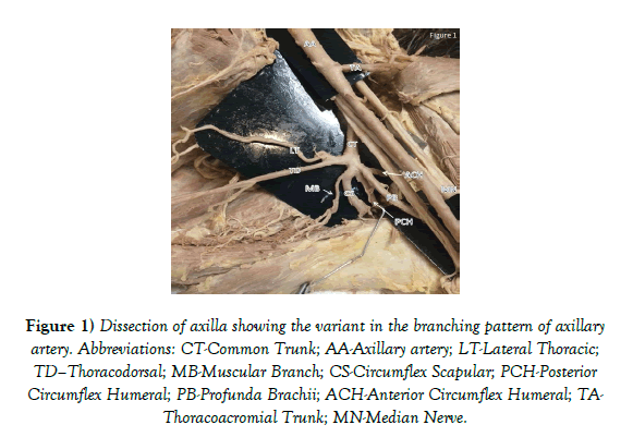
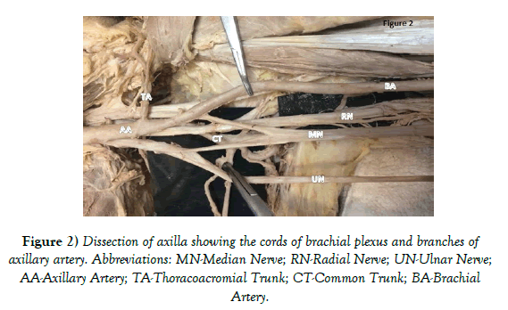
During a routine dissection of the axilla on a formalin preserved 69-year-old female cadaver, variation was observed in the branching pattern of the axillary artery bilaterally. The axillary artery was the continuation of the subclavian artery had a normal course along the region of axilla. Superior thoracic artery was seen arising from the first part of axillary artery. Thoraco-acromial trunk, branch from the second part was seen along the medial margin of pectoralis minor muscle (Figure 1). As the main trunk of axillary artery was traced further, a large branch was noted arising from the inferior aspect of second part of axillary artery, which was initially mistaken as subscapular artery. This large branch was seen passing between the lateral and posterior cord of brachial plexus. Further exploration of axillary artery was alarming as no branches of the third part were found (Figure 2). Therefore, we shifted our focus on the large branch found in the second part, on dissection, this branch divided into five branches after a short course (Figure 1). The first branch from this unusual large trunk seen splinting into two one among those was seen running on the lateral thoracic wall and was named as lateral thoracic artery (LT) and the other was traced along the latissimus dorsi and named as thoraco-dorsal artery (TD). Second branch had large in diameter and seen entering the upper triangular space, which was circumflex scapular artery (CS). Third and the fourth branches were encircling the Humerus therefore were named as anterior and posterior circumflex arteries (ACH and PCH). The fifth branch from the variant branch was profunda brachii artery (PB) which was found running along with radial nerve towards the spiral groove. In present case, lateral thoracic and thoracodorsal arteries arise from a single branch of the common trunk; also, that the circumflex scapular artery was a separate branch from the common trunk (Figure 1). No subscapular artery is observed. Beyond the teres major muscle, the axillary artery continued as the brachial artery to supply the upper limb. Therefore, the large common trunk which was seen originating from the second part of axillary artery, replaced some of the branches arising from the trunk of axillary artery and even found that the profunda brachii artery, normally a branch of the brachial artery, was arising from the common trunk.
Figure 1) Dissection of axilla showing the variant in the branching pattern of axillary artery. Abbreviations: CT-Common Trunk; AA-Axillary artery; LT-Lateral Thoracic; TD–Thoracodorsal; MB-Muscular Branch; CS-Circumflex Scapular; PCH-Posterior Circumflex Humeral; PB-Profunda Brachii; ACH-Anterior Circumflex Humeral; TA-Thoracoacromial Trunk; MN-Median Nerve.
Discussion
Anatomical variations in the axilla are common and widespread. Variation in the branching pattern of axillary artery is not an uncommon finding. Many authors have reported such findings, but the significance of any anatomical variations in the clinical scenario is assessed by the fact that the variation does require surgical or therapeutic intervention (4). Venieratos and Lolis discovered a common trunk which gave the subscapular artery, anterior and posterior circumflex humeral arteries, profunda brachii, and ulnar collateral arteries (5). They named this trunk the common subscapular trunk and evaluated it to have an occurrence of 0.45%. Karambelkar et al. found variations in 35% of cadavers (6). The most common variation they observed was a common trunk for the anterior and posterior circumflex humeral arteries. Another study of 40 cadavers by Astik and Dave (7) found axillary artery variations in 62.5% of the limbs. A total of 6 different types of variations were found in that study. Nkomozepi et al. explains about the branching pattern of axillary artery into superficial and deep brachial artery and discusses its close relation to median nerve, leading to idiopathic entrapment neuropathy (8). Stanchev et al. and Fontes et al. reported the presence of common trunk for anterior and posterior circumflex humeral artery in 2 of 24 cases (8.3%), wherein compression of posterior circumflex humeral artery was observed in the quadrangular space. This condition is known as quadrangular space syndrome and is associated with paresthesia and pain radiating to the arm. Furthermore, they have even described the risk of injury to the common trunk during fractures of the proximal Humerus (9,10). A branching pattern identical to the one presented in this report was not found in the available literature.
In the recent times, Axillary artery has gained more clinical importance for various reasons;
1. The axillary artery has become an increasingly popular site for cannulation in patients who are at high risk of developing atherosclerotic emboli and the evidence in support of axillary cannulation is growing (11).
2. Increased number of vascular injuries seen in athletes with performing forceful, repetitive, high velocity arm movements.
3. Variations in the branches and course can alter the collateral flow between the subclavian and axillary artery or between axillary and brachial artery.
Fong et al. (12) showed that axillary artery cannulation for cardiopulmonary bypass surgeries greatly reduced the morbidity rate and risk of stroke after the procedure. Just 3.3% of patients suffered minor complications as a direct result of axillary cannulation. Fong et al. (12) also showed that axillary artery cannulation had to be abandoned in 8 patients due to inadequate size of the blood vessel. This is a very important point because abnormal branching and especially bifurcation of the axillary artery into a large trunk as seen in our case can reduce the size of the axillary artery making it inadequate for cannulation. Surgeons should be well aware of the possible branching patterns and their relative frequencies in the general population in order to be well prepared for surgery, especially when operating on the types of high-risk patients which require axillary cannulation. The axillary artery is often used as an alternative access for endovascular procedures in treatment of thoracic aortic diseases.
The preferred access is through the femoral artery; however this may be contraindicated due to factors such as size of the vessel, obstruction, calcification, dissection or extreme tortuosity. In the case that the femoral artery is contraindicated, the axillary artery serves as a popular alternative. Awareness of abnormal branching patterns is of interest to vascular and cardiothoracic surgeons during preoperative planning and assessment (13,14). Furthermore, the surgeon’s knowledge of variant branching patterns of the axillary artery is of strategic importance for the identification and preservation of these structures in axillary lymph node dissection and sentinel lymph node biopsy procedures (15).
Conclusion
Awareness of all possible variations of major arteries is important to physicians and anatomists, given the growing popularity of utilizing the axillary artery in major cardiovascular surgical procedures. With this case report we hope to shed light on a previously unreported variation in order to increase the body of knowledge regarding variant branching patterns of the axillary artery.
REFERENCES
- Drake RL, Vogl AW, Mitchell AWM. Upper limb, in: Gray’s anatomy for students. Philadelphia. 2015;pp:733-5.
- Mc Carthy WJ, Yao JST, Schafer MF, et al. Upper extremity arterial injury in athletes. J Vasc Surg. 1989;9:317-27.
- Durham JR, Yao JST, Pearce WH, et al. Arterial injuries in the thoracic outlet syndrome. J Vasc Surg. 1995;21:57-70.
- Georgiev GP. Significance of anatomical variations for clinical practice. Inter J Anat Var. 2017; 10:43-4.
- Venieratos D, Lolis ED. Abnormal ramification of the axillary artery: subscapular common trunk. Morphologie. 2001;85:23-4.
- Karambelkar RR, Shewale AD, Umarji BN. Variations in branching pattern of axillary artery and its clinical significance. Anatomica Karnataka. 2011;5:47-51.
- Astik R, Dave U. Variations in branching patterns of the axillary artery: a study in 40 human cadavers. J Vasc Bras. 2012;11:12-17.
- Nkomozepi P, Xhakaza N, Swanepoel E. Superficial brachial artery: A possible cause for idiopathic median nerve entrapment neuropathy. Folia Morphol. 2017.
- Stanchev S, Iliev A, Georgiev GP, et al. А case of bilateral variations in the arterial branching in the upper limb and clinical implications. Chro J Cli Case Reports. 2017;1:006.
- Fontes EB, Precht BLC, Andrade RCL, et al. Output Relations of Humeral Circumflex Arteries and its Variations. Int J Morphol. 2015;33:1171-5.
- Calvaruso D, Voisine P, Mohammadi S, et al. Axillary artery cannulation. Multimed Man Cardiothorac Surg. 2012;p:004.
- Fong LS, Bassin L, Mathur MN. Liberal use of axillary artery cannulation for aortic and complex cardiac surgery. Interact CardioVasc Thorac Surg. 2013;16:755-8.
- Saadi EK, Dussin LH, Moura L, et al. The axillary artery-a new approach for endovascular treatment of thoracic aortic diseases. Interact CardioVasc Thorac Surg. 2010;11:617-9.
- Sabik JF, Lytle BW, McCarthy PM, et al. Axillary artery: an alternative site of arterial cannulation for patients with extensive aortic and peripheral vascular disease. J Thorac Cardiovasc Surg. 1995;109:885-90.
- Soares EWS. Anatomical variations of the axilla. SpringerPlus. 2014;3:306.


