A Rare Case: Duplication of Lungs on Left Side in Human Cadaveric Study
2 Assistant professor, D Y Patil Medical college, Pimpri, India, Email: Melissa.Quinn@osumc.edu
3 Assistant Professor MGM Medical College Kamothe, India, Email: prakashmane@gmail.com
Received: 26-May-2021 Accepted Date: Jun 21, 2021; Published: 28-Jun-2021, DOI: 10.37532/1308-4038.14(6).134-135
Citation: Anjali Sabnis, Mrunal K Muley, Prakash Mane. MA Rare Case: Dupication of Lungs on Left Side in Human Cadaveric Study. Int J Anat Var. 2021;14(6):101-102.
This open-access article is distributed under the terms of the Creative Commons Attribution Non-Commercial License (CC BY-NC) (http://creativecommons.org/licenses/by-nc/4.0/), which permits reuse, distribution and reproduction of the article, provided that the original work is properly cited and the reuse is restricted to noncommercial purposes. For commercial reuse, contact reprints@pulsus.com
Abstract
Knowledge of lung variation is essential to all medical professionals to exactly interpret radiographs, computed tomography scans, to diagnose, plan and modify a surgical procedure.
In this case, a very rare variation has been studied, where three lungs were present in a cadaver. There was a duplication of lungs on left side, none of them had a fissure. Whereas, right lung had normal gross anatomical features. Such variation has not been reported till date. It should be taken into consideration while interpreting radiological variations and during surgeries like lobectomy, segmentectomies.
Keywords
Lung; Variation; Duplication of Lung; Three Lungs
Introduction
Lungs are essential paired respiratory organs situated in thoracic cavity on either side of heart. The right lung has oblique and horizontal fissures dividing it into superior, middle, and inferior lobes; whereas the left lung has superior and inferior lobes separated by an oblique fissure [1]
The fissures facilitate a uniform expansion of whole lung for more air intake during respiration. As the fissures form boundaries for lobes of the lungs, knowledge of their position is necessary to appreciate lobar anatomy and locating the bronchopulmonary segments which is significant both anatomically and clinically [2].
Case Report
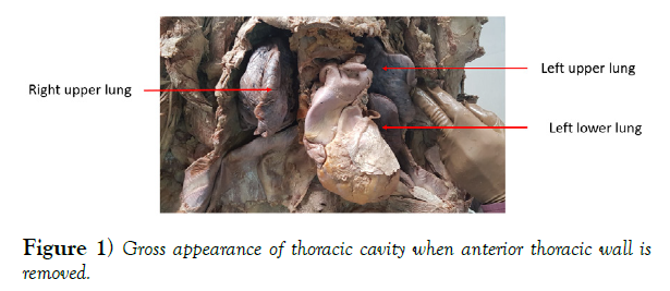
During regular cadaveric dissection of thorax region (1st MBBS batch 2017- 2018 MGM medical college, Kamothe), a rare variation in lungs was discovered. While studying lungs in situ, it appeared that there were two separate lung segments on left side. To study it further, lungs on either side were separated and removed by cutting their attachments at hila. It was confirmed that on left side there were two lungs. The gross appearance of two lungs in situ was as shown in (Figure 1).
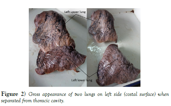
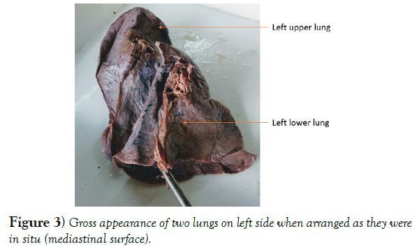
Each of two lungs on left side presented an apex and a base, a hilum with its own bronchus, a branch of pulmonary artery and pulmonary veins (Figure 2). The left lower lung presented a lingula. No fissure was observed in either of two lungs on left side (Figure 3).
The lung on right side was normal and did not show any variation. Gross anatomical features and dimensions in 3 lungs were as follows (Table 1).
| Rt. Lung | Lt. upper lung | Lt. lower lung | |
|---|---|---|---|
| Length | 9 cm | 11 cm | 15 cm |
| Width | 14 cm | 11 cm | 11 cm |
| Thickness | 6 cm | 3 cm | 3 cm |
| Lobes/ fissures | 3 lobes / 2 fissures | 1 lobe/ no fissure | 1 lobe with lingula / no fissure |
Table 1 Comparison of gross anatomical features of 3 lungs in a cadaver.
Discussion
Number of studies have been done in past pertaining to variations in gross Anatomy of lungs. A single lung extending uniformly throughout the thoracic cavity was detected in a 35year old male cadaver [3]. However, a rare variation procedure like segmentectomies, lobectomies. This will help to reduce the morbidity and mortality associated with lung surgeries. Also, it would be of immense help in case of lung donation and lung transplantation surgeries.
Acknowledgement
The authors are highly thankful to Mahatma Gandhi Medical College, Kamothe for providing the necessary support. Authors also acknowledge the immense help received from the scholars whose articles and books cited and included in discussion and references of this manuscript.
REFERENCES
- Shah P, Johnson D, Standring S. Thorax Standring S. Gray's Anatomy: The Anatomical Basis of Clinical Practice. 39th ed. Edinburgh: Churchill Livingstone. 2005; 1068-1069.
- Meenakshi S, Manjunath KY, Balasubramanyam V. Morphological variations of the lung fissures and lobes. Indian J Chest Dis Allied Sci. 2004; 46:179-182.
- Prakash, Bhardwaj AK, Shashirekha M, et al. Lung morphology: a cadaver study in Indian population. Ital J Anat Embryol. 2010;115(3):235-4
- Sadlar TW. Langman’s medical embryology. 9th ed. Baltimore, MD: Lippincott Williams & Wilkins. 2004:223-84.
- Sudikshya KC, Pragya Shreshtha, Aashish Kumar Shah, et al. Variations in human pulmonary fissures and lobes: a study conducted in Nepalese cadavers. Anatomy & Cell Biology 2018;51(2): 85-92.


