A rare case of internal small intestinal strangulation
2 Department of Hospital Surgery, South Ural State Medical University, Chelyabinsk, Russia, Email: garb@inbox.ru
3 Federal Center of Cardiovascular Surgery, Chelyabinsk, Russia, Email: nikolai.arefyev@gmail.com
Received: 10-Jan-2018 Accepted Date: Jan 20, 2018; Published: 16-Feb-2018
Citation: Garbuzenko D.V., Belov D.V., Arefyev N.O. A rare case of internal small intestinal strangulation. Gen Surg: Open Access. 2018;1(1):3-4.
This open-access article is distributed under the terms of the Creative Commons Attribution Non-Commercial License (CC BY-NC) (http://creativecommons.org/licenses/by-nc/4.0/), which permits reuse, distribution and reproduction of the article, provided that the original work is properly cited and the reuse is restricted to noncommercial purposes. For commercial reuse, contact reprints@pulsus.com
Abstract
Although internal abdominal hernias cause acute intestinal obstruction in no more than 6% of cases, the lethality associated with them reaches 50% in case of untimely diagnosed strangulation and gangrene of the intestine. The rarest type of such hernias is a transomental hernia, which occurs in 1-4% of cases. The diagnosis is difficult when such hernias are strangulated; therefore, they require an emergency operation at the slightest suspicion. In this report, we describe a case of internal small intestinal strangulation in the abnormal transomental aperture, accompanied by clinical features of strangulation intestinal obstruction without any findings during an instrumental examination.
Introduction
Internal hernias occur in the peritoneal recesses or folds when a hollow organ, most commonly the small intestine, protrudes through a normal or abnormal aperture in the peritoneum or mesentery. Such hernias do not have specific clinical features and, as a rule, are diagnosed when acute intestinal obstruction occurs. The most common types of internal hernias are paraduodenal (53%), pericecal (13%), foramen of Winslow (8%), transmesenteric (8%), and intersigmoid (6%) [1]. Transomental hernias are the rarest (1-4%) [2]. We describe an internal small intestinal strangulation in the abnormal aperture of the greater omentum without any findings during an instrumental examination.
Case Report
A 22-year-old man arrived at the Emergency Department of South Ural State Medical University Hospital on 07/14/2017. He reported acute cramping left-sided abdominal pain and vomiting with stomach contents once. He fell ill about 6 h ago when he picked up a bucket of cement while working on a construction site. He had not noticed such symptoms before and never had any surgical procedures on the organs of the abdominal cavity.
The patient’s condition was fair. The body type was asthenic. He behaved restlessly and could not find a comfortable position. The skin color was normal. The tongue was moist and clean. Body temperature was normal. Arterial blood pressure was 110/70 mmHg and heart rate was 76 beats per minute. The abdomen was asymmetric due to the area of limited bloating on the left side near the navel and took part in the act of breathing but moved unevenly at the expense of the left half. On palpation, the abdomen was soft and painful on the left flank. On auscultation, there were no succession splashes. Symptoms of peritonitis were absent. Peristaltic sounds were weakened at the place of limited bloating. There were no signs of external hernias. Gas passed out normally. Diuresis was within normal limits. Blood test: HGB 153 g/l, RBC 5.22 • 012/l, WBC 7.9 × 10⁹/l; ESR 23 mm/h. The analysis of urine was within normal limits.
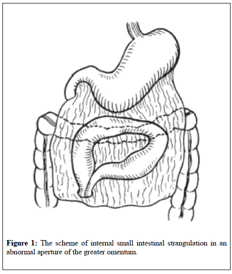
Ultrasound examination and radiography of abdominal organs did not reveal any pathology. Multi-slice helical computed tomography was not performed due to a lack of technical capability. The preliminary diagnosis was as follows: strangulation intestinal obstruction due to the volvulus of the small intestine. In this connection, we established the indications for an emergency surgery and performed a midline laparotomy. There were up to 200 ml of serous hemorrhagic effusion in the abdominal cavity, mainly in the small pelvis. During the inspection, in approximately 1 meter distally from the Treitz ligament, we revealed a strangulation of about 20 cm of the small intestine in the 4.0 cm abnormal aperture located in the thinnest portion of the greater omentum (Figure 1). These findings attested the presence of a type A strangulated transomental hernia in the patient. The vitality of the strangulated small intestine was not disturbed. The defect was sutured after the reduction of small intestinal loops. The postoperative period proceeded without complications. The patient was discharged on 07/24/2017.
Discussion
Although internal abdominal hernias cause acute intestinal obstruction in no more than 6% of cases, the lethality associated with them reaches 50% in case of untimely diagnosed strangulation and gangrene of the intestine [3]. The rarest type of such hernias is a transomental hernia, which occurs either spontaneously or as a result of tension and increased intraabdominal pressure. Such defects in the greater omentum can be congenital, traumatic, or due to inflammatory changes, atrophy, etc. [4]. Perhaps, in our case, the cause of its formation in the thinnest portion of the greater omentum was abdominal hypertension due to the lifting of the heavy object by the emaciated person.
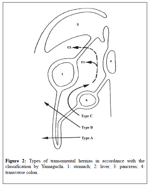
Yamaguchi [5] classified transomental hernias into three types: A, B, and C (Figure 2). Strangulated internal hernias are difficult to diagnose against a background of acute intestinal obstruction because clinical manifestations are not specific. At the same time, taking into account the life-threatening situation, the early identification of internal intestinal strangulation is important. In the presence of strangulation signs, an internal hernia can be suspected in patients who did not have abdominal operations, when excluding external abdominal hernias [6]. Ultrasound examination and radiography of the abdominal cavity, although not always, may show a picture of intestinal obstruction [7]. The introduction of multi-slice helical computed tomography into the urgent surgical practice increased the accuracy of diagnosis. The characteristic features of a strangulated transomental hernia are mesenteric vascular congestion and the beak-like triangular configuration of the proximal and distal small bowel loops in the anomalous aperture of the greater omentum. Small intestinal loops are filled with fluid contents and located in specific areas of the abdominal cavity according to each type of transomental hernias [8]. At the same time, unfortunately, the round-the-clock possibility to perform multi-slice helical computed tomography is not always present in the usual clinical conditions, as it was in our case.
The slightest suspicion of internal strangulation serves as an absolute indication for an emergency operation, which can be performed using video laparoscopy under certain conditions [9]. The type of surgical intervention depends on the vitality of the small intestine.
REFERENCES
- Akyildiz H, Artis T, Sozuer E, et al. Internal hernia: complex diagnostic and therapeutic problem. Int J Surg 2009;7:334-7.
- Rudroff C, Balogh A, Hilswicht S. Internal transomental herniation with a trapped small bowel mimicking acute appendicitis. Int J Surg Case Rep 2013;4:1153-5.
- Martin LC, Merkle EM, Thompson WM. Review of internal hernias: radiographic and clinical findings. AJR Am J Roentgenol 2006;186:703-17.
- Hull JD. Transomental hernia. Am Surg 1976;42:278-84.
- Yamaguchi T. A case of incarceration of sigmoid colon into hiatus of greater omentum. Rinsho Geka 1978;33:1041-5.
- Garbuzenko DV. Selected lectures in emergency abdominal surgery. Saarbrücken, Germany: LAP LAMBERT Academic Publishing GmbH& Co. 2012; 212.
- Lassandro F, Iasiello F, Pizza NL, et al. Abdominal hernias: Radiological features. World J Gastrointest Endosc 2011;3:110-7.
- Сamera L, De Gennaro A, Longobardi M, et al. A spontaneous strangulated transomental hernia: Prospective and retrospective multi-detector computed tomography findings. World J Radiol 2014;6:26-30.
- Talebpour M, Habibi GR, Bandarian F. Laparoscopic management of an internal double omental hernia: a rare cause of intestinal obstruction. Hernia 2005;9:195-7.


