A rare case of occurrence of fleshy muscle fibers in the anterior wall of rectus sheath
Mohandas Rao KG1* Venkata Ramana V2 and Vincent R1
1Asian Institute of Medicine, Science and Technology, Semeling, Bedong, Kedah, Malaysia
2Department of Anatomy, Melaka Manipal Medical College (Manipal Campus) Manipal, Karnataka, India
- *Corresponding Author:
- Pasura Subbaiah Sujan Ganapathy
Centre for Vrikshayurveda, A Division of Centre for Advanced Studies in Biosciences, Jain University, Chamrajpet- 560019, Karnataka, India.
E-mail: sujan.ganapathy@gmail.com
Date of Received: November 10th, 2008
Date of Accepted: March 8th, 2009
Published Online: March 10th, 2009
© Int J Anat Var (IJAV). 2009; 1: 31–32.
[ft_below_content] =>Keywords
internal oblique, external oblique, inguinal hernia, conjoint tendon
Introduction
Generally, the anterior wall of the lower part rectus sheath is formed by the contribution of aponeuroses of the three flat muscles of the anterior abdominal wall; namely, external oblique internal oblique and transverse abdominis. Contributions of these aponeuroses for the formation of rectus sheath vary at different levels. Anatomical knowledge of the rectus sheath below the arcuate line is surgically important because of its close relationship with inguinal canal. Normally, below the arcuate line, the anterior wall is formed by the aponeurosis of the external oblique, and an aponeurosis formed by the fusion of internal oblique and transverse abdominis muscles. In the lowermost part, this aponeurosis passes behind the superficial inguinal ring to reach the pubic crest and pectenial line to form conjoint tendon, which strengthens the medial portion of the posterior wall of inguinal canal [1].
Case Report
During the routine dissections for the first MBBS students of Melaka Manipal Medical College, Manipal, a case of unusual presence of fleshy muscle fibers in the lower part of the anterior wall of the rectus sheath was noted. The variation was observed only on the left half of the anterior abdominal wall of an approximately 50-year-old male cadaver.
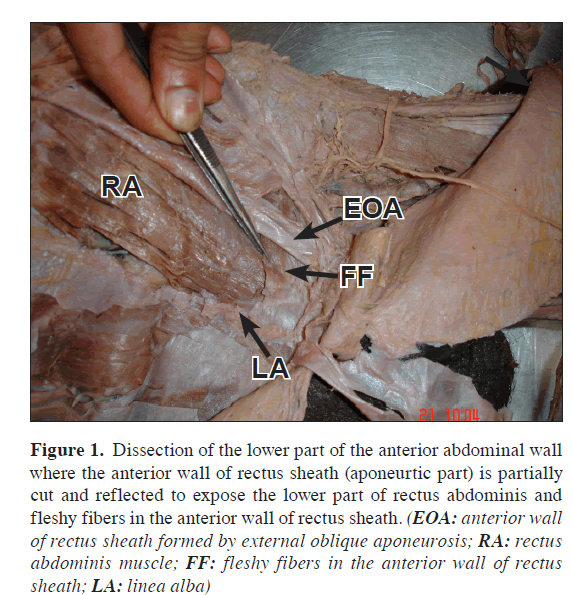
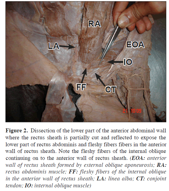
It was observed that, the anterior wall of the rectus sheath below the arcuate line was partially aponeurotic and partially fleshy. Contribution from the external oblique was aponeurotic and considered as normal. However, posterior to it, fleshy fibers of internal oblique and transverse abdominis continued beyond the semilunar line to reach the linea alba (Figure 1). When traced below, these fleshy fibers were continuing with the conjoint tendon, which was much thinner and weaker than normal (Figure 2).
Figure 1: Dissection of the lower part of the anterior abdominal wall where the anterior wall of rectus sheath (aponeurtic part) is partially cut and reflected to expose the lower part of rectus abdominis and fleshy fibers in the anterior wall of rectus sheath. (EOA: anterior wall of rectus sheath formed by external oblique aponeurosis; RA: rectus abdominis muscle; FF: fleshy fibers in the anterior wall of rectus sheath; LA: linea alba)
Figure 2: Dissection of the lower part of the anterior abdominal wall where the rectus sheath is partially cut and reflected to expose the lower part of rectus abdominis and fleshy fibers fibers in the anterior wall of rectus sheath. Note the fleshy fibers of the internal oblique continuing on to the anterior wall of rectus sheath. (EOA: anterior wall of rectus sheath formed by external oblique aponeurosis; RA: rectus abdominis muscle; FF: fleshy fibers of the internal oblique in the anterior wall of rectus sheath; LA: linea alba; CT: conjoint tendon; IO: internal oblique muscle)
Discussion
Variation in the formation of rectus sheath is not common. In the present case we observed:
1. Presence of fleshy muscle fibers in the anterior wall of rectus sheath, which is being reported for the first time.
2. A slender conjoint tendon which is much thinner and weaker than normal.
During Lichtenstein’s technique of inguinal hernia repair normally a polypropylene mesh is used to reinforce the posterior wall of inguinal canal making a new internal ring for spermatic cord. The mesh is fixed inferiorly to the shelving part of inguinal ligament and superiorly to the conjoint tendon [2]. Similarly, while repairing recurrent inguinal hernia a mersilene plate is usually fixed to the conjoint tendon and inguinal ligament [3]. During such surgical procedures, awareness of possibility of the fleshy fibers in the anterior wall of rectus sheath or a weak conjoint tendon –as observed in this case– may be useful for surgeons.
Moreover, the conjoint tendon is the main support for the anterior abdominal wall, where it is weakened by superficial inguinal ring. Weakening of the conjoint tendon may lead to inguinal hernia [4]. Similarly congenital variations in the conjoint tendon as reported in this case may also be a cause for inguinal hernia.
We conclude by stating that, awareness of variations in the inguinal region is of importance during the inguinal hernia surgery. Hence we are reporting one such variation.
References
- Standring S, ed. Gray’s Anatomy, The Anatomical basis for clinical practice. 39th Ed., New York, Elsevier Churchill Livingstone. 2005: 1101–1112.
- Sidhu BS, Mishra L. Evaluation of Lichtenstein’s technique of inguinal hernia repair under local analgesia. J Indian Med Assoc. 2004; 102: 314–316.
- Bourde J. The repair of inguinal hernia by insertion of prosthetic material with tunnelisation of the spermatic cord. Nouv Presse Med. 1979; 8: 3047–3048. French.
- Jacobs CJ, Steyn WH, Boon JM. Segmental nerve damage during a McBurney’s incision: a cadaveric study. Surg Radiol Anat. 2004; 26: 66–69.
Mohandas Rao KG1* Venkata Ramana V2 and Vincent R1
1Asian Institute of Medicine, Science and Technology, Semeling, Bedong, Kedah, Malaysia
2Department of Anatomy, Melaka Manipal Medical College (Manipal Campus) Manipal, Karnataka, India
- *Corresponding Author:
- Pasura Subbaiah Sujan Ganapathy
Centre for Vrikshayurveda, A Division of Centre for Advanced Studies in Biosciences, Jain University, Chamrajpet- 560019, Karnataka, India.
E-mail: sujan.ganapathy@gmail.com
Date of Received: November 10th, 2008
Date of Accepted: March 8th, 2009
Published Online: March 10th, 2009
© Int J Anat Var (IJAV). 2009; 1: 31–32.
Abstract
Normally, the anterior wall of the lower part rectus sheath is formed by the contribution of aponeuroses of the three flat muscles of the anterior abdominal wall; namely, external oblique internal oblique and transverse abdominis. Here, we report a rare case of fleshy fibers in the lower part anterior wall of rectus sheath. Its surgical importance is also discussed.
-Keywords
internal oblique, external oblique, inguinal hernia, conjoint tendon
Introduction
Generally, the anterior wall of the lower part rectus sheath is formed by the contribution of aponeuroses of the three flat muscles of the anterior abdominal wall; namely, external oblique internal oblique and transverse abdominis. Contributions of these aponeuroses for the formation of rectus sheath vary at different levels. Anatomical knowledge of the rectus sheath below the arcuate line is surgically important because of its close relationship with inguinal canal. Normally, below the arcuate line, the anterior wall is formed by the aponeurosis of the external oblique, and an aponeurosis formed by the fusion of internal oblique and transverse abdominis muscles. In the lowermost part, this aponeurosis passes behind the superficial inguinal ring to reach the pubic crest and pectenial line to form conjoint tendon, which strengthens the medial portion of the posterior wall of inguinal canal [1].
Case Report
During the routine dissections for the first MBBS students of Melaka Manipal Medical College, Manipal, a case of unusual presence of fleshy muscle fibers in the lower part of the anterior wall of the rectus sheath was noted. The variation was observed only on the left half of the anterior abdominal wall of an approximately 50-year-old male cadaver.
It was observed that, the anterior wall of the rectus sheath below the arcuate line was partially aponeurotic and partially fleshy. Contribution from the external oblique was aponeurotic and considered as normal. However, posterior to it, fleshy fibers of internal oblique and transverse abdominis continued beyond the semilunar line to reach the linea alba (Figure 1). When traced below, these fleshy fibers were continuing with the conjoint tendon, which was much thinner and weaker than normal (Figure 2).
Figure 1: Dissection of the lower part of the anterior abdominal wall where the anterior wall of rectus sheath (aponeurtic part) is partially cut and reflected to expose the lower part of rectus abdominis and fleshy fibers in the anterior wall of rectus sheath. (EOA: anterior wall of rectus sheath formed by external oblique aponeurosis; RA: rectus abdominis muscle; FF: fleshy fibers in the anterior wall of rectus sheath; LA: linea alba)
Figure 2: Dissection of the lower part of the anterior abdominal wall where the rectus sheath is partially cut and reflected to expose the lower part of rectus abdominis and fleshy fibers fibers in the anterior wall of rectus sheath. Note the fleshy fibers of the internal oblique continuing on to the anterior wall of rectus sheath. (EOA: anterior wall of rectus sheath formed by external oblique aponeurosis; RA: rectus abdominis muscle; FF: fleshy fibers of the internal oblique in the anterior wall of rectus sheath; LA: linea alba; CT: conjoint tendon; IO: internal oblique muscle)
Discussion
Variation in the formation of rectus sheath is not common. In the present case we observed:
1. Presence of fleshy muscle fibers in the anterior wall of rectus sheath, which is being reported for the first time.
2. A slender conjoint tendon which is much thinner and weaker than normal.
During Lichtenstein’s technique of inguinal hernia repair normally a polypropylene mesh is used to reinforce the posterior wall of inguinal canal making a new internal ring for spermatic cord. The mesh is fixed inferiorly to the shelving part of inguinal ligament and superiorly to the conjoint tendon [2]. Similarly, while repairing recurrent inguinal hernia a mersilene plate is usually fixed to the conjoint tendon and inguinal ligament [3]. During such surgical procedures, awareness of possibility of the fleshy fibers in the anterior wall of rectus sheath or a weak conjoint tendon –as observed in this case– may be useful for surgeons.
Moreover, the conjoint tendon is the main support for the anterior abdominal wall, where it is weakened by superficial inguinal ring. Weakening of the conjoint tendon may lead to inguinal hernia [4]. Similarly congenital variations in the conjoint tendon as reported in this case may also be a cause for inguinal hernia.
We conclude by stating that, awareness of variations in the inguinal region is of importance during the inguinal hernia surgery. Hence we are reporting one such variation.
References
- Standring S, ed. Gray’s Anatomy, The Anatomical basis for clinical practice. 39th Ed., New York, Elsevier Churchill Livingstone. 2005: 1101–1112.
- Sidhu BS, Mishra L. Evaluation of Lichtenstein’s technique of inguinal hernia repair under local analgesia. J Indian Med Assoc. 2004; 102: 314–316.
- Bourde J. The repair of inguinal hernia by insertion of prosthetic material with tunnelisation of the spermatic cord. Nouv Presse Med. 1979; 8: 3047–3048. French.
- Jacobs CJ, Steyn WH, Boon JM. Segmental nerve damage during a McBurney’s incision: a cadaveric study. Surg Radiol Anat. 2004; 26: 66–69.


