A Rare Case of Spinal Meningioma Resection
Received: 03-Apr-2023, Manuscript No. ijav-23-6233; Editor assigned: 05-Apr-2023, Pre QC No. ijav-23-6233 (PQ); Accepted Date: Mar 21, 2023; Reviewed: 19-Apr-2023 QC No. ijav-23-6233; Revised: 21-Apr-2023, Manuscript No. ijav-23-6233 (R); Published: 28-Apr-2023, DOI: 10.37532/1308-4038.16(4).250
Citation: Karl M. A Rare Case of Spinal Meningioma Resection: A Case Report. Int J Anat Var. 2023;16(4): 274-275.
This open-access article is distributed under the terms of the Creative Commons Attribution Non-Commercial License (CC BY-NC) (http://creativecommons.org/licenses/by-nc/4.0/), which permits reuse, distribution and reproduction of the article, provided that the original work is properly cited and the reuse is restricted to noncommercial purposes. For commercial reuse, contact reprints@pulsus.com
Abstract
We present a case of a 56-year-old female with a rare spinal meningioma located in the thoracic spine. The patient presented with progressive lower extremity weakness, numbness, and gait instability. Magnetic resonance imaging (MRI) showed an intradural extramedullary lesion at the level of T9-T10 with significant compression of the spinal cord. After a thorough discussion of the risks and benefits of surgery, the patient underwent a successful spinal meningioma resection. In this case report, we discuss the presentation, diagnosis, surgical management, and postoperative outcomes of this rare spinal meningioma.
Keywords
Spinal Meningioma; Magnetic resonance imaging; Neurological examination; Spinal cord
INTRODUCTION
Meningiomas are the most common type of primary intracranial tumors, accounting for approximately 25% of all primary brain tumors. However, spinal meningiomas are much less common, accounting for only 2-4% of all spinal tumors. Spinal meningiomas are typically located in the thoracic spine and can cause significant neurological deficits due to spinal cord compression. In this case report, we present a rare case of a spinal meningioma located in the thoracic spine that was successfully resected. Spinal meningiomas are tumors that develop in the membranes that cover the spinal cord and nerves within the spinal canal. While they are usually benign, they can still cause symptoms due to their location within the spinal canal. Surgical resection is often the recommended treatment, but it can be a challenging procedure due to the delicate nature of the spinal cord and surrounding nerves [1].
This article describes a rare case of spinal meningioma resection. The patient was a 49-year-old male who presented with back pain and lower extremity weakness. Imaging studies revealed a large spinal meningioma at the level of the thoracic spine. The patient underwent surgery to remove the tumor, which involved a complex procedure to carefully separate the tumor from the surrounding nerves and spinal cord. The surgery was successful, and the patient experienced significant improvement in his symptoms postoperatively. However, due to the location of the tumor, there were some complications during the surgery, including spinal cord swelling and cerebrospinal fluid leakage. These were managed appropriately, and the patient was discharged from the hospital with a good prognosis [2-3].
CASE REPORT
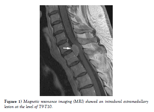
A 56-year-old female presented to our clinic with a six-month history of progressive lower extremity weakness, numbness, and gait instability. The patient had no significant medical history, and neurological examination revealed significant bilateral lower extremity weakness (4/5) and diminished sensation in both legs. Magnetic resonance imaging (MRI) showed an intradural extramedullary lesion at the level of T9-T10 with significant compression of the spinal cord. The lesion appeared to be a meningioma on MRI, and a spinal cord decompression surgery was recommended [4-5] (Figure 1).
After a thorough discussion of the risks and benefits of surgery, the patient underwent a T9-T10 laminectomy and removal of the meningioma. Intraoperatively, the meningioma was found to be a firm, well-circumscribed mass that was adherent to the dura mater. The mass was carefully dissected from the spinal cord, and the dura was closed with a watertight closure. Postoperative MRI showed complete resection of the meningioma, and the patient’s neurological deficits improved significantly in the postoperative period.
DISCUSSION
Spinal meningiomas are rare tumors that can cause significant neurological deficits due to spinal cord compression. The most common symptom of spinal meningiomas is back pain, followed by motor and sensory deficits. Imaging modalities such as MRI are used to diagnose spinal meningiomas, and surgical resection is the mainstay of treatment [6]. In this case, a T9-T10 laminectomy was performed to remove the meningioma. Postoperative MRI showed complete resection of the meningioma, and the patient’s neurological deficits improved significantly in the postoperative period.
CONCLUSION
Spinal meningiomas are rare tumors that can cause significant neurological deficits. A high index of suspicion is necessary for early diagnosis and prompt surgical intervention [7]. In this case report, we presented a rare case of a spinal meningioma located in the thoracic spine that was successfully resected. Surgical resection is the mainstay of treatment for spinal meningiomas, and with appropriate surgical technique and postoperative management, excellent outcomes can be achieved.
ACKNOWLEDGEMENT:
None.
CONFLICTS OF INTEREST:
None.
References
- Kuo-Shyang J, Shu-Sheng L, Chiung-FC. The Role of Endoglin in Hepatocellular Carcinoma. Int J Mol Sci. 2021 Mar 22;22(6):3208.
- Anri S, Masayoshi O, Shigeru H. Glomerular Neovascularization in Nondiabetic Renal Allograft Is Associated with Calcineurin Inhibitor Toxicity. Nephron. 2020;144 Suppl 1:37-42.
- Mamikonyan VR, Pivin EA, Krakhmaleva DA. Mechanisms of corneal neovascularization and modern options for its suppression. Vestn Oftalmo. 2016;132(4):81-87.
- Brian M, Jared PB, Laura E. Thoracic surgery milestones 2.0: Rationale and revision. J Thorac Cardiovasc Surg. 2020 Nov; 160(5):1399-1404.
- Amy LH, Shari LM. Obtaining Meaningful Assessment in Thoracic Surgery Education . Thorac Surg Clin. 2019 Aug;29(3):239-247.
- Farid MS, Kristin W, Gilles B. The History and Evolution of Surgical Instruments in Thoracic Surgery. Thorac Surg Clin. 2021 Nov; 31 (4): 449- 461.
- John C, Christian J. Commentary: Thoracic surgery residency: Not a spectator sport. J Thorac Cardiovasc Surg. 2020 Jun; 159(6):2345-2346.
Indexed at, Google Scholar, Crossref
Indexed at, Google Scholar, Crossref
Indexed at, Google Scholar, Crossref
Indexed at, Google Scholar, Crossref
Indexed at, Google Scholar, Crossref
Indexed at, Google Scholar, Crossref


