A rare coexistence of axillo-brachial neurovascular variations with embryological review
Hironmoy Roy1*, Parthapratim Pradhan2, and Samar Deb3
1Department of Anatomy, North Bengal Medical College & Hospital, Siliguri, WB, India
2Department of Anatomy, Midnapore Medical College & Hospital, Paschim Medinipur, WB, India
3Department of Anatomy, College of Medicine &JNM Hospital, WBUHS, Kalyani, Nadia, WB, India
- *Corresponding Author:
- Dr. Hironmoy Roy, MD
Department of Anatomy, North Bengal Medical College & Hospital, Sushrutanagar, Siliguri, WB, 734012, India
Tel: +91 974 8828588
E-mail: hironmoy19@gmail.com
Date of Received: August 8th, 2010
Date of Accepted: January 6th, 2011
Published Online: January 14th, 2011
IJAV. 2011; 4: 5–8.
[ft_below_content] =>Keywords
axillary artery, median nerve, musculocutaneous nerve
Introduction
The axillary neurovascular structures are one of the potential anatomical fields to show variations in many ways. The axillary artery gets divided in three parts by the fore lying pectoralis minor and provides the superior thoracic artery from first part, thoraco-acromial artery, lateral thoracic artery from second part and the third part gives anterior circumflex humeral, posterior circumflex humeral and subscapular arteries. After the distal border of teres major the axillary artery is continued in arm as brachial artery, from which the branches in the arm get distributed.
To form the median nerve trunk from lateral (C6,7) and medial (C8, T1) cords of brachial plexus the two roots of median nerve emerge and unite embracing the third part of axillary artery, either anterior or lateral to it. The lateral root often being smaller than medial one, the median nerve usually passes lateral to axillary artery and continues in upper arm as lateral to brachial artery [1].
On the other hand the musculocutaneous nerve (C5,6,7) initially accompanies the third part of the axillary artery and pierces the coracobrachialis muscle after supplying it. Next it appears in between biceps brachii and brachialis, supplies them and just below the elbow it pierces the deep fascia lateral to the tendon of biceps brachii and extends further downward as the lateral cutaneous nerve of the forearm [2].
Case Report
During routine dissection on a 60-year-old male cadaver, multiple variations were noted in both left axillo-brachial vessels and nerves, which as a combination was unique in presentation till searched for. Surprisingly the fellow side showed no obvious variation.
The variations noted are described as below:
Variations in arterial arcade
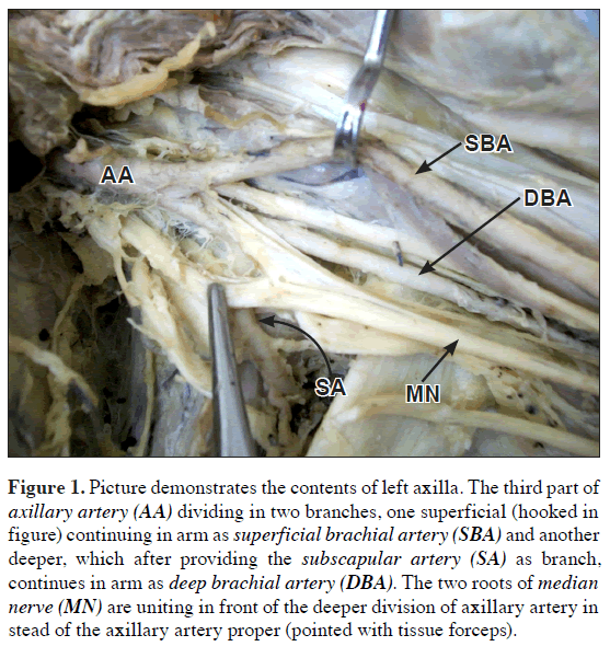
1. The third part of the axillary artery got divided into a thick superficial and a thin deep branches (Figure 1).
Figure 1: Picture demonstrates the contents of left axilla. The third part of axillary artery (AA) dividing in two branches, one superficial (hooked in figure) continuing in arm as superficial brachial artery (SBA) and another deeper, which after providing the subscapular artery (SA) as branch, continues in arm as deep brachial artery (DBA). The two roots of median nerve (MN) are uniting in front of the deeper division of axillary artery in stead of the axillary artery proper (pointed with tissue forceps).
2. The superficial one continued in the arm without providing any branch and finally in the cubital fossa got terminated by dividing in two branches corresponding as radial and ulnar arteries. The arterial pattern in the forearm and in palm was absolutely as usual.
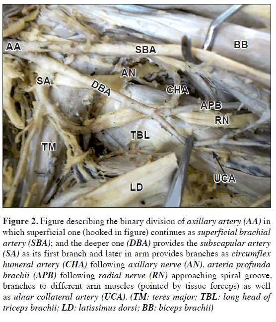
3. On the other hand the deeper one with its thinner caliber gave off the subscapular artery as its first branch from which the circumflex scapular and thoracodorsal arteries branched out (Figure 2).
Figure 2: Figure describing the binary division of axillary artery (AA) in which superficial one (hooked in figure) continues as superficial brachial artery (SBA); and the deeper one (DBA) provides the subscapular artery (SA) as its first branch and later in arm provides branches as circumflex humeral artery (CHA) following axillary nerve (AN), arteria profunda brachii (APB) following radial nerve (RN) approaching spiral groove, branches to different arm muscles (pointed by tissue forceps) as well as ulnar collateral artery (UCA). (TM: teres major; TBL: long head of triceps brachii; LD: latissimus dorsi; BB: biceps brachii)
4. Later continued in arm in parallel with the superficial counterpart to provide the branches as (i) a slender one, which following the axillary nerve entering trough the quadrangular space recognized as the circumflex humeral artery, (ii) another one entering in the spiral groove accompanying the radial nerve recognized as the deep artery of arm, (iii) branches to the muscles of the flexor compartment of arm, and (iv) ulnar collateral branches descending parallel with the ulnar nerve. It has finished by supplying the biceps brachii near the elbow joint (Figure 2).
Variations among the nerves
1. The roots of the median nerve united in front of the deeper branch of brachial artery instead of third part of axillary artery proper, so that the superficial branch was in front of the median nerve and the deeper one was posterior to it (Figure 1).
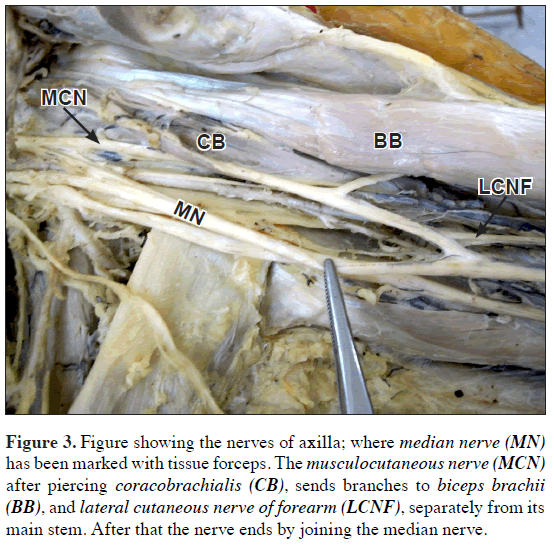
2. Again in the musculocutaneous nerve an unusual connection with median nerve was noted almost 5 cm proximal to elbow joint, after the emergence of lateral cutaneous branch for forearm (Figure 3).
Figure 3: Figure showing the nerves of axilla; where median nerve (MN) has been marked with tissue forceps. The musculocutaneous nerve (MCN) after piercing coracobrachialis (CB), sends branches to biceps brachii (BB), and lateral cutaneous nerve of forearm (LCNF), separately from its main stem. After that the nerve ends by joining the median nerve.
Cubital fossa onwards the nerves and the vessels were distributed in their usual course without any gross change. Even no gross variation was noted in fellow side.
Discussion
Such a rare combined variation in axillo-brachial neurovascular arcade can be a result of the unusual path chosen by the axial artery of upper limb. As described in literature, the development of the entire arterial tree of the upper limb occurs in stage-wise fashion [3].
At first (Stage 1) the lateral branch of seventh intersegmental artery, i.e., the subclavian artery extends up to the wrist as the axis artery of upper limb, where it terminates by dividing into terminal branches for the fingers forming a capillary plexus. The proximal portion of it forms axillary and brachial arteries respectively, whereas distal portion persists as the ‘anterior interosseous artery’ of forearm. Subsequently (Stage II) a ‘median artery’ arises from the anterior interosseous artery, grows along the median nerve to communicate with palmar capillary plexus to feed it. By this time the anterior interosseous artery undergoes regression. Later (Stage III) the ‘ulnar artery’ arises from brachial artery and unites distally with the existing median artery to form superficial palmar arch.
Side by side at Stage IV a ‘superficial brachial artery’ develops in the axillary region from the axial trunk and traverses the medial surface of the arm, runs diagonally from the ulnar to the radial side of the forearm to the posterior surface of the wrist to divide over the carpus into digital branches.
Finally at Stage V three changes occur simultaneously as:
(1) The ‘median artery’ regresses to a small slender vessel, familiar in adult life as arteria nervi mediana.
(2) The superficial brachial artery gives off a distal branch anastomosing with the superficial palmar arch formed already, and
(3) at the elbow an anastomotic branch develops between the main trunk of brachial artery and existent superficial brachial artery, which shortly enlarges to form the ‘radial artery’ with the distal portion of the superficial brachial artery. Where as the proximal portion of the superficial brachial artery atrophies correspondingly.
For these multiple plexiform sources of sequential appearance and disappearance of the arteries of upper limb, it may often get finalized by an unusual path, maintained throughout the life [2], as reported in pertinent literature [4–6]. The arterial variation takes place when the vessel to get obliterated either persists or the one to persist get obliterated. Even the incomplete development of the vessel or fusions and absorption of their parts in different combinations may lead to the unusual course [7].
Here, instead of regression of the distal part of the embryonic superficial brachial artery, the distal part of the main brachial artery gets atrophied to become the slender ‘deep brachial artery’. So why, both the radial and ulnar arteries had their origin from the superficial brachial artery retaining their embryonic communications.
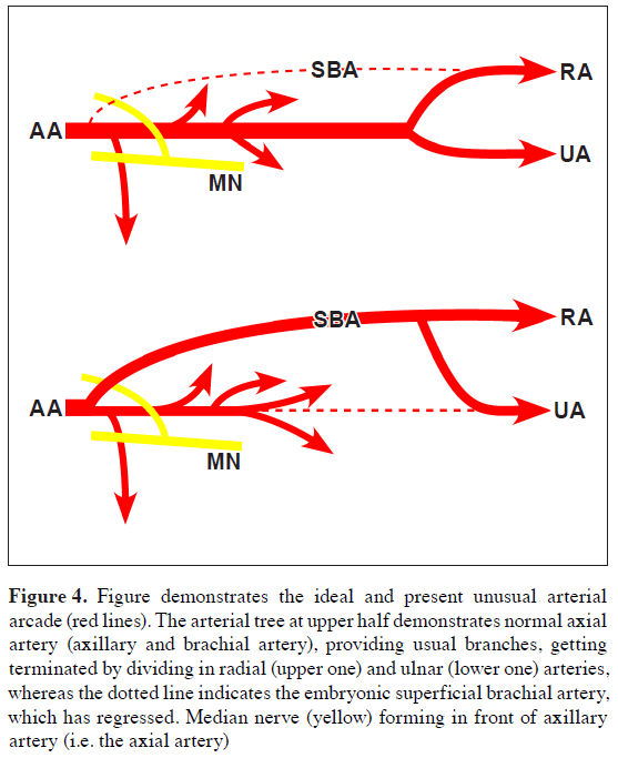
Since here the deep brachial artery represented the true axial artery of upper limb, so the scheduled branches of the third part of axillary artery (axial artery) also appeared from this deep brachial artery (as subscapular and circumflex humeral artery). For the same reason the two roots of median nerve, which to be formed in front of the axillary artery (i.e. the axial artery of upper limb), here forms in front of the deep brachial artery (Figure 4).
Figure 4: Figure demonstrates the ideal and present unusual arterial arcade (red lines). The arterial tree at upper half demonstrates normal axial artery (axillary and brachial artery), providing usual branches, getting terminated by dividing in radial (upper one) and ulnar (lower one) arteries, whereas the dotted line indicates the embryonic superficial brachial artery, which has regressed. Median nerve (yellow) forming in front of axillary artery (i.e. the axial artery)
The lower half demonstrates the present case, where the embryonic superficial brachial artery persists, continues in forearm to provide the radial and ulnar arteries; whereas the original axial artery (forming the deep brachial artery) get regressed (dotted line) after providing scheduled branches in axilla and arm. But the median nerve as a rule forms in front of the axial artery, i.e. here the deep brachial artery. (AA: axial artery; SBA: superficial brachial artery; RA: radial artery; UA: ulnar artery; MN: median nerve)
On the other hand it is not uncommon to find a nerve trunk of considerable size leaving the musculocutaneous, while lying behind the biceps, passes distally and medially to join the median nerve. This is regarded as due fibers for median nerve from the lateral cord passes aberrantly through the musculocutaneous returns to join the median nerve later, where incidence of it often found to vary as 8.1% to 36.19% [8]. Chouhan et al. also demonstrated such communications between median and musculocutaneous nerves in their studies [9].
Existence of such a communication here also likes to demonstrate the delivery of median nerve fibers originating from lateral cord, to it, bypassing the proximal union of the two roots.
Conclusion
Though individual variation patterns in arterial and neural arcades were reported previously but their co-existence was not found earlier as searched for. These may be asymptomatic totally but definitely having immense practical importance for the surgery of axillary region. Probability of variant branches should be kept in mind to prevent any unwanted damage. Therefore both the normal and variant anatomy of this region should be well known for accurate diagnostic interpretation and surgical intervention.
Acknowledgement
Sincere thanks and regards to all the faculty members of the Department of Anatomy and the respected Principal and Chairman of Ethical Committee of the North Bengal Medical College & Hospital.
References
- Johnson D. Axillary artery. In: Standring S, Borley N, Collin P, eds. Gray’s Anatomy. The Anatomical basis of Clinical Practice. 40th Ed., Edinburgh, Churchill Livingstone-Elsevier. 2008: 814.
- Williams P, Bannister L, Collins P, Dyson M, Dussek J, Ferguson M, eds. Gray’s Anatomy. The Anatomical basis of Medicine and Surgery. 38th Ed., Edinburgh, Churchill Livingstone. 1995; 319, 1269.
- Singer E. Embryological patterns persisting in the arteries of the arm. Anat Rec. 1933; 55: 406–413.
- Tan CB, Tan CK. An unusual course and relations of the human axillary artery. Singapore Med J. 1994; 35: 263–264.
- Jurjus AR, Correa-De-Aruaujo R, Bohn RC. Bilateral double axillary artery: embryological basis and clinical implications. Clin Anat. 1999; 12: 135–140.
- Patnaik VVG, Kalsey G, Singla RK. Bifurcation of axillary Artery in its 3rd part – a case report. J Anat. Soc India. 2001; 50: 166–169.
- Arey LB. Developmental Anatomy. A Textbook and Laboratory Manual of Embryology. 7th Ed., Philadelphia, W.B. Saunders. 1965; 358–359.
- Kerr AT. The brachial plexus of nerves in man, the variation in its formation and branches. Am J Anat. 1918; 23: 285–395.
- Chauhan R, Roy TS. Communication between the median and musculocutaneous nerve – a case report. J Anat Soc India. 2002; 51: 72–75.
Hironmoy Roy1*, Parthapratim Pradhan2, and Samar Deb3
1Department of Anatomy, North Bengal Medical College & Hospital, Siliguri, WB, India
2Department of Anatomy, Midnapore Medical College & Hospital, Paschim Medinipur, WB, India
3Department of Anatomy, College of Medicine &JNM Hospital, WBUHS, Kalyani, Nadia, WB, India
- *Corresponding Author:
- Dr. Hironmoy Roy, MD
Department of Anatomy, North Bengal Medical College & Hospital, Sushrutanagar, Siliguri, WB, 734012, India
Tel: +91 974 8828588
E-mail: hironmoy19@gmail.com
Date of Received: August 8th, 2010
Date of Accepted: January 6th, 2011
Published Online: January 14th, 2011
IJAV. 2011; 4: 5–8.
Abstract
Nerves and vessels as reach their target area sometimes violate their usual course, demonstrated as variant distribution. During routine dissection multiple such variations were found in a 60-year-old male cadaver unilaterally showing (a) third part of axillary artery dividing in superficial and deep branches as continued in arm, (b) the median nerve forming in unusual place and (c) the musculocutaneous nerve providing extra communicating branch to median nerve. Such a combined coexistent variation being extremely rare in literature, studied with probable embryological explanation.
-Keywords
axillary artery, median nerve, musculocutaneous nerve
Introduction
The axillary neurovascular structures are one of the potential anatomical fields to show variations in many ways. The axillary artery gets divided in three parts by the fore lying pectoralis minor and provides the superior thoracic artery from first part, thoraco-acromial artery, lateral thoracic artery from second part and the third part gives anterior circumflex humeral, posterior circumflex humeral and subscapular arteries. After the distal border of teres major the axillary artery is continued in arm as brachial artery, from which the branches in the arm get distributed.
To form the median nerve trunk from lateral (C6,7) and medial (C8, T1) cords of brachial plexus the two roots of median nerve emerge and unite embracing the third part of axillary artery, either anterior or lateral to it. The lateral root often being smaller than medial one, the median nerve usually passes lateral to axillary artery and continues in upper arm as lateral to brachial artery [1].
On the other hand the musculocutaneous nerve (C5,6,7) initially accompanies the third part of the axillary artery and pierces the coracobrachialis muscle after supplying it. Next it appears in between biceps brachii and brachialis, supplies them and just below the elbow it pierces the deep fascia lateral to the tendon of biceps brachii and extends further downward as the lateral cutaneous nerve of the forearm [2].
Case Report
During routine dissection on a 60-year-old male cadaver, multiple variations were noted in both left axillo-brachial vessels and nerves, which as a combination was unique in presentation till searched for. Surprisingly the fellow side showed no obvious variation.
The variations noted are described as below:
Variations in arterial arcade
1. The third part of the axillary artery got divided into a thick superficial and a thin deep branches (Figure 1).
Figure 1: Picture demonstrates the contents of left axilla. The third part of axillary artery (AA) dividing in two branches, one superficial (hooked in figure) continuing in arm as superficial brachial artery (SBA) and another deeper, which after providing the subscapular artery (SA) as branch, continues in arm as deep brachial artery (DBA). The two roots of median nerve (MN) are uniting in front of the deeper division of axillary artery in stead of the axillary artery proper (pointed with tissue forceps).
2. The superficial one continued in the arm without providing any branch and finally in the cubital fossa got terminated by dividing in two branches corresponding as radial and ulnar arteries. The arterial pattern in the forearm and in palm was absolutely as usual.
3. On the other hand the deeper one with its thinner caliber gave off the subscapular artery as its first branch from which the circumflex scapular and thoracodorsal arteries branched out (Figure 2).
Figure 2: Figure describing the binary division of axillary artery (AA) in which superficial one (hooked in figure) continues as superficial brachial artery (SBA); and the deeper one (DBA) provides the subscapular artery (SA) as its first branch and later in arm provides branches as circumflex humeral artery (CHA) following axillary nerve (AN), arteria profunda brachii (APB) following radial nerve (RN) approaching spiral groove, branches to different arm muscles (pointed by tissue forceps) as well as ulnar collateral artery (UCA). (TM: teres major; TBL: long head of triceps brachii; LD: latissimus dorsi; BB: biceps brachii)
4. Later continued in arm in parallel with the superficial counterpart to provide the branches as (i) a slender one, which following the axillary nerve entering trough the quadrangular space recognized as the circumflex humeral artery, (ii) another one entering in the spiral groove accompanying the radial nerve recognized as the deep artery of arm, (iii) branches to the muscles of the flexor compartment of arm, and (iv) ulnar collateral branches descending parallel with the ulnar nerve. It has finished by supplying the biceps brachii near the elbow joint (Figure 2).
Variations among the nerves
1. The roots of the median nerve united in front of the deeper branch of brachial artery instead of third part of axillary artery proper, so that the superficial branch was in front of the median nerve and the deeper one was posterior to it (Figure 1).
2. Again in the musculocutaneous nerve an unusual connection with median nerve was noted almost 5 cm proximal to elbow joint, after the emergence of lateral cutaneous branch for forearm (Figure 3).
Figure 3: Figure showing the nerves of axilla; where median nerve (MN) has been marked with tissue forceps. The musculocutaneous nerve (MCN) after piercing coracobrachialis (CB), sends branches to biceps brachii (BB), and lateral cutaneous nerve of forearm (LCNF), separately from its main stem. After that the nerve ends by joining the median nerve.
Cubital fossa onwards the nerves and the vessels were distributed in their usual course without any gross change. Even no gross variation was noted in fellow side.
Discussion
Such a rare combined variation in axillo-brachial neurovascular arcade can be a result of the unusual path chosen by the axial artery of upper limb. As described in literature, the development of the entire arterial tree of the upper limb occurs in stage-wise fashion [3].
At first (Stage 1) the lateral branch of seventh intersegmental artery, i.e., the subclavian artery extends up to the wrist as the axis artery of upper limb, where it terminates by dividing into terminal branches for the fingers forming a capillary plexus. The proximal portion of it forms axillary and brachial arteries respectively, whereas distal portion persists as the ‘anterior interosseous artery’ of forearm. Subsequently (Stage II) a ‘median artery’ arises from the anterior interosseous artery, grows along the median nerve to communicate with palmar capillary plexus to feed it. By this time the anterior interosseous artery undergoes regression. Later (Stage III) the ‘ulnar artery’ arises from brachial artery and unites distally with the existing median artery to form superficial palmar arch.
Side by side at Stage IV a ‘superficial brachial artery’ develops in the axillary region from the axial trunk and traverses the medial surface of the arm, runs diagonally from the ulnar to the radial side of the forearm to the posterior surface of the wrist to divide over the carpus into digital branches.
Finally at Stage V three changes occur simultaneously as:
(1) The ‘median artery’ regresses to a small slender vessel, familiar in adult life as arteria nervi mediana.
(2) The superficial brachial artery gives off a distal branch anastomosing with the superficial palmar arch formed already, and
(3) at the elbow an anastomotic branch develops between the main trunk of brachial artery and existent superficial brachial artery, which shortly enlarges to form the ‘radial artery’ with the distal portion of the superficial brachial artery. Where as the proximal portion of the superficial brachial artery atrophies correspondingly.
For these multiple plexiform sources of sequential appearance and disappearance of the arteries of upper limb, it may often get finalized by an unusual path, maintained throughout the life [2], as reported in pertinent literature [4–6]. The arterial variation takes place when the vessel to get obliterated either persists or the one to persist get obliterated. Even the incomplete development of the vessel or fusions and absorption of their parts in different combinations may lead to the unusual course [7].
Here, instead of regression of the distal part of the embryonic superficial brachial artery, the distal part of the main brachial artery gets atrophied to become the slender ‘deep brachial artery’. So why, both the radial and ulnar arteries had their origin from the superficial brachial artery retaining their embryonic communications.
Since here the deep brachial artery represented the true axial artery of upper limb, so the scheduled branches of the third part of axillary artery (axial artery) also appeared from this deep brachial artery (as subscapular and circumflex humeral artery). For the same reason the two roots of median nerve, which to be formed in front of the axillary artery (i.e. the axial artery of upper limb), here forms in front of the deep brachial artery (Figure 4).
Figure 4: Figure demonstrates the ideal and present unusual arterial arcade (red lines). The arterial tree at upper half demonstrates normal axial artery (axillary and brachial artery), providing usual branches, getting terminated by dividing in radial (upper one) and ulnar (lower one) arteries, whereas the dotted line indicates the embryonic superficial brachial artery, which has regressed. Median nerve (yellow) forming in front of axillary artery (i.e. the axial artery)
The lower half demonstrates the present case, where the embryonic superficial brachial artery persists, continues in forearm to provide the radial and ulnar arteries; whereas the original axial artery (forming the deep brachial artery) get regressed (dotted line) after providing scheduled branches in axilla and arm. But the median nerve as a rule forms in front of the axial artery, i.e. here the deep brachial artery. (AA: axial artery; SBA: superficial brachial artery; RA: radial artery; UA: ulnar artery; MN: median nerve)
On the other hand it is not uncommon to find a nerve trunk of considerable size leaving the musculocutaneous, while lying behind the biceps, passes distally and medially to join the median nerve. This is regarded as due fibers for median nerve from the lateral cord passes aberrantly through the musculocutaneous returns to join the median nerve later, where incidence of it often found to vary as 8.1% to 36.19% [8]. Chouhan et al. also demonstrated such communications between median and musculocutaneous nerves in their studies [9].
Existence of such a communication here also likes to demonstrate the delivery of median nerve fibers originating from lateral cord, to it, bypassing the proximal union of the two roots.
Conclusion
Though individual variation patterns in arterial and neural arcades were reported previously but their co-existence was not found earlier as searched for. These may be asymptomatic totally but definitely having immense practical importance for the surgery of axillary region. Probability of variant branches should be kept in mind to prevent any unwanted damage. Therefore both the normal and variant anatomy of this region should be well known for accurate diagnostic interpretation and surgical intervention.
Acknowledgement
Sincere thanks and regards to all the faculty members of the Department of Anatomy and the respected Principal and Chairman of Ethical Committee of the North Bengal Medical College & Hospital.
References
- Johnson D. Axillary artery. In: Standring S, Borley N, Collin P, eds. Gray’s Anatomy. The Anatomical basis of Clinical Practice. 40th Ed., Edinburgh, Churchill Livingstone-Elsevier. 2008: 814.
- Williams P, Bannister L, Collins P, Dyson M, Dussek J, Ferguson M, eds. Gray’s Anatomy. The Anatomical basis of Medicine and Surgery. 38th Ed., Edinburgh, Churchill Livingstone. 1995; 319, 1269.
- Singer E. Embryological patterns persisting in the arteries of the arm. Anat Rec. 1933; 55: 406–413.
- Tan CB, Tan CK. An unusual course and relations of the human axillary artery. Singapore Med J. 1994; 35: 263–264.
- Jurjus AR, Correa-De-Aruaujo R, Bohn RC. Bilateral double axillary artery: embryological basis and clinical implications. Clin Anat. 1999; 12: 135–140.
- Patnaik VVG, Kalsey G, Singla RK. Bifurcation of axillary Artery in its 3rd part – a case report. J Anat. Soc India. 2001; 50: 166–169.
- Arey LB. Developmental Anatomy. A Textbook and Laboratory Manual of Embryology. 7th Ed., Philadelphia, W.B. Saunders. 1965; 358–359.
- Kerr AT. The brachial plexus of nerves in man, the variation in its formation and branches. Am J Anat. 1918; 23: 285–395.
- Chauhan R, Roy TS. Communication between the median and musculocutaneous nerve – a case report. J Anat Soc India. 2002; 51: 72–75.


