A rare instance of an accessory long flexor to the second toe
Georgi P. Georgiev1*, Lazar Jelev1, Plamen Kinov2 and Nikolai K. Vidinov1
1Department of Anatomy, Histology and Embryology, Medical University Sofia, Sofia, Bulgaria
2Department of Orthopedics and Traumatology, University Hospital Queen Giovanna, ISUL, Sofia, Bulgaria
- *Corresponding Author:
- Georgi P. Georgiev, MD
Department of Anatomy, Histology and Embryology, Medical University Sofia, Blvd. Sv. Georgi Sofiiski 1, BG-1431 Sofia, Bulgaria
Tel: +359 2 9172636
E-mail: georgievgp@yahoo.com
Date of Received: June 1st, 2009
Date of Accepted: September 10th, 2009
Published Online: September 15th, 2009
© IJAV. 2009; 2: 108–110.
[ft_below_content] =>Keywords
flexor digitorum accessorius longus, foot, clinical significance
Introduction
A number of variant or accessory muscles in and around the human foot and ankle are seen occasionally [1]. Four of them could be discovered in the region of the tarsal tunnel – the peroneocalcaneus internus, the tibiocalcaneus internus, the accessory soleus and the flexor digitorum accessorius longus (FDAL) [1]. The last mentioned muscle, also named “accessorius ad accessorium”, “accessorius secundus” and “pronator pedis”, is the most common and has been described as a very variable slip, both in its origin and insertion [2]. Despite the fact, that first anatomical descriptions of the FDAL exits at the beginning of the 19th century, it is not so popular and continues to be unduly ignored in anatomical and surgical textbooks and therefore poorly characterized [3]. However, after the report of Sammarco and Stephens, in which they first described tarsal tunnel syndrome due to the presence of FDAL, this interesting variation has attracted renewed attention not only to anatomists, but also to clinicians [1].
In this report we describe a rare case of FDAL, review the current literature and emphasize on its possible clinical significance. This is the first report of such a variation in Bulgarian population.
Case Report
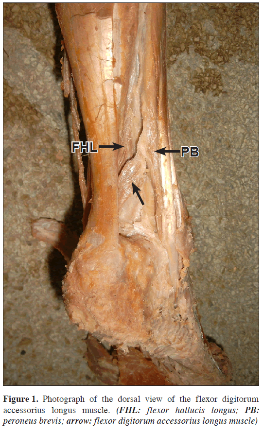
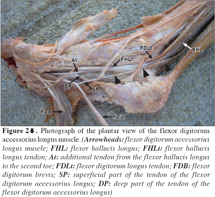
A case of an accessory long flexor (FDAL) has been encountered during routine anatomical dissections of a right lower extremity of a 57-year-old female cadaver. This was the only instance among 63 dissected lower extremities from the autopsy material available at the Department of Anatomy, Histology and Embryology at the Medical University of Sofia (Figures 1,2). The aberrant muscle arose from the lower third of the posterior crural intermuscular septum, between the flexor hallucis longus and the peroneus brevis muscles, 50 mm superior to the lateral malleolus (Figure 1). The FDAL had a well-formed muscular body (length – 55 mm, width – 11 mm) that passed obliquely downward and medially deep to the calcaneal tendon. Passing beneath the flexor retinaculum in the canal of the flexor hallucis longus tendon, the aberrant muscle reached the sole region where it prolonged in a thin tendon (length – 52 mm, width 1.4 mm). Cut and retraction of the flexor digitorum longus tendon revealed that at the end, the tendon of the FDAL divided into two parts – superficial and deep. The superficial part was attached to the second toe tendon of the flexor digitorum longus. The deep part of the tendon joined the tendon of the quadratus plantae and also an additional portion of the flexor hallucis longus tendon to the second toe (Figure 2). The innervation of the variant muscle was from the tibial nerve and the blood supply from the posterior tibial artery. Due to its morphological characteristic and possible mechanics, the described variant muscle could be termed in Latin “musculus flexor digiti secundi accessorius”.
Figure 2: Photograph of the plantar view of the flexor digitorum accessorius longus muscle. (Arrowheads: flexor digitorum accessorius longus muscle; FHL: flexor hallucis longus; FHLt: flexor hallucis longus tendon; At: additional tendon from the flexor hallucis longus to the second toe; FDLt: flexor digitorum longus tendon; FDB: flexor digitorum brevis; SP: superficial part of the tendon of the flexor digitorum accessorius longus; DP: deep part of the tendon of the flexor digitorum accessorius longus)
Discussion
FDAL is described to be a variation among the flexor muscle in the leg and its incidence vary between 5-13% in different populations [2,4–7]. However, there are several articles that present very low incidence for this muscle – 1-2% [3,8,9]. According to Hwang and Hill, the presence of the FDAL varies greatly from population to population, and the overall incidence is realistically at the lower end of the ranges previously reported [3]. This is also confirmed from our investigation, where the variant muscle was the only instance among the 63 lower extremities examined (incidence of approximately 1.6%).
Morphologically, there have been described accessory long flexors with one or two heads arising from different bone or fibrous points of the leg [3,8,9]. Among them, the FDAL with one head is the most common case [3]. It can arise from the deep part of the leg, from tibial and/or fibular side, but more commonly from the fibular side [2,3, 6–8]. In most of the reported cases, the FDAL originated several centimeters inferior to the popliteal fossa, near the origins of the other long digital flexors [3]. In a very few cases this variant muscle arises close to the ankle joint, as in our case [2,7,10].
The accessory tendons of the leg muscles have definite clinical significance [3,11–13]. In some cases, they may hamper arthroscopy and cause difficulties in radiographic interpretations [14,15]. Moreover, the variant muscles, especially FDAL has been included in two significant syndromes of the lower limb: the tarsal tunnel syndrome (TTS) and flexor hallucis syndrome (FHS) [1,3,12,13]. The TTS and FHS are entrapment neuropathies of the lower extremity, similar to the carpal tunnel syndrome at the distal arm [3]. The both syndromes are characterized by pain and paresthesia of the limb [3]. They could be provoked by trauma, space-occupying lesion, or skeletal deformities [1]. The presence of a FDAL could be a predisposing factor that may contribute to both TTS and FHS [1,5,13]. In cases of symptomatic patient, the presence of this muscle could be found during physical examination of the tarsal tunnel as an abnormal fullness between the Achilles tendon and the tibia [5]. In these patients when the ankle is held in dorsiflexion, there are limited dorsiflexion, which is tethered by the “cork-in-a-bottle” effect of the distal muscle mass of the FDAL at the flexor retinaculum and fibro-osseous canal of the flexor hallucis longus [5]. The MRI examination could confirmed the presence of an abnormal muscle mass around the ankle joint and the foot [5].
In this report, we present a rare variation of FDAL in a lower limb. In the last two decades, similar variations have attracted clinical attention, because of their possible role in TTS and FHS. Therefore, the described here aberrant muscle should be always in mind for orthopedics and radiologists in differential diagnosis of compression neuropathies of the lower limb.
Acknowledgments
The authors thank to Dr. Sunny Hwang for finding some of the references.
References
- Sammarco GJ, Stephens MM. Tarsal tunnel syndrome caused by the flexor digitorum accessorius longus. A case report. J Bone Joint Surg Am. 1990; 72: 453–454.
- Jaijesh P, Shenoy M, Anuradha L, Chithralekha KK. Flexor accessorius longus: A rare variation of the deep extrinsic digital flexors of the leg and its phylogenetic significance. Indian J Plast Surg. 2006; 39: 169–171.
- Hwang SH, Hill RV. An unusual variation of the flexor digitorum accessorius longus muscle-its anatomy and clinical significance. Anat Sci Int. 2009; 84: 257–263.
- Deroy AR, Clause CC, Baskin ES, Bauer GR. Recognition of the flexor digitorum accessorius longus. J Am Podiatr Med Assoc. 2002; 92: 463–466.
- Eberle CF, Moran B, Gleason T. The accessory flexor digitorum longus as a cause of Flexor Hallucis Syndrome. Foot Ankle Int. 2002; 23: 51–55.
- Peterson DA, Stinson W, Lairmore JR. The long accessory flexor muscle: an anatomical study. Foot Ankle Int. 1995; 16: 637–640.
- VanCourt RB, Siesel KJ. Flexor digitorum accessorius longus muscle. J Am Podiatr Med Assoc. 1996; 86: 559–560.
- Gumusalan Y, Kalaycioglu A. Bilateral accessory flexor digitorium longus muscle in man. Ann Anat. 2000; 182: 573–576.
- Wood J. Variations in human myology observed during the winter session of 1867–68 at King’s college, London. Proc R Soc Lond. 1868, 16: 483–525.
- Nathan H, Gloobe H, Yosipovitch Z. Flexor digitorum accessorius longus. Clin Orthop Relat Res. 1975; 113: 158–161.
- Dobbs MB, Walton T, Gordon JE, Schoenecker PL, Gurnett CA. Flexor digitorum accessorius longus muscle is associated with familial idiopathic clubfoot. J Pediatr Orthop. 2005; 25: 357–359.
- Grogono BJ, Jowsey J. Flexor accessorius longus: an unusual muscle anomaly. J Bone Joint Surg Br. 1965; 47: 118–119.
- Sammarco GJ, Conti SF. Tarsal tunnel syndrome caused by an anomalous muscle. J Bone Joint Surg Am. 1994; 76: 1308–1314.
- Buckingham RA, Winson IG, Kelly AJ. An anatomical study of a new portal for ankle arthroscopy. J Bone Joint Surg Br. 1997; 79: 650–652.
- Wittmayer BC, Freed L. Diagnosis and surgical management of flexor digitorum accessorius longus-induced tarsal tunnel syndrome. J Foot Ankle Surg. 2007; 46: 484–487.
Georgi P. Georgiev1*, Lazar Jelev1, Plamen Kinov2 and Nikolai K. Vidinov1
1Department of Anatomy, Histology and Embryology, Medical University Sofia, Sofia, Bulgaria
2Department of Orthopedics and Traumatology, University Hospital Queen Giovanna, ISUL, Sofia, Bulgaria
- *Corresponding Author:
- Georgi P. Georgiev, MD
Department of Anatomy, Histology and Embryology, Medical University Sofia, Blvd. Sv. Georgi Sofiiski 1, BG-1431 Sofia, Bulgaria
Tel: +359 2 9172636
E-mail: georgievgp@yahoo.com
Date of Received: June 1st, 2009
Date of Accepted: September 10th, 2009
Published Online: September 15th, 2009
© IJAV. 2009; 2: 108–110.
Abstract
During routine anatomical dissection a rare case of a flexor digitorum accessorius longus muscle was observed. This muscle arose with a well-formed muscular belly from the lower part of the lateral intermuscular septum, and then passed obliquely downward and medially deep to the calcaneal tendon in the canal of the flexor hallucis longus. In the sole region the aberrant muscle prolonged in a thin tendon that divided into two parts: superficial and deep. The superficial part attached to the tendon of the flexor digitorum longus for the second toe; the deep part of the tendon joined the tendon of the quadratus plantae and also an additional portion of the flexor hallucis longus tendon to the second toe. The possible clinical implications of this muscle in practical surgery and also in imaging radiology are reviewed.
-Keywords
flexor digitorum accessorius longus, foot, clinical significance
Introduction
A number of variant or accessory muscles in and around the human foot and ankle are seen occasionally [1]. Four of them could be discovered in the region of the tarsal tunnel – the peroneocalcaneus internus, the tibiocalcaneus internus, the accessory soleus and the flexor digitorum accessorius longus (FDAL) [1]. The last mentioned muscle, also named “accessorius ad accessorium”, “accessorius secundus” and “pronator pedis”, is the most common and has been described as a very variable slip, both in its origin and insertion [2]. Despite the fact, that first anatomical descriptions of the FDAL exits at the beginning of the 19th century, it is not so popular and continues to be unduly ignored in anatomical and surgical textbooks and therefore poorly characterized [3]. However, after the report of Sammarco and Stephens, in which they first described tarsal tunnel syndrome due to the presence of FDAL, this interesting variation has attracted renewed attention not only to anatomists, but also to clinicians [1].
In this report we describe a rare case of FDAL, review the current literature and emphasize on its possible clinical significance. This is the first report of such a variation in Bulgarian population.
Case Report
A case of an accessory long flexor (FDAL) has been encountered during routine anatomical dissections of a right lower extremity of a 57-year-old female cadaver. This was the only instance among 63 dissected lower extremities from the autopsy material available at the Department of Anatomy, Histology and Embryology at the Medical University of Sofia (Figures 1,2). The aberrant muscle arose from the lower third of the posterior crural intermuscular septum, between the flexor hallucis longus and the peroneus brevis muscles, 50 mm superior to the lateral malleolus (Figure 1). The FDAL had a well-formed muscular body (length – 55 mm, width – 11 mm) that passed obliquely downward and medially deep to the calcaneal tendon. Passing beneath the flexor retinaculum in the canal of the flexor hallucis longus tendon, the aberrant muscle reached the sole region where it prolonged in a thin tendon (length – 52 mm, width 1.4 mm). Cut and retraction of the flexor digitorum longus tendon revealed that at the end, the tendon of the FDAL divided into two parts – superficial and deep. The superficial part was attached to the second toe tendon of the flexor digitorum longus. The deep part of the tendon joined the tendon of the quadratus plantae and also an additional portion of the flexor hallucis longus tendon to the second toe (Figure 2). The innervation of the variant muscle was from the tibial nerve and the blood supply from the posterior tibial artery. Due to its morphological characteristic and possible mechanics, the described variant muscle could be termed in Latin “musculus flexor digiti secundi accessorius”.
Figure 2: Photograph of the plantar view of the flexor digitorum accessorius longus muscle. (Arrowheads: flexor digitorum accessorius longus muscle; FHL: flexor hallucis longus; FHLt: flexor hallucis longus tendon; At: additional tendon from the flexor hallucis longus to the second toe; FDLt: flexor digitorum longus tendon; FDB: flexor digitorum brevis; SP: superficial part of the tendon of the flexor digitorum accessorius longus; DP: deep part of the tendon of the flexor digitorum accessorius longus)
Discussion
FDAL is described to be a variation among the flexor muscle in the leg and its incidence vary between 5-13% in different populations [2,4–7]. However, there are several articles that present very low incidence for this muscle – 1-2% [3,8,9]. According to Hwang and Hill, the presence of the FDAL varies greatly from population to population, and the overall incidence is realistically at the lower end of the ranges previously reported [3]. This is also confirmed from our investigation, where the variant muscle was the only instance among the 63 lower extremities examined (incidence of approximately 1.6%).
Morphologically, there have been described accessory long flexors with one or two heads arising from different bone or fibrous points of the leg [3,8,9]. Among them, the FDAL with one head is the most common case [3]. It can arise from the deep part of the leg, from tibial and/or fibular side, but more commonly from the fibular side [2,3, 6–8]. In most of the reported cases, the FDAL originated several centimeters inferior to the popliteal fossa, near the origins of the other long digital flexors [3]. In a very few cases this variant muscle arises close to the ankle joint, as in our case [2,7,10].
The accessory tendons of the leg muscles have definite clinical significance [3,11–13]. In some cases, they may hamper arthroscopy and cause difficulties in radiographic interpretations [14,15]. Moreover, the variant muscles, especially FDAL has been included in two significant syndromes of the lower limb: the tarsal tunnel syndrome (TTS) and flexor hallucis syndrome (FHS) [1,3,12,13]. The TTS and FHS are entrapment neuropathies of the lower extremity, similar to the carpal tunnel syndrome at the distal arm [3]. The both syndromes are characterized by pain and paresthesia of the limb [3]. They could be provoked by trauma, space-occupying lesion, or skeletal deformities [1]. The presence of a FDAL could be a predisposing factor that may contribute to both TTS and FHS [1,5,13]. In cases of symptomatic patient, the presence of this muscle could be found during physical examination of the tarsal tunnel as an abnormal fullness between the Achilles tendon and the tibia [5]. In these patients when the ankle is held in dorsiflexion, there are limited dorsiflexion, which is tethered by the “cork-in-a-bottle” effect of the distal muscle mass of the FDAL at the flexor retinaculum and fibro-osseous canal of the flexor hallucis longus [5]. The MRI examination could confirmed the presence of an abnormal muscle mass around the ankle joint and the foot [5].
In this report, we present a rare variation of FDAL in a lower limb. In the last two decades, similar variations have attracted clinical attention, because of their possible role in TTS and FHS. Therefore, the described here aberrant muscle should be always in mind for orthopedics and radiologists in differential diagnosis of compression neuropathies of the lower limb.
Acknowledgments
The authors thank to Dr. Sunny Hwang for finding some of the references.
References
- Sammarco GJ, Stephens MM. Tarsal tunnel syndrome caused by the flexor digitorum accessorius longus. A case report. J Bone Joint Surg Am. 1990; 72: 453–454.
- Jaijesh P, Shenoy M, Anuradha L, Chithralekha KK. Flexor accessorius longus: A rare variation of the deep extrinsic digital flexors of the leg and its phylogenetic significance. Indian J Plast Surg. 2006; 39: 169–171.
- Hwang SH, Hill RV. An unusual variation of the flexor digitorum accessorius longus muscle-its anatomy and clinical significance. Anat Sci Int. 2009; 84: 257–263.
- Deroy AR, Clause CC, Baskin ES, Bauer GR. Recognition of the flexor digitorum accessorius longus. J Am Podiatr Med Assoc. 2002; 92: 463–466.
- Eberle CF, Moran B, Gleason T. The accessory flexor digitorum longus as a cause of Flexor Hallucis Syndrome. Foot Ankle Int. 2002; 23: 51–55.
- Peterson DA, Stinson W, Lairmore JR. The long accessory flexor muscle: an anatomical study. Foot Ankle Int. 1995; 16: 637–640.
- VanCourt RB, Siesel KJ. Flexor digitorum accessorius longus muscle. J Am Podiatr Med Assoc. 1996; 86: 559–560.
- Gumusalan Y, Kalaycioglu A. Bilateral accessory flexor digitorium longus muscle in man. Ann Anat. 2000; 182: 573–576.
- Wood J. Variations in human myology observed during the winter session of 1867–68 at King’s college, London. Proc R Soc Lond. 1868, 16: 483–525.
- Nathan H, Gloobe H, Yosipovitch Z. Flexor digitorum accessorius longus. Clin Orthop Relat Res. 1975; 113: 158–161.
- Dobbs MB, Walton T, Gordon JE, Schoenecker PL, Gurnett CA. Flexor digitorum accessorius longus muscle is associated with familial idiopathic clubfoot. J Pediatr Orthop. 2005; 25: 357–359.
- Grogono BJ, Jowsey J. Flexor accessorius longus: an unusual muscle anomaly. J Bone Joint Surg Br. 1965; 47: 118–119.
- Sammarco GJ, Conti SF. Tarsal tunnel syndrome caused by an anomalous muscle. J Bone Joint Surg Am. 1994; 76: 1308–1314.
- Buckingham RA, Winson IG, Kelly AJ. An anatomical study of a new portal for ankle arthroscopy. J Bone Joint Surg Br. 1997; 79: 650–652.
- Wittmayer BC, Freed L. Diagnosis and surgical management of flexor digitorum accessorius longus-induced tarsal tunnel syndrome. J Foot Ankle Surg. 2007; 46: 484–487.


