A rare occurrence of an accessory thyroid artery
Sara Doll*
Ruprecht-Karls-University Heidelberg, Institute of Anatomy and Cell Biology, Heidelberg, Germany
- *Corresponding Author:
- Sara Doll
Ruprecht-Karls-University Heidelberg, Institute of Anatomy and Cell Biology, Im Neuenheimer Feld 307, 69120 Heidelberg, Germany
Tel: +49 6221 5638078
E-mail: doll@ana.uni-heidelberg.de
Date of Received : March 16th, 2009
Date of Accepted : July 8th, 2009
Published Online : July 9th, 2009
© IJAV. 2009; 2: 71–72.
[ft_below_content] =>Keywords
inferior thyroid artery, variation, accessory
Introduction
As most anatomy atlases will illustrate, thyroid glands receive their blood supply from one superior thyroid artery arising from the external carotid artery, and from one inferior thyroid artery, branching off the thyrocervical trunk, which also gives rise to the suprascapular, transverse cervical and ascending cervical arteries. In addition to its branches to the inferior region of the thyroid gland, the inferior thyroid artery also vascularizes the parathyroid glands, pharynx, larynx and esophagus. Relatively few textbooks include descriptions of variations in the vessels supplying structures in the neck. Some mention variations in branches of the subclavian artery or the presence of an variant branch directly off the common carotid artery. The most detailed description of arterial variations associated with the recurrent laryngeal nerve, for example, can be found in an anatomy textbook written by Lanz-Wachsmuth [1]. Although this information is very important for surgeons, newer textbooks seldom report cervical vascular variations and only a few articles have been published that describe such variations.
Case Report
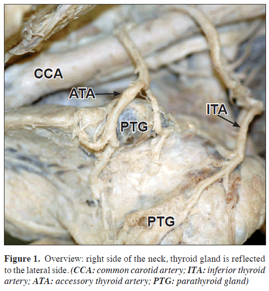
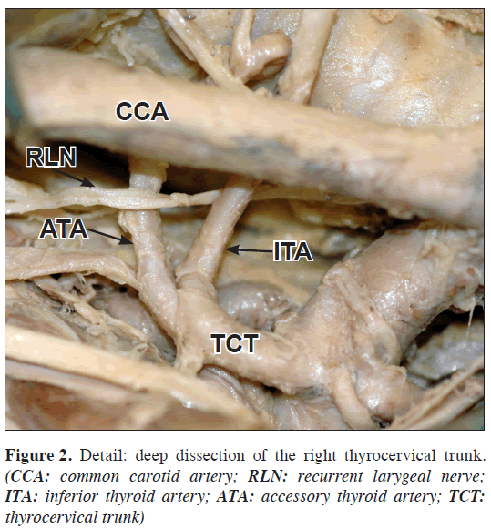
During the traditional human anatomy dissecting course for first year medical students, we discovered an extremely large thyroid gland on a 81-year-old female cadaver. In addition, enlarged parathyroid glands were observed on the dorsal side of each lobe. Upon bisection of the enlarged thyroid gland, and lateral reflection of each lobe, we identified two inferior thyroid arteries on the right side of the neck. Each artery bifurcated to provide blood to the middle region of the thyroid gland and to the parathyroid glands (Figure 1). Each branch supplied roughly the same outside diameter of the gland as the superior thyroid artery (4.4 mm). The arteries appeared to originate from the medial aspect of the common carotid artery however upon manual elevation of the carotid artery we found that indeed both arteries branched off the thyrocervical trunk, and then wound 20 mm cranial. The two branches then formed loops which continued along the dorsal aspect of the gland. The recurrent laryngeal nerve crossed under one and above the other thyroid inferior artery (Figure 2). The other vessels of the right thyrocervical trunk showed a regular course as did those on the left side of the neck. Unfortunately, the veins had already been removed and therefore cannot be discussed in this case report.
Discussion
Variations of the superior and inferior thyroid arteries are rarely described in anatomy textbooks, although knowledge of such variations is very important for surgical procedures in this region. The superior thyroid artery generally follows the more reliable course. In contrast, the inferior thyroid artery, which typically arises off the thyrocervical trunk, less frequently off the subclavian artery, is often observed to meander considerably. Descriptions of these meanderings usually include the extent, the course taken, the diameter of the vessels, and their anatomical relationship to the recurrent laryngeal nerve. One of the typically occurring variations of inferior thyroid artery is its absence, which was reportedly observed in 0.27% (n = 276) [2], and 6% (n = 100) of dissected cadavers [3]. In each case the gland was supplied with blood by either a “least thyroid artery” or a variant descending branch of the superior thyroid artery. Another variation described is a double artery. Specifically, Ziolkowski et al. dissected 276 fetuses without gross abnormalities and found that 1.08% of the cadavers possessed an additional thyroid artery [2]. They interpreted this observation as representative of an accessory and a middle thyroid artery. Another rare variation was that of an additional artery branching off the suprascapular artery or common carotid artery. This was observed only once. Accessory arteries have also been reported branching off either the thyrocervical trunk, the vertebral or subclavian artery, or the costocervical trunk (n=2). On one cadaver, one branch arose from the right subclavian artery and the other from the right costocervical trunk. Faller and Schaerer [3], dissected 100 cadavers and found smaller diameters of the variant vessels (average = 3 mm). Lang et al. [4], examined 73 cadavers, and reported a long thyrocervical trunk (average = 4.9 mm) with an outside diameter between 2.8 and 9.5 mm (average = 5.5 mm). In 70% of the inspected cadavers (n = 51) the recurrent laryngeal nerve found between the normal branch and the accessory thyroid artery (Lanz-Wachsmuth observed this variation in only 32% of their cadavers). The most commonly observed arterial variation was an ascending cranial loop, giving rise to a double inferior thyroid artery (9.5%), followed by the ramification that we also observed (7.5%). The most infrequent observation (1%) was a second, caudal loop of the artery, ending in a bifurcation. The significance of these variations is obvious; during thyroidectomy or parathyroidectomy the thyroid vessels must be ligated and the recurrent laryngeal nerve spared to prevent vocal cord disturbances. Furthermore, medical students should be made taught to expect anatomical variability during their brief exposure to cadaveric material. They must understand that the images they see in their atlases represent those most commonly observed, but definitely not the only ones to which they will be exposed during their careers as physicians. Indeed, our finding is an uncommon variation, however we consider it important to share our observation with others and encourage our colleagues to also report their findings to a platform such as the IJAV.
References
- Lanz v T, Wachsmuth W. Praktische Anatomie. Heidelberg, Springer. 1955; 252–255. (German)
- Ziolkowski M, Bieganska-Dembowska D, Kurlej W. Variations in the number and in origin of the thyroid arteries. Folia Morphol (Warsz). 1994; 53: 105–110.
- Faller A, Scharer O. Über die Variabilität der Arteriae thyroideae. Acta Anatomica. 1947; 4: 119–122. (German)
- Lang J, Fischer K, Nachbaur S, Meuer H-W. Über den Verlauf und die Zweige des N. laryngeus reccurens, der A. thyroidea inferior und der A. laryngea inferior. [Course and branches of the recurrent laryngeal nerve, inferior thyroid artery and inferior laryngeal artery]. Gegenbaurs Morphol Jahrb. 1986;132: 617–643. (German)
Sara Doll*
Ruprecht-Karls-University Heidelberg, Institute of Anatomy and Cell Biology, Heidelberg, Germany
- *Corresponding Author:
- Sara Doll
Ruprecht-Karls-University Heidelberg, Institute of Anatomy and Cell Biology, Im Neuenheimer Feld 307, 69120 Heidelberg, Germany
Tel: +49 6221 5638078
E-mail: doll@ana.uni-heidelberg.de
Date of Received : March 16th, 2009
Date of Accepted : July 8th, 2009
Published Online : July 9th, 2009
© IJAV. 2009; 2: 71–72.
Abstract
During our traditional human cadaveric dissecting course we found a rare variation of the inferior thyroid artery off the thyrocervical trunk on the right side of the neck. The right superior thyroid artery was normal as was the vascular supply to the left lobe of the thyroid gland. This variation is not common, although it has been described in more detailed textbooks and case reports. However, it is important to be aware of the frequency of occurrence of arterial variations, because surgeons conducting thyroid gland surgery need to know which variations to anticipate.
-Keywords
inferior thyroid artery, variation, accessory
Introduction
As most anatomy atlases will illustrate, thyroid glands receive their blood supply from one superior thyroid artery arising from the external carotid artery, and from one inferior thyroid artery, branching off the thyrocervical trunk, which also gives rise to the suprascapular, transverse cervical and ascending cervical arteries. In addition to its branches to the inferior region of the thyroid gland, the inferior thyroid artery also vascularizes the parathyroid glands, pharynx, larynx and esophagus. Relatively few textbooks include descriptions of variations in the vessels supplying structures in the neck. Some mention variations in branches of the subclavian artery or the presence of an variant branch directly off the common carotid artery. The most detailed description of arterial variations associated with the recurrent laryngeal nerve, for example, can be found in an anatomy textbook written by Lanz-Wachsmuth [1]. Although this information is very important for surgeons, newer textbooks seldom report cervical vascular variations and only a few articles have been published that describe such variations.
Case Report
During the traditional human anatomy dissecting course for first year medical students, we discovered an extremely large thyroid gland on a 81-year-old female cadaver. In addition, enlarged parathyroid glands were observed on the dorsal side of each lobe. Upon bisection of the enlarged thyroid gland, and lateral reflection of each lobe, we identified two inferior thyroid arteries on the right side of the neck. Each artery bifurcated to provide blood to the middle region of the thyroid gland and to the parathyroid glands (Figure 1). Each branch supplied roughly the same outside diameter of the gland as the superior thyroid artery (4.4 mm). The arteries appeared to originate from the medial aspect of the common carotid artery however upon manual elevation of the carotid artery we found that indeed both arteries branched off the thyrocervical trunk, and then wound 20 mm cranial. The two branches then formed loops which continued along the dorsal aspect of the gland. The recurrent laryngeal nerve crossed under one and above the other thyroid inferior artery (Figure 2). The other vessels of the right thyrocervical trunk showed a regular course as did those on the left side of the neck. Unfortunately, the veins had already been removed and therefore cannot be discussed in this case report.
Discussion
Variations of the superior and inferior thyroid arteries are rarely described in anatomy textbooks, although knowledge of such variations is very important for surgical procedures in this region. The superior thyroid artery generally follows the more reliable course. In contrast, the inferior thyroid artery, which typically arises off the thyrocervical trunk, less frequently off the subclavian artery, is often observed to meander considerably. Descriptions of these meanderings usually include the extent, the course taken, the diameter of the vessels, and their anatomical relationship to the recurrent laryngeal nerve. One of the typically occurring variations of inferior thyroid artery is its absence, which was reportedly observed in 0.27% (n = 276) [2], and 6% (n = 100) of dissected cadavers [3]. In each case the gland was supplied with blood by either a “least thyroid artery” or a variant descending branch of the superior thyroid artery. Another variation described is a double artery. Specifically, Ziolkowski et al. dissected 276 fetuses without gross abnormalities and found that 1.08% of the cadavers possessed an additional thyroid artery [2]. They interpreted this observation as representative of an accessory and a middle thyroid artery. Another rare variation was that of an additional artery branching off the suprascapular artery or common carotid artery. This was observed only once. Accessory arteries have also been reported branching off either the thyrocervical trunk, the vertebral or subclavian artery, or the costocervical trunk (n=2). On one cadaver, one branch arose from the right subclavian artery and the other from the right costocervical trunk. Faller and Schaerer [3], dissected 100 cadavers and found smaller diameters of the variant vessels (average = 3 mm). Lang et al. [4], examined 73 cadavers, and reported a long thyrocervical trunk (average = 4.9 mm) with an outside diameter between 2.8 and 9.5 mm (average = 5.5 mm). In 70% of the inspected cadavers (n = 51) the recurrent laryngeal nerve found between the normal branch and the accessory thyroid artery (Lanz-Wachsmuth observed this variation in only 32% of their cadavers). The most commonly observed arterial variation was an ascending cranial loop, giving rise to a double inferior thyroid artery (9.5%), followed by the ramification that we also observed (7.5%). The most infrequent observation (1%) was a second, caudal loop of the artery, ending in a bifurcation. The significance of these variations is obvious; during thyroidectomy or parathyroidectomy the thyroid vessels must be ligated and the recurrent laryngeal nerve spared to prevent vocal cord disturbances. Furthermore, medical students should be made taught to expect anatomical variability during their brief exposure to cadaveric material. They must understand that the images they see in their atlases represent those most commonly observed, but definitely not the only ones to which they will be exposed during their careers as physicians. Indeed, our finding is an uncommon variation, however we consider it important to share our observation with others and encourage our colleagues to also report their findings to a platform such as the IJAV.
References
- Lanz v T, Wachsmuth W. Praktische Anatomie. Heidelberg, Springer. 1955; 252–255. (German)
- Ziolkowski M, Bieganska-Dembowska D, Kurlej W. Variations in the number and in origin of the thyroid arteries. Folia Morphol (Warsz). 1994; 53: 105–110.
- Faller A, Scharer O. Über die Variabilität der Arteriae thyroideae. Acta Anatomica. 1947; 4: 119–122. (German)
- Lang J, Fischer K, Nachbaur S, Meuer H-W. Über den Verlauf und die Zweige des N. laryngeus reccurens, der A. thyroidea inferior und der A. laryngea inferior. [Course and branches of the recurrent laryngeal nerve, inferior thyroid artery and inferior laryngeal artery]. Gegenbaurs Morphol Jahrb. 1986;132: 617–643. (German)


