A rare variation in the mode of termination of posterior cord of brachial plexusaxilla
Jamuna M*
Department of Anatomy, PSG Institute of Medical Sciences and Research, Coimbatore, India
- *Corresponding Author:
- Dr. Jamuna M, MS
Assistant Professor, Department of Anatomy, PSG Institute of Medical Sciences and Research, Peelamedu, Coimbatore, Tamilnadu, India
Tel: +91 94437 37586
E-mail: drjamunam@gmail.com
Date of Received: November 18th, 2009
Date of Accepted: June 21st, 2010
Published Online: July 9th, 2010
© IJAV. 2010; 3: 95–96.
[ft_below_content] =>Keywords
brachial plexus, posterior cord variation, radial nerve
Introduction
The brachial plexus is an ordered network of large nerves through which the sensory and motor nerve supply is distributed to all structures that constitute the upper limb. It is formed by the anterior rami of C5 to T1 spinal nerves. They unite to form the upper, middle and lower trunks. Each trunk splits into anterior and posterior divisions. All three posterior divisions merge to form the posterior cord; in it are gathered all C5 to T1 nerves fibers for the extensor compartment. It lies behind the axillary artery [1].
The posterior cord terminates in the axillary and radial nerves. The three minor branches of the posterior cord are the two subscapular nerves and the thoracodorsal nerve between them [1]. Variations in the formation of brachial plexus and its terminal branches has been reported in 65.3% of cases [2]. Here, a rare variation in the branching pattern of the terminal branches of the posterior cord of the brachial plexus is reported.
Case Report
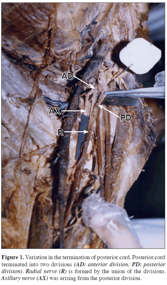
The study of 50 brachial plexuses was done in 25 embalmed adult cadavers during routine gross anatomical dissection in Madurai Medical College, Madurai. Dissection of the brachial plexus was done on right and left sides of the cadaver. The pectoral region, axilla and the arm were dissected. The axillary artery, the cords and the branches of the cords of the brachial plexus were identified. In one infraclavicular part of the brachial plexus in the left sided plexusaxilla, the present variation was noted. The posterior cord was splitting into anterior and posterior divisions and the radial nerve was formed by the union of the two divisions and the axillary nerve was arising from the posterior division. In the right side brachial plexus the posterior cord was showing usual branching pattern.
Discussion
Gupta et al. described the variations in plexus patterns may be due to unusual formation during the development of trunks, divisions or cords [3]. Kumar Megur has reported that the posterior cord divided into two roots, enclosing the sub scapular artery and the two roots fused to continue as radial nerve. The axillary nerve was originating from the thick posterior root of the cord [4]. The formation of radial nerve by the union of the two divisions from the posterior cord coincides with the above report but the two roots which enclosed the subscapular artery in the above report is contrary to the present report, where the two divisions were not enclosing the subscapular artery. Bertha et al. reported in one case instead of a single entity, the posterior cord was formed of two parts, upper and lower. The upper part was the continuation of posterior division of upper trunk. The lower part was formed by the union of the posterior divisions of middle and lower trunks. The upper posterior cord continued as axillary nerve after giving off upper and lower sub scapular nerves and upper root of radial nerve. The lower posterior cord after giving off thoracodorsal nerve continued as lower root of radial nerve and joined with the upper root to form radial nerve [5]. In the case reported here, the formation of posterior cord was not coinciding with the above report but the posterior cord was splitting into two divisions and the radial nerve formation by the union of those divisions coincides with the above report. The axillary nerve arising from the posterior division in the case reported here also doesn’t coincide with the report by Bertha et al., where the axillary nerve was the continuation of the upper posterior cord. In one brachial plexus, two cords as medial and lateral and three abnormal communications were observed. The lateral cord sent an unusual branch to the medial cord as the medial root of median nerve emerged from the latter. A branch from the posterior aspect of the medial cord divided into the radial and axillary nerves [6]. Pandey and Shukla also had reported absence of the posterior cord in 3.5% of cadavers [7]. These variations were not observed in the present study. The presence of this variation may be due to factors which influence the formation of limb muscles and peripheral nerves during embryonic period. Embryologically, the brachial plexus appears as a single radicular cone of axons of spinal nerves, growing distally to reach the muscles and skin of the upper limb; later these axons divide to form ventral and dorsal divisions [8]. Sannes et al. suggested that the guidance of the developing axons is regulated by expression of chemo-attractants and chemo-repulsants in a highly coordinated site-specific fashion [9]. Anatomical brachial plexus variation knowledge is helpful for shoulder joint traumatology, axilla and shoulder repair operations, radical neck dissections and for the therapy of humerus collum chirurgicum fracture displacements [10]. It is also important to be aware of these variations during infraclavicular brachial plexus block.
References
- Rosse C, Gaddum-Rosse P. Hollinshead’s textbook of anatomy. 5th Ed., Philadelphia: Lippincott-Raven. 1997; 217–221.
- Buch-Hansen K. [Variations of the median nerve and the musculocutaneous nerve and their connections.] Anat Anz. 1955; 102:187–203. (German)
- Gupta M, Goyal N, Harjeet. Anomalous communications in the branches of brachial plexus. J Anat Soc India. 2005; 54: 22–25.
- Bhat KMR, Grijavallabhan V. Variation in the branching pattern of posterior cord of brachial plexus. Neuroanatomy. 2008; 7: 10–11.
- Bertha A, Kulkarni NV, Maria A, Jestin O, Joseph K. Entrapment of deep axillary arch by two roots of radial nerve - an anatomical variation. J Anat Soc India. 2009; 58: 40–43.
- Oluyemi KA, Adesanya OA, Ofusori DA, Okwuonu CU, Ukwenya VO, Om’iniabohs FA, Odion BI. Abnormal pattern of brachial plexus formation: an original case report. The Internet Journal of Neurosurgery. 2007; Volume 4: Number 2.
- Pandey SK, Shukla VK. Anatomical variations of the cords of brachial plexus and the median nerve. Clin Anat. 2007; 20: 150–156.
- Iwata H. Studies on the development of the brachial plexus in Japanese embryo. Rep Dept Anat Mie Prefect Univ Sch Med. 1960; 13: 129–144.
- Sannes HD, Reh TA, Harris WA. Development of the nervous system: Axon growth and guidance. New York, Academic Press. 2000; 189–197.
- Benjamin A, Hirschowitz D, Arden GP, Blackburn N. [Double osteotomy of the shoulder joint (author’s transl)] Orthopade. 1981; 10: 245–249. (German)
Jamuna M*
Department of Anatomy, PSG Institute of Medical Sciences and Research, Coimbatore, India
- *Corresponding Author:
- Dr. Jamuna M, MS
Assistant Professor, Department of Anatomy, PSG Institute of Medical Sciences and Research, Peelamedu, Coimbatore, Tamilnadu, India
Tel: +91 94437 37586
E-mail: drjamunam@gmail.com
Date of Received: November 18th, 2009
Date of Accepted: June 21st, 2010
Published Online: July 9th, 2010
© IJAV. 2010; 3: 95–96.
Abstract
Anatomical variations in the formation of brachial plexus and its terminal branches have been reported in the literature. During the routine dissection of embalmed adult cadavers in the Institute of Anatomy, MMC, Madurai, a rare variation in the mode of termination of the posterior cord of the brachial plexus was noted. The posterior cord was terminating into two divisions, and the radial nerve was formed by the union of those divisions and the axillary nerve was arising from one of those divisions. The clinical implications of this variation are discussed.
-Keywords
brachial plexus, posterior cord variation, radial nerve
Introduction
The brachial plexus is an ordered network of large nerves through which the sensory and motor nerve supply is distributed to all structures that constitute the upper limb. It is formed by the anterior rami of C5 to T1 spinal nerves. They unite to form the upper, middle and lower trunks. Each trunk splits into anterior and posterior divisions. All three posterior divisions merge to form the posterior cord; in it are gathered all C5 to T1 nerves fibers for the extensor compartment. It lies behind the axillary artery [1].
The posterior cord terminates in the axillary and radial nerves. The three minor branches of the posterior cord are the two subscapular nerves and the thoracodorsal nerve between them [1]. Variations in the formation of brachial plexus and its terminal branches has been reported in 65.3% of cases [2]. Here, a rare variation in the branching pattern of the terminal branches of the posterior cord of the brachial plexus is reported.
Case Report
The study of 50 brachial plexuses was done in 25 embalmed adult cadavers during routine gross anatomical dissection in Madurai Medical College, Madurai. Dissection of the brachial plexus was done on right and left sides of the cadaver. The pectoral region, axilla and the arm were dissected. The axillary artery, the cords and the branches of the cords of the brachial plexus were identified. In one infraclavicular part of the brachial plexus in the left sided plexusaxilla, the present variation was noted. The posterior cord was splitting into anterior and posterior divisions and the radial nerve was formed by the union of the two divisions and the axillary nerve was arising from the posterior division. In the right side brachial plexus the posterior cord was showing usual branching pattern.
Discussion
Gupta et al. described the variations in plexus patterns may be due to unusual formation during the development of trunks, divisions or cords [3]. Kumar Megur has reported that the posterior cord divided into two roots, enclosing the sub scapular artery and the two roots fused to continue as radial nerve. The axillary nerve was originating from the thick posterior root of the cord [4]. The formation of radial nerve by the union of the two divisions from the posterior cord coincides with the above report but the two roots which enclosed the subscapular artery in the above report is contrary to the present report, where the two divisions were not enclosing the subscapular artery. Bertha et al. reported in one case instead of a single entity, the posterior cord was formed of two parts, upper and lower. The upper part was the continuation of posterior division of upper trunk. The lower part was formed by the union of the posterior divisions of middle and lower trunks. The upper posterior cord continued as axillary nerve after giving off upper and lower sub scapular nerves and upper root of radial nerve. The lower posterior cord after giving off thoracodorsal nerve continued as lower root of radial nerve and joined with the upper root to form radial nerve [5]. In the case reported here, the formation of posterior cord was not coinciding with the above report but the posterior cord was splitting into two divisions and the radial nerve formation by the union of those divisions coincides with the above report. The axillary nerve arising from the posterior division in the case reported here also doesn’t coincide with the report by Bertha et al., where the axillary nerve was the continuation of the upper posterior cord. In one brachial plexus, two cords as medial and lateral and three abnormal communications were observed. The lateral cord sent an unusual branch to the medial cord as the medial root of median nerve emerged from the latter. A branch from the posterior aspect of the medial cord divided into the radial and axillary nerves [6]. Pandey and Shukla also had reported absence of the posterior cord in 3.5% of cadavers [7]. These variations were not observed in the present study. The presence of this variation may be due to factors which influence the formation of limb muscles and peripheral nerves during embryonic period. Embryologically, the brachial plexus appears as a single radicular cone of axons of spinal nerves, growing distally to reach the muscles and skin of the upper limb; later these axons divide to form ventral and dorsal divisions [8]. Sannes et al. suggested that the guidance of the developing axons is regulated by expression of chemo-attractants and chemo-repulsants in a highly coordinated site-specific fashion [9]. Anatomical brachial plexus variation knowledge is helpful for shoulder joint traumatology, axilla and shoulder repair operations, radical neck dissections and for the therapy of humerus collum chirurgicum fracture displacements [10]. It is also important to be aware of these variations during infraclavicular brachial plexus block.
References
- Rosse C, Gaddum-Rosse P. Hollinshead’s textbook of anatomy. 5th Ed., Philadelphia: Lippincott-Raven. 1997; 217–221.
- Buch-Hansen K. [Variations of the median nerve and the musculocutaneous nerve and their connections.] Anat Anz. 1955; 102:187–203. (German)
- Gupta M, Goyal N, Harjeet. Anomalous communications in the branches of brachial plexus. J Anat Soc India. 2005; 54: 22–25.
- Bhat KMR, Grijavallabhan V. Variation in the branching pattern of posterior cord of brachial plexus. Neuroanatomy. 2008; 7: 10–11.
- Bertha A, Kulkarni NV, Maria A, Jestin O, Joseph K. Entrapment of deep axillary arch by two roots of radial nerve - an anatomical variation. J Anat Soc India. 2009; 58: 40–43.
- Oluyemi KA, Adesanya OA, Ofusori DA, Okwuonu CU, Ukwenya VO, Om’iniabohs FA, Odion BI. Abnormal pattern of brachial plexus formation: an original case report. The Internet Journal of Neurosurgery. 2007; Volume 4: Number 2.
- Pandey SK, Shukla VK. Anatomical variations of the cords of brachial plexus and the median nerve. Clin Anat. 2007; 20: 150–156.
- Iwata H. Studies on the development of the brachial plexus in Japanese embryo. Rep Dept Anat Mie Prefect Univ Sch Med. 1960; 13: 129–144.
- Sannes HD, Reh TA, Harris WA. Development of the nervous system: Axon growth and guidance. New York, Academic Press. 2000; 189–197.
- Benjamin A, Hirschowitz D, Arden GP, Blackburn N. [Double osteotomy of the shoulder joint (author’s transl)] Orthopade. 1981; 10: 245–249. (German)


