A rare variation of the round ligament of the liver*
Aysin Kale*, Ozcan Gayretli, Adnan Ozturk, Bulent Bayraktar, Ahmet Usta and Ilke Ali Gurses
Istanbul University Istanbul Faculty of Medicine, Department of Anatomy, Istanbul, Turkey
- *Corresponding Author:
- Aysin Kale, MD
Department of Anatomy, Istanbul University, Istanbul Faculty of Medicine, 34390, Capa, Istanbul, Turkey
Tel: +90 212 4142000 / 33083
Fax: +90 212 4142176
E-mail: aysinckale@yahoo.com
Date of Received: January 26th, 2009
Date of Accepted: June 15th, 2009
Published Online: June 30th, 2009
© IJAV. 2009; 2: 62–64.
[ft_below_content] =>Keywords
variation, round ligament of liver, umbilical vein
Introduction
During routine dissections, we encountered an unusual structure over the rectus abdominis sheath of a 63-year-old male cadaver, which was supposed to be the round ligament of liver (RLOL). As it was more superficial than it normally should be, we decided to examine this structure.
Case Report
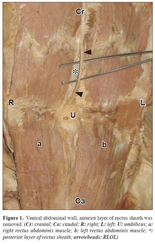
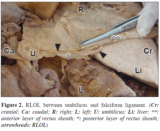
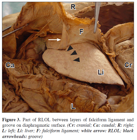
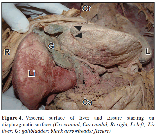
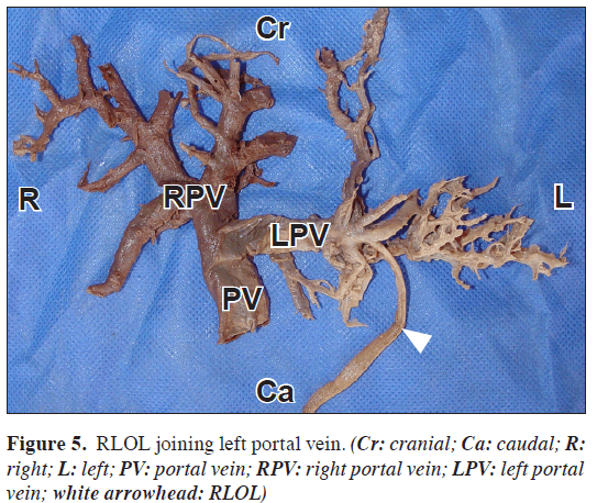
We found a cord like structure between the layers of the rectus sheath, medial to the left rectus abdominis muscle of a 63-year-old cadaver (Figure 1). Considering its course with careful dissection, we identified this structure as RLOL. In this case, the ligament was more superficial than it was supposed to be. It was running between the two layers of the rectus sheath from the umbilicus until it reached the two sheaths of the falciform ligament (Figure 2). Due to the curved shape of the ligament, we firstly placed a rope segment and then used a digital caliper to have the exact measurements of the ligament. Length of RLOL at the ventral abdominal wall was 83.97 mm. While lying between the two sheaths of the falciform ligament, RLOL coursed in a curved groove on the diaphragmatic surface of the liver (Figure 3). The length of this groove was measured as 50.07 mm with the method of measurement with a rope segment. Then, RLOL was embedded inside the parenchyma. From this point, a fissure was lying inferolaterally and reached the inferior margin of the liver but it did not extend onto the visceral surface. The length of this fissure was measured as 31.87 mm with the method of measurement with a rope segment. After examining the diaphragmatic surface, we evaluated the visceral surface. At the visceral surface, the fissure for the round ligament (fissura ligamenti teretis), the ventral component of the left sagittal fissure, was not present, and the anatomic differentiation between the left and quadrate lobes could not be done (Figure 4). Afterwards in order to determine the course of RLOL in the parenchyma of liver, we removed the parenchyma of liver by delicate dissection. Exposing the distributions of the portal and hepatic veins and the hepatic artery, we encountered that RLOL joined the left portal vein (Figure 5).
Discussion
RLOL extends in the free edge of the falciform ligament and it is formed when the left umbilical vein obliterates after birth and becomes a fibrous cord [1]. At the 4th week of the embryologic life, the blood from the placenta reaches the capillary network in the liver by using both left and right umbilical veins. During the 5th week, the right umbilical vein becomes completely obliterated, whereas the left umbilical vein persists. The oxygenated blood from the placenta thus reaches the heart via the single umbilical vein and ductus venosus [2,3]. Therefore, this ligament extends from the umbilicus cranially in between two peritoneal sheaths of the falciform ligament at the free caudal margin, to a notch on the inferior border of the liver named notch for round ligament of the liver. Then it passes dorsally to the visceral surface of the liver and runs in the floor of the ventral part of the left sagittal fissure, which is also called fissure for round ligament of the liver [2]. At the porta hepatis, it joins the umbilical portion of the left portal vein. The ligamentum venosum initiates from this connection point and extends to the left hepatic vein in the fissure for the ligamentum venosum [4]. RLOL is normally accepted as completely closed and avascular. There are accompanying paraumbilical veins around it, representing a potential circulation between the portal vein and systemic venous system on the ventral abdominal wall. However, the lumen of the left umbilical vein does not completely disappear [5].
The reported variations of RLOL are limited. Yılmaz et al. reported a case in 1993, in which RLOL was found between the layers of the rectus sheath. In their case, RLOL lied in an unusual groove on the diaphragmatic surface of liver like our case. They added that the RLOL then coursed between the aponeurosis of obliquus externus abdominis muscle and anterior lamina of the aponeurosis of the obliquus internus abdominis muscle. It lied between the layers of the rectus sheath, and was located medial and anterior to the right rectus muscle. They reported that the macroscopic differentiation between right and left lobes could be done on the visceral surface [5]. In our case, macroscopic differentiation between the left and quadrate lobes could not be done. This might be explained by the following information: the liver is a single unit in its earliest embryonic stages; it is usually separated into the left and right lobes by the falciform and round ligaments in the second month of the gestation, and a lack of separation at this stage might result in lobar fusion [6].
Çolak et al. had defined some unusual attachment points for the greater omentum and they reported that one of these was the right upper part of the greater omentum combined with RLOL [7]. We did not observe this kind of variation.
After performing laparoscopy on 1802 patients, Sato et al. reported the incidence of high insertion of RLOL as 2.8 % [6]. In our case, RLOL also had a high insertion, but unlike Sato et al., we observed a distinct groove on the diaphragmatic surface of the liver.
Sanli et al. reported a case of a 70-year-old male cadaver in which the fissure for the round ligament on the visceral surface was absent like our case, and RLOL was embedded inside the parenchyma on the diaphragmatic surface [4]. Nevertheless they did not mention whether RLOL was regular or variant at the ventral abdominal wall.
The residual lumen of RLOL has clinical importance for left umbilical vein catheterization, though this procedure can be used for emergency extracorporeal shunt, portal hepatography or for chemotherapy of hepatic neoplasms [1,8]. Ying et al. mentioned that they had successfully used RLOL as a vascularised pedicled flap for the surgical repair of the extrahepatic bile passages [9]. Soon et al. reported that, as RLOL connected the umbilicus to the left lobe of the liver, a hepatic lesion could spread to the umbilicus and ventral abdominal wall. They added that this ligament was very important because of this reason [10]. In the light of these all, we believe that the variation of RLOL we reported here, may provide additional contribution to the literature.
References
- Storer EH, Akin TJ. Chemotherapy of hepatic neoplasms via the umblical-portal vein. Am J Surg. 1966; 111: 56–65.
- Standring S, ed. Gray’s Anatomy. 39th Ed., Spain, Churchill Livingstone 2005; 1047, 1054, 1106, 1127, 1128, 1213.
- Larsen WJ. Human Embryology. 3rd Ed., Philadelphia, Churchill Livingstone. 2001; 212–213, 245.
- Sanli EC, Kurtoglu Z, Kara A, Uzmansel D. Left sided-right portal joined round ligament with an anomaly of the intrahepatic portal vein. Saudi Med J. 2006; 27: 1897–1900.
- Yilmaz E, Celik HH, Atasever A, Durgun B. Ligamentum teres hepatis: an unusual variation. Bull Assoc Anat (Nancy). 1993; 77: 31–32.
- Sato S, Watanabe M, Nagasawa S, Niigaki M, Sakai S, Akagi S. Laparoscopic observations of congenital anomalies of the liver. Gastrointest Endosc. 1998; 47: 136–140.
- Colak T, Dalcik C, Ozbek A, Filiz S, Sahin M, Dalcik H. A rare multiple variation of the greater omentum. Okajimas Folia Anat Jpn. 2002; 79: 159–162.
- Bayly JH, Carbalhaes OG. The umblical vein in the adult: diagnosis, treatment and research. Am Surg. 1964; 30: 56–60.
- Ying DJ, Ho GT, Cai JX. Anatomic bases of the vascularized hepatic teres ligament flap. Surg Radiol Anat. 1997; 19: 293–294.
- Kim SY, Lim JH. Extension of hepatoma to the rectus abdominis muscle via ligamentum teres hepatis. Gastrointest Radiol. 1985; 10: 119–121.
Aysin Kale*, Ozcan Gayretli, Adnan Ozturk, Bulent Bayraktar, Ahmet Usta and Ilke Ali Gurses
Istanbul University Istanbul Faculty of Medicine, Department of Anatomy, Istanbul, Turkey
- *Corresponding Author:
- Aysin Kale, MD
Department of Anatomy, Istanbul University, Istanbul Faculty of Medicine, 34390, Capa, Istanbul, Turkey
Tel: +90 212 4142000 / 33083
Fax: +90 212 4142176
E-mail: aysinckale@yahoo.com
Date of Received: January 26th, 2009
Date of Accepted: June 15th, 2009
Published Online: June 30th, 2009
© IJAV. 2009; 2: 62–64.
Abstract
The round ligament of liver is formed by obliteration of the umblical vein, which exists in embryological life. We report an unusual variation of the round ligament of liver. During routine dissections, we encountered an unusual structure over rectus abdominis sheath of a 63-year-old male cadaver. This structure was determined to be the round ligament of liver. This ligament was not only more superficial than it normally should be, but also in an unusual manner: it reached the liver’s visceral surface after running on its way on the diaphragmatic surface of liver, thus the fissure for round ligament was not observed. Consequently this cadaver’s liver was not divided into lobes on the visceral surface. The residual lumen of the round ligament of liver is important for umbilical vein catheterization. We conclude that this rare variation may be important for catheterization and abdominal surgical procedures.
-Keywords
variation, round ligament of liver, umbilical vein
Introduction
During routine dissections, we encountered an unusual structure over the rectus abdominis sheath of a 63-year-old male cadaver, which was supposed to be the round ligament of liver (RLOL). As it was more superficial than it normally should be, we decided to examine this structure.
Case Report
We found a cord like structure between the layers of the rectus sheath, medial to the left rectus abdominis muscle of a 63-year-old cadaver (Figure 1). Considering its course with careful dissection, we identified this structure as RLOL. In this case, the ligament was more superficial than it was supposed to be. It was running between the two layers of the rectus sheath from the umbilicus until it reached the two sheaths of the falciform ligament (Figure 2). Due to the curved shape of the ligament, we firstly placed a rope segment and then used a digital caliper to have the exact measurements of the ligament. Length of RLOL at the ventral abdominal wall was 83.97 mm. While lying between the two sheaths of the falciform ligament, RLOL coursed in a curved groove on the diaphragmatic surface of the liver (Figure 3). The length of this groove was measured as 50.07 mm with the method of measurement with a rope segment. Then, RLOL was embedded inside the parenchyma. From this point, a fissure was lying inferolaterally and reached the inferior margin of the liver but it did not extend onto the visceral surface. The length of this fissure was measured as 31.87 mm with the method of measurement with a rope segment. After examining the diaphragmatic surface, we evaluated the visceral surface. At the visceral surface, the fissure for the round ligament (fissura ligamenti teretis), the ventral component of the left sagittal fissure, was not present, and the anatomic differentiation between the left and quadrate lobes could not be done (Figure 4). Afterwards in order to determine the course of RLOL in the parenchyma of liver, we removed the parenchyma of liver by delicate dissection. Exposing the distributions of the portal and hepatic veins and the hepatic artery, we encountered that RLOL joined the left portal vein (Figure 5).
Discussion
RLOL extends in the free edge of the falciform ligament and it is formed when the left umbilical vein obliterates after birth and becomes a fibrous cord [1]. At the 4th week of the embryologic life, the blood from the placenta reaches the capillary network in the liver by using both left and right umbilical veins. During the 5th week, the right umbilical vein becomes completely obliterated, whereas the left umbilical vein persists. The oxygenated blood from the placenta thus reaches the heart via the single umbilical vein and ductus venosus [2,3]. Therefore, this ligament extends from the umbilicus cranially in between two peritoneal sheaths of the falciform ligament at the free caudal margin, to a notch on the inferior border of the liver named notch for round ligament of the liver. Then it passes dorsally to the visceral surface of the liver and runs in the floor of the ventral part of the left sagittal fissure, which is also called fissure for round ligament of the liver [2]. At the porta hepatis, it joins the umbilical portion of the left portal vein. The ligamentum venosum initiates from this connection point and extends to the left hepatic vein in the fissure for the ligamentum venosum [4]. RLOL is normally accepted as completely closed and avascular. There are accompanying paraumbilical veins around it, representing a potential circulation between the portal vein and systemic venous system on the ventral abdominal wall. However, the lumen of the left umbilical vein does not completely disappear [5].
The reported variations of RLOL are limited. Yılmaz et al. reported a case in 1993, in which RLOL was found between the layers of the rectus sheath. In their case, RLOL lied in an unusual groove on the diaphragmatic surface of liver like our case. They added that the RLOL then coursed between the aponeurosis of obliquus externus abdominis muscle and anterior lamina of the aponeurosis of the obliquus internus abdominis muscle. It lied between the layers of the rectus sheath, and was located medial and anterior to the right rectus muscle. They reported that the macroscopic differentiation between right and left lobes could be done on the visceral surface [5]. In our case, macroscopic differentiation between the left and quadrate lobes could not be done. This might be explained by the following information: the liver is a single unit in its earliest embryonic stages; it is usually separated into the left and right lobes by the falciform and round ligaments in the second month of the gestation, and a lack of separation at this stage might result in lobar fusion [6].
Çolak et al. had defined some unusual attachment points for the greater omentum and they reported that one of these was the right upper part of the greater omentum combined with RLOL [7]. We did not observe this kind of variation.
After performing laparoscopy on 1802 patients, Sato et al. reported the incidence of high insertion of RLOL as 2.8 % [6]. In our case, RLOL also had a high insertion, but unlike Sato et al., we observed a distinct groove on the diaphragmatic surface of the liver.
Sanli et al. reported a case of a 70-year-old male cadaver in which the fissure for the round ligament on the visceral surface was absent like our case, and RLOL was embedded inside the parenchyma on the diaphragmatic surface [4]. Nevertheless they did not mention whether RLOL was regular or variant at the ventral abdominal wall.
The residual lumen of RLOL has clinical importance for left umbilical vein catheterization, though this procedure can be used for emergency extracorporeal shunt, portal hepatography or for chemotherapy of hepatic neoplasms [1,8]. Ying et al. mentioned that they had successfully used RLOL as a vascularised pedicled flap for the surgical repair of the extrahepatic bile passages [9]. Soon et al. reported that, as RLOL connected the umbilicus to the left lobe of the liver, a hepatic lesion could spread to the umbilicus and ventral abdominal wall. They added that this ligament was very important because of this reason [10]. In the light of these all, we believe that the variation of RLOL we reported here, may provide additional contribution to the literature.
References
- Storer EH, Akin TJ. Chemotherapy of hepatic neoplasms via the umblical-portal vein. Am J Surg. 1966; 111: 56–65.
- Standring S, ed. Gray’s Anatomy. 39th Ed., Spain, Churchill Livingstone 2005; 1047, 1054, 1106, 1127, 1128, 1213.
- Larsen WJ. Human Embryology. 3rd Ed., Philadelphia, Churchill Livingstone. 2001; 212–213, 245.
- Sanli EC, Kurtoglu Z, Kara A, Uzmansel D. Left sided-right portal joined round ligament with an anomaly of the intrahepatic portal vein. Saudi Med J. 2006; 27: 1897–1900.
- Yilmaz E, Celik HH, Atasever A, Durgun B. Ligamentum teres hepatis: an unusual variation. Bull Assoc Anat (Nancy). 1993; 77: 31–32.
- Sato S, Watanabe M, Nagasawa S, Niigaki M, Sakai S, Akagi S. Laparoscopic observations of congenital anomalies of the liver. Gastrointest Endosc. 1998; 47: 136–140.
- Colak T, Dalcik C, Ozbek A, Filiz S, Sahin M, Dalcik H. A rare multiple variation of the greater omentum. Okajimas Folia Anat Jpn. 2002; 79: 159–162.
- Bayly JH, Carbalhaes OG. The umblical vein in the adult: diagnosis, treatment and research. Am Surg. 1964; 30: 56–60.
- Ying DJ, Ho GT, Cai JX. Anatomic bases of the vascularized hepatic teres ligament flap. Surg Radiol Anat. 1997; 19: 293–294.
- Kim SY, Lim JH. Extension of hepatoma to the rectus abdominis muscle via ligamentum teres hepatis. Gastrointest Radiol. 1985; 10: 119–121.


