A strange case involving partial fracture of percutaneous transluminal coronary angioplasty wire and its successful retrieval
- *Corresponding Author:
- Dr Sinha Santosh Kumar
Faculty Department of Cardiology LPS Institute of Cardiology, GSVM Medical College, Kanpur, Uttar Pradesh, India 208002
Tel: 91-9670220088
Fax: 91-0512-2556199,2556521
E-mail: fionasan@rediffmail.com
This open-access article is distributed under the terms of the Creative Commons Attribution Non-Commercial License (CC BY-NC) (http:// creativecommons.org/licenses/by-nc/4.0/), which permits reuse, distribution and reproduction of the article, provided that the original work is properly cited and the reuse is restricted to noncommercial purposes. For commercial reuse, contact reprints@pulsus.com
[ft_below_content] =>Keywords
Partial fracture; Rescue wire; Shoulder point; Spring coil; Springtype wire
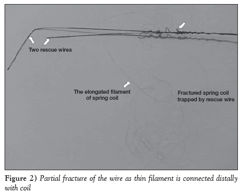
Coronary angioplasty devices are moved within the coronary system using a guidewire; the guidewire is usually of a spring-type design. The soft, atraumatic tip of coronary wires have been to known to fracture if manipulated excessively. This most frequently occurs when the shapeable wire tip becomes lodged in atheromatous plaque and separates from the body of the wire when the wire is retracted. Entrapment, overcoiling and excessive traction of the guidewire can cause it to fracture. Coronary wires currently used have a proximal stainless steel shaft, a nitinol distal shaft with a middle-weight balance and a hydrophilic-coated radiopaque coil at the tip. The incidence of wire fractures are minimized in contemporary wire design. Nevertheless, it still occurs with an incidence of approximately 0.2% to 0.8% [1]. Most of the fractures are complete, which means complete separation of the spring tip from the shoulder. There is a likelihood of acute embolization leading to acute coronary thrombus if the fragment of guidewire is not removed. In the present case, a partial fracture occurred at the junction of the shaft and coil due to excessive manipulation to unloop the wire. As the proximal section of the wire was retrieved, a thin elongated filament of spring coil also came out from the Y connector, with a portion of the coil left trapped in the lesion. This thin filament was lying across the left main artery to the left anterior descending (LAD) artery from the Y connector. It was successfully retrieved using tangling technique with the assistance of two balance middle-weight (BMW) wires acting as rescue wires.
Case Presentation
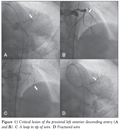
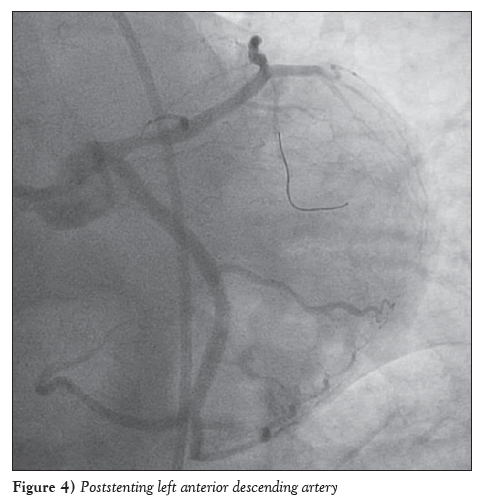
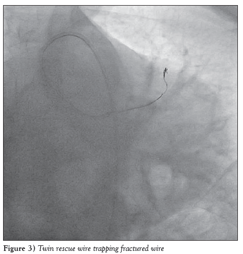
A 43-year-old woman, with a three-year history of hypertension was admitted with Canadian Cardiovascular Society grade III angina despite guideline-directed medical treatment for the past two years. Her treadmill test was strongly positive for reversible myocardial ischemia. She underwent coronary artery angiography after proper consent, revealing a critical lesion of the proximal LAD artery (Figures 1A and 1B). Percutaneous coronary intervention was subsequently scheduled. Intravenous heparin (100 U/kg) was administered. The left main artery was hooked using a 6 F JL Launcher Coronary Guide Catheter (Medtronic, USA). The lesion was crossed using a 0.014 inch 190 cm Hi-Torque BMW wire (Abbott Vascular, Santa Clara, USA) with some difficulty due to calcium. A loop was formed distally, which could not be further advanced (Figure 1C). Due to excessive manipulation when attempting to unloop it, a fracture occurred at the shoulder region (Figure 1D). As the proximal end was pulled, a thin filament of the spring also came out of the Y-connector confirming partial fracture of the wire (Figure 2). Two BMW wires were placed distally and the proximal ends of both wires were inserted together in a torque device, which were firmly screwed and rotated 45 to 55 times in a circular pattern to catch hold of the wire (Figure 3). During this rotational motion, the broken segment, along with thin filament, were tangled within these rescue wires and all three wires were removed together. Following this, the lesion was recrossed with a 0.014 inch 190 cm Hi-Torque BMW wire. The lesion was predilated with a 2×10 and 2.5×10 Sprinter Legend Rx balloon (Medtronic, USA). Xience Prime 3.0 mm × 28 mm ( Abbott Vascular, USA) was deployed in the proximal LAD at 12 atm with postprocedural thrombolysis in myocardial infarction (TIMI) III flow (Figure 4). The patient was discharged in stable condition with appropriate advice.
Discussion
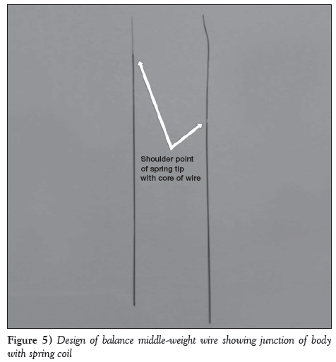
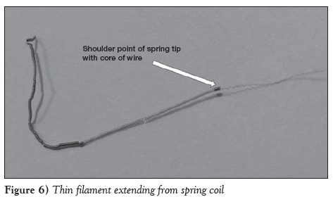
It is generally believed that the retained guidewire in the coronary tree may result in complications, such as emboli, thrombosis, dissection and rupture, and should be removed from the coronary system [2,3]. Most fractures are complete; the tip completely separates from the body of the wire. Various solutions have been described to solve the problem of a broken guidewire in a coronary artery, ranging from surgery to leaving the segment of wire in place in cases where it cannot be removed, such as in vessels that are already occluded or in very small branches; however, all efforts should be made to remove the guidewire percutaneously [4,5]. In our case, a partial fracture occurred because thin filament was still attached to the distal spring coil while the proximal end was removed from the Y-connector (Figures 5 and 6). This thin filament was lying across the left main to the mid LAD artery. Therefore, we decided to retrieve it. A simple technique was used because snare loop wire was not available. Two wires were used for the retrieval. Usually wire breaks at the shoulder point of the body with spring coil; however, our case was unique because it was a partial fracture. In BMW wire, the core does not extend up to the tip, so excessive turning may cause a fracture or, less commonly, a manufacturing defect may play a role. Vigorous twisting of the wire should always be avoided. In our case, the partial fracture was caused by trying to unloop the wire. To our knowledge, this is the first report of a partial fracture.
Disclosures
The authors have no financial relationships or conflicts of interest to declare.
References
- Chang TM, Pellegrini D, Ostrovsky A, Marrangoni AG. Surgical management of entrapped percutaneous transluminal coronary angioplasty hardware. Tex Heart Inst J 2002;29:329-32.
- Hartzler GO, Rutherford BD, McConahay DR. Retained percutaneous transluminal coronary angioplasty equipment components and their management. Am J Cardiol 1987;60:1260-4.
- Sethi GK, Ferguson TB Jr, Miller G, Scott SM. Entrapment of broken guidewire in the left main coronary artery during percutaneous transluminal coronary angioplasty.Ann Thorac Surg 1989;47:455-7.
- Patel T, Shah S, Pandya R, Sanghvi K, Fonseca K. Broken guidewire fragment: A simplified retrieval technique. Catheter Cardiovasc Interv 2000;51:483-6.
- Demircan S, Yazici M, Durna K, Yasar E. Intracoronary guidewire emboli: A unique complication and retrieval of the wire. Cardiovasc Revasc Med 2008;9:278-80.
- *Corresponding Author:
- Dr Sinha Santosh Kumar
Faculty Department of Cardiology LPS Institute of Cardiology, GSVM Medical College, Kanpur, Uttar Pradesh, India 208002
Tel: 91-9670220088
Fax: 91-0512-2556199,2556521
E-mail: fionasan@rediffmail.com
This open-access article is distributed under the terms of the Creative Commons Attribution Non-Commercial License (CC BY-NC) (http:// creativecommons.org/licenses/by-nc/4.0/), which permits reuse, distribution and reproduction of the article, provided that the original work is properly cited and the reuse is restricted to noncommercial purposes. For commercial reuse, contact reprints@pulsus.com
Abstract
angina of a critical lesion of the proximal left anterior descending artery. After negotiating with spring-type wire (balance middle-weight wire), a loop was formed. Due to excessive manipulation to unloop the wire, it partially fractured at the shaft and coil junction. As the proximal section was retrieved, a thin elongated filament of spring coil also came out from the Y-connector while the remainder of the coil was still trapped in the lesion. Two balance middle-weight wires were then placed distally, and the proximal ends of both wires were inserted together in a torque device, which were firmly screwed and rotated 45 to 55 times in a circular pattern. During this rotational motion, the broken segment along with thin filament was tangled within these rescue wires and all three wires were removed together using tangling technique. Following this, angioplasty was performed successfully. After reviewing the literature using PubMed, to the authors’ knowledge, this is the first incident of its kind to be reported.
-Keywords
Partial fracture; Rescue wire; Shoulder point; Spring coil; Springtype wire
Coronary angioplasty devices are moved within the coronary system using a guidewire; the guidewire is usually of a spring-type design. The soft, atraumatic tip of coronary wires have been to known to fracture if manipulated excessively. This most frequently occurs when the shapeable wire tip becomes lodged in atheromatous plaque and separates from the body of the wire when the wire is retracted. Entrapment, overcoiling and excessive traction of the guidewire can cause it to fracture. Coronary wires currently used have a proximal stainless steel shaft, a nitinol distal shaft with a middle-weight balance and a hydrophilic-coated radiopaque coil at the tip. The incidence of wire fractures are minimized in contemporary wire design. Nevertheless, it still occurs with an incidence of approximately 0.2% to 0.8% [1]. Most of the fractures are complete, which means complete separation of the spring tip from the shoulder. There is a likelihood of acute embolization leading to acute coronary thrombus if the fragment of guidewire is not removed. In the present case, a partial fracture occurred at the junction of the shaft and coil due to excessive manipulation to unloop the wire. As the proximal section of the wire was retrieved, a thin elongated filament of spring coil also came out from the Y connector, with a portion of the coil left trapped in the lesion. This thin filament was lying across the left main artery to the left anterior descending (LAD) artery from the Y connector. It was successfully retrieved using tangling technique with the assistance of two balance middle-weight (BMW) wires acting as rescue wires.
Case Presentation
A 43-year-old woman, with a three-year history of hypertension was admitted with Canadian Cardiovascular Society grade III angina despite guideline-directed medical treatment for the past two years. Her treadmill test was strongly positive for reversible myocardial ischemia. She underwent coronary artery angiography after proper consent, revealing a critical lesion of the proximal LAD artery (Figures 1A and 1B). Percutaneous coronary intervention was subsequently scheduled. Intravenous heparin (100 U/kg) was administered. The left main artery was hooked using a 6 F JL Launcher Coronary Guide Catheter (Medtronic, USA). The lesion was crossed using a 0.014 inch 190 cm Hi-Torque BMW wire (Abbott Vascular, Santa Clara, USA) with some difficulty due to calcium. A loop was formed distally, which could not be further advanced (Figure 1C). Due to excessive manipulation when attempting to unloop it, a fracture occurred at the shoulder region (Figure 1D). As the proximal end was pulled, a thin filament of the spring also came out of the Y-connector confirming partial fracture of the wire (Figure 2). Two BMW wires were placed distally and the proximal ends of both wires were inserted together in a torque device, which were firmly screwed and rotated 45 to 55 times in a circular pattern to catch hold of the wire (Figure 3). During this rotational motion, the broken segment, along with thin filament, were tangled within these rescue wires and all three wires were removed together. Following this, the lesion was recrossed with a 0.014 inch 190 cm Hi-Torque BMW wire. The lesion was predilated with a 2×10 and 2.5×10 Sprinter Legend Rx balloon (Medtronic, USA). Xience Prime 3.0 mm × 28 mm ( Abbott Vascular, USA) was deployed in the proximal LAD at 12 atm with postprocedural thrombolysis in myocardial infarction (TIMI) III flow (Figure 4). The patient was discharged in stable condition with appropriate advice.
Discussion
It is generally believed that the retained guidewire in the coronary tree may result in complications, such as emboli, thrombosis, dissection and rupture, and should be removed from the coronary system [2,3]. Most fractures are complete; the tip completely separates from the body of the wire. Various solutions have been described to solve the problem of a broken guidewire in a coronary artery, ranging from surgery to leaving the segment of wire in place in cases where it cannot be removed, such as in vessels that are already occluded or in very small branches; however, all efforts should be made to remove the guidewire percutaneously [4,5]. In our case, a partial fracture occurred because thin filament was still attached to the distal spring coil while the proximal end was removed from the Y-connector (Figures 5 and 6). This thin filament was lying across the left main to the mid LAD artery. Therefore, we decided to retrieve it. A simple technique was used because snare loop wire was not available. Two wires were used for the retrieval. Usually wire breaks at the shoulder point of the body with spring coil; however, our case was unique because it was a partial fracture. In BMW wire, the core does not extend up to the tip, so excessive turning may cause a fracture or, less commonly, a manufacturing defect may play a role. Vigorous twisting of the wire should always be avoided. In our case, the partial fracture was caused by trying to unloop the wire. To our knowledge, this is the first report of a partial fracture.
Disclosures
The authors have no financial relationships or conflicts of interest to declare.
References
- Chang TM, Pellegrini D, Ostrovsky A, Marrangoni AG. Surgical management of entrapped percutaneous transluminal coronary angioplasty hardware. Tex Heart Inst J 2002;29:329-32.
- Hartzler GO, Rutherford BD, McConahay DR. Retained percutaneous transluminal coronary angioplasty equipment components and their management. Am J Cardiol 1987;60:1260-4.
- Sethi GK, Ferguson TB Jr, Miller G, Scott SM. Entrapment of broken guidewire in the left main coronary artery during percutaneous transluminal coronary angioplasty.Ann Thorac Surg 1989;47:455-7.
- Patel T, Shah S, Pandya R, Sanghvi K, Fonseca K. Broken guidewire fragment: A simplified retrieval technique. Catheter Cardiovasc Interv 2000;51:483-6.
- Demircan S, Yazici M, Durna K, Yasar E. Intracoronary guidewire emboli: A unique complication and retrieval of the wire. Cardiovasc Revasc Med 2008;9:278-80.


