A study of fissures and lobes of lungs from clinical perspective
Received: 22-Aug-2018 Accepted Date: Sep 04, 2018; Published: 15-Sep-2018
Citation: Mote D, Awari P, Bharambe V. A study of fissures and lobes of lungs from clinical perspective. Pulsus J Surg Res. 2018;2(2):41-43.
This open-access article is distributed under the terms of the Creative Commons Attribution Non-Commercial License (CC BY-NC) (http://creativecommons.org/licenses/by-nc/4.0/), which permits reuse, distribution and reproduction of the article, provided that the original work is properly cited and the reuse is restricted to noncommercial purposes. For commercial reuse, contact reprints@pulsus.com
Abstract
Aim: The present study was taken up to study variations in fissures and lobes of lung and to assess their clinical and surgical importance.
Materials and Methods: Thirty lungs were obtained from formalin fixed cadavers and the lobes and fissures were studied. Any variations were photographed and noted. These were then classified using Craig and Walker classification of lung fissures.
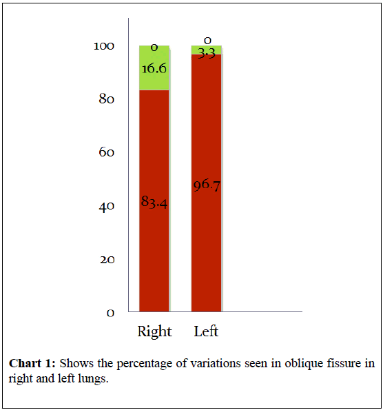
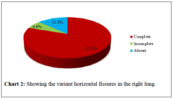
Results: The right lung showed incomplete oblique fissure in five lungs (16.6%). The horizontal fissure was incomplete in two right lungs (6.6%) and it was absent in four right lungs (13.3%). The left lung showed incomplete oblique fissure in one lung (3.3%). No other variations were observed in the left lungs studied. No accessory fissures of either right or left lungs were observed.
Conclusion: Lungs develop by division and re-division of lung buds arising from endodermal foregut. Defective development from the lung buds will give rise to variations such as incomplete or absence of fissures of right or left lung. The structure of the fissure is of importance while planning operative steps in thoracoscopic pulmonary resection. An incomplete fissure could cause postoperative air leakage in case of lung lobectomy. They can also allow spread of disease within the lung tissue that would otherwise be separated due to presence of the complete fissure.
Keywords
Lobes of lung; Complete oblique fissure; Incomplete oblique fissure; Absent fissure; Horizontal fissure; Surgery
Introduction
Exchange of respiratory gases is a necessary life process in all the organisms. In living organisms respiratory system has undergone various modifications and in human beings it developed into a system of branching network of bronchial tree.
Respiratory system is important for living organisms to retain their life. It includes the nasal cavity, larynx which continues as trachea, lungs, bronchus which further divides into bronchioles and diaphragm. Right lung has two fissures oblique and horizontal which divides it into 3 lobes: upper, middle and lower; and the left lung has two lobes divided by oblique fissure. The lungs expand maximally in the inferior direction because movements of the thoracic wall and diaphragm are maximal towards the base of the lung [1].
The bronchial tree, also known as main or primary bronchus is a passage of airway in respiratory tract that conducts air into lungs. There is a right bronchus and a left bronchus, these bronchi branch into smaller secondary and tertiary bronchi which branch into smaller tubes, known as bronchioles [2]. The branching pattern of the bronchial tree is not uniform throughout in all mammals. It is also not uniform among all humans, but shows variations [3].
The knowledge of variations in fissures and lobes of lung, is useful in various aspects such as to thoracic surgeons for doing difficult maneuvers such as surgical resection of segments or lobes in diseases of lungs, to spread the importance of pathological conditions like bronchiectasis, to know the pathway for entry of foreign particles and to maintain posture for patients suffering from suppurative lung disorders.
Aim and objectives: The present study was taken up to study variations in fissures and lobes of lung and to assess their clinical and surgical importance.
Materials and Methods
Twenty formalin (4%) fixed cadavers were obtained from the dissection room of the Department of Anatomy of our Medical College. They were dissected meticulously to obtain 1 pair of lungs from each cadaver.
The removal of lungs was carried out by following procedure: With the cadaver in supine position, with the help of a scalpel an incision was made from acromion process of scapula to the manubrium sterni to the opposite side acromion process. A vertical incision was made from the manubrium sterni to the xiphoid process. A horizontal incision was made from the xiphoid process to the midaxillary line on both right and left sides.
The skin and superficial fascia were removed over this region of the thoracic cage. The mammary glands in case of female cadavers were also being removed. The attachments of various heads of the pectoralis major were cut and reflected towards its insertion. Similarly the pectoralis minor muscle was also reflected to the coracoid process. The digitations of insertion of serratus anterior muscle were also detached from the ribcage and the subclavius muscle was cut.
This way the intercostal muscles, the costal cartilage and the ribs were visualized clearly. The manubrium sterni was cut with a saw transversely. The internal thoracic vessels were also cut. The ribs were cut in the midaxillary line till the 7th rib. The superior part of the ribcage was gently reflected downwards hinged on the superior part of anterior abdominal wall, to expose the contents of the thoracic cavities after cutting the sternopericardial ligaments. The pleura on both the sides were cut along its line of reflection from sternum to mediastinum and reflected away to expose the lungs. Each lung was pulled from the mediastinum and its root was defined. The pulmonary ligament was delineated. Incision was made in the anterior layer of pulmonary ligament and structures at the hilum of the lung were identified. Each of these was dissected clean and cut 1 cm away from the hilum.
These lungs were then kept in pairs and the pairs were labeled from 1 to 15. Thus 30 lungs were obtained.
Each lung was studied meticulously. Presence of any extra lobes or fissures (complete, incomplete, absent) was noted. The Anatomical classification proposed by Craig and Walker was followed to determine the presence and completeness of fissures.
Results
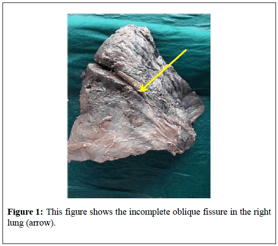
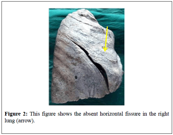
A total of 15 pairs of lungs were studied i.e. 30 lungs. The right lung showed incomplete oblique fissure in five lungs (16.6%) (Figure 1 and Chart 1). The horizontal fissure was incomplete in two right lungs (6.6%) and it was absent in four right lungs (13.3%) (Figure 2 and Chart 2).
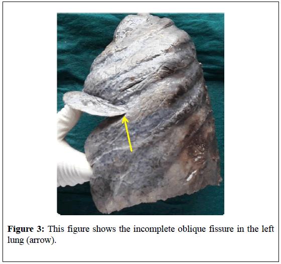
The left lung showed incomplete oblique fissure in one lung (3.3%) (Figure 3). No other variations were observed in the left lungs studied. No accessory fissures of either right or left lungs were observed.
Craig and Walker classified fissures of lungs in grades as Grade 1 wherein the lungs had complete fissures and entirely separate lobes, grade 2 wherein there was complete visceral cleft but parenchymal fusion at the base of the fissure could be observed, grade 3 wherein the there was visceral cleft evident for part of the fissure and grade 4 wherein there was complete fusion of the lobes with no evident fissure line [3]. Applying this classification to the horizontal and oblique fissures in the present set of lungs the grades are depicted in Table 1.
| Grade 1 | Grade 2 | Grade 3 | Grade 4 | ||
|---|---|---|---|---|---|
| Right lung | Horizontal fissure | 43.3%(13) | 36.7%(11) | 7%(2) | 13.3%(4) |
| Oblique fissure | 76.7%(23) | 17%(5) | 6%(2) | 0 | |
| Left lung | Oblique fissure | 83%(25) | 14%(4) | 3%(1) | 0 |
Table 1: This table depicts the classification of the fissures as observed in the present study as per the Craig and Walker classification.
Discussion and Conclusion
The lungs are made of bronchopulmonary segments which form lobes which are separated from each other by fissures. While the right lung has 10 lobes, the left has 8 [2]. Each segment has its own tertiary or segmental bronchus. Often in case of infection or cancer, initially it gets limited to its own bronchopulmonary segment and then its lobe [1].
In the present study 16.6% of the oblique fissures of the right lung were found to be incomplete. In a similar study by Bhimadevi et al. 9% of the right lungs studied showed incomplete oblique fissure while in the study by Minakshi et al. this was reported as 36.6% and Nene et al as 6% [4-7]. In a study carried out by Prakash et al., 39.3% of the right lungs showed incomplete oblique fissure while 7.1% showed absence of the oblique fissure in the right lung [8]. Thus the range of incidence of incomplete oblique fissure lies between 6% to 39.3%. The finding reported by present study lies within this range. The range of incidence of absence of oblique fissure of right lung was reported between 2 to 7.1% [5-8]. No such finding was reported in present study.
The present study reports 13.3% incidence of absence of the horizontal fissure of the right lung while 6.6% incidence of incomplete horizontal fissure of the right lung. The study by Prakash et al. reported the incidence of absence of the horizontal fissure of right lung as 7.1% while 50% of the horizontal fissures were found to be incomplete [8]. Minakshi et al. reported absence as 16.6% and incomplete as 63.3% [6]. Studies by Bhimadevi et al., Nene et al and Lukose et al reported absent horizontal fissure of right lung as 9%, 14% and 10.5% respectively while the incidence of incomplete horizontal fissure of right lung was reported by the same authors as 18%, 8% and 21% respectively. [4,5,7]. The study by Radha et al. reported the highest incidence of absent horizontal fissure of right lung [9]. The present study reports the lowest incidence of 6.6% incomplete horizontal fissure of right lung.
The present study reports 3.3% of incomplete oblique fissure of the left lung and no incidence of absence of this fissure was reported by present study. Minakshi et al., Prakash et al., Bhimadevi et al. reported high incidence (46.6%, 35.7% and 36.3% respectively) of incomplete oblique fissure of left lung [5,6,8]. Prakash et al. and Bhimadevi et al. also reported 10.7% and 9% incidence of absence of oblique fissure of left lung which was not observed at all in the present study [5,8].
Lungs develop by division and re-division of lung buds arising from endodermal foregut. Defective development from the lung buds will give rise to variations such as incomplete or absence of fissures of right or left lung due to defect in obliteration of the prenatal fissures. While incomplete obliteration would be seen as incomplete fissure, nonobliteration of earlier fissures could result in accessory fissures which were not reported in present study [10].
The structure of the fissure is of importance while planning operative steps in thoracoscopic pulmonary resection. An incomplete fissure could cause postoperative air leakage in case of lung lobectomy. They can also allow spread of disease within the lung tissue that would otherwise be separated due to presence of the complete fissure. Pneumonia in a lobe of lung lies within its confines due to the complete fissures. However it can spread to adjacent lung tissue due to the parenchymal continuation resulting from incomplete fissures [11].
Incomplete fissure may alter the pattern of lung collapse due to lesions or may give rise to an abnormal distribution of pleural effusion [12,13].
In the present study higher incidence of incomplete oblique fissure was observed in right lung as compared to left lung. However study by Bhimadevi et al and Minakshi et al reported higher incidence of incomplete oblique fissure in left lung as compared to the right [5,6].
A normal pattern of distribution of fissures and lobes of lung could be taken to indicate uniform expansion of lobes of lung. This pattern is used to decide position of the patient using gravity assisted lung drainage. For example when the patient is placed flat on his back in a head low position, the anterior segments of both lower lobes can be drained easily. Similarly a patient placed in left lateral position with the head placed low could assist in drainage of lateral segment of right lower lobe while a patient in left lateral position would drain the lateral segment of left lower lobe. Thus variable positions of patient are used to bring about gravity assisted lung drainage. However a variation in the fissures and lobes of lung would affect this drainage adversely [1,14].
Thus it can be concluded that a thorough knowledge of fissures and lobes of lung with the possible variation is essential for radiologists, cardiothoracic surgeons, physiotherapists among other practitioners of general medicine.
This how we do huddle in SLMC, customized as to the needs of our patients. Because at SLMC, the needs of our patients come first. As we present the emerging model of communication, we are anticipating for opportunities of growth and its application makes us happy in sharing it.
The lessons it taught us, opens more opportunities for strengthen partnerships and active involvement of our physician partnerships in our daily huddle. The testing of the model likewise is a rich area for data analysis, this time in big and group huddle.
REFERENCES
- Jennifer A Pryor. Physiotherapy for respiratory and cardiac problems. 3rd edi. Churchill Livingstone Elsevier 2004;202-204.
- Strandring S. Grays Anatomy.40edi.spain:churchill living stone Elsevier 2008;945.
- Walker WS, Craig SR. A proposed anatomical classification of the pulmonary fissures. J R Coll Surg Edinberg 1997;42:233-234.
- Lukose R, Paul S, Sunitha, et al. Morphology of the lungs: Variations in the lobes and fissures.Biomedians 1999;19:227-232.
- Bhimai DN, Narasinga RB, Sunitha V. Morphological valuations of lung A cadaveric study in north costal Andhra pradesh. Int J Biol Med Res 2011;2:1149-1152.
- Meenakshi S, Manjunah K, Balasubramnaysam V. Morphological variations of lung fissures and lobes. Indian J Chest Dis Allied Sci 2004;46:179-182.
- Nene AJ, Gajendra KS, Sarma MVR. A variant oblique fissure of left lung. Int J Anat Var 2010;3:125-127.
- Prakash, Bhardwaj AK, Shashirekha M, et al. Lung morphology: cadaver study in Indian population. Ital J Anat Embryol 2010;115:235-240.
- Radha K, Durian Pandian K. Fissures and Lobes of Lungs: A Morphological and anatomical study. Int J Anat Res 2015;3:995-998.
- Moore KL, Persaud TVN, Torchia MG The developing human: Clinically oriented embryology (10th edition.). Philadelphia, PA: Elsevier 2016.
- Glazer H, Anderson D, Dicroce JJ. Anatomy of the major fissure; evaluation with standard and thin section CT. Radiology 1991;180;839-844.
- Kent EM, Blades B. The Surgical anatomy of the pulmonary lobes. J Thoracic Surg 1942;12:18-30.
- Tarver RD. How common are incomplete pulmonary fissures, and what is their clinical significance? American Journal of Roentgenology 1995;164:761.
- Datta AK. Essentials of human anatomy. 9th edn. Current books 35-46.


