A study of horseshoe kidney: A rare anatomical variant
Received: 10-Nov-2017 Accepted Date: Nov 27, 2017; Published: 04-Dec-2017
Citation: Dimple M, Vaishaly B, Manvikar PR, et al. A study of horseshoe kidney: A rare anatomical variant. Int J Anat Var. 2017;10(4): 103-105.
This open-access article is distributed under the terms of the Creative Commons Attribution Non-Commercial License (CC BY-NC) (http://creativecommons.org/licenses/by-nc/4.0/), which permits reuse, distribution and reproduction of the article, provided that the original work is properly cited and the reuse is restricted to noncommercial purposes. For commercial reuse, contact reprints@pulsus.com
Abstract
Sacral During dissection of abdomen in a male cadaver at the medical college, a horseshoe shaped kidney mass was observed. It was dissected meticulously. The right and left kidneys were found to be fused at the lower end by the parenchymatous isthmus. The right kidney was supplied by right renal artery and three accessory arteries arising from abdominal aorta. The left kidney was supplied by left renal artery and two accessory renal arteries. The isthmus was supplied by a variant artery taking origin from abdominal aorta also giving branch to left kidney. Ureters ran downwards anterior to isthmus. The inferior mesenteric artery ran over the mass of tissue joining the lower poles of two kidneys.
Horseshoe kidney is a rare congenital malformation which can be asymptomatic or can predispose the patient to ureteric obstruction, infection, malignancies, hydronephrosis and posing technical difficulties during renal surgeries, renal transplants and endovascular procedures.
Keywords
Horseshoe kidney; Accessory renal artery; Major calyses of kidney
Introduction
Horseshoe kidney (HSK) is a rare congenital developmental malformation of the kidney [1]. It occurs with an incidence of 0.25% in general population. It is more frequent in males than females (2:1) [2,3]. The anomaly is a fusion of two kidneys on either side of midline joined by isthmus that can be composed of parenchyma or fibrous tissue.
The ureters usually are directed anteriorly and pass over the isthmus. In most cases this HSK mass functions as a normal kidney and the condition is noticed only during some investigations. However when symptomatic, it is usually because of obstruction of outflow tract, infection, stone formation etc. Rarely there may be other associated congenital anomalies [4].
In the present article the authors describe a rare finding of horseshoe kidney with associated renal arterial variations. The article also discusses the development of horseshoe kidney and attempts to explain the present findings.
Case Report
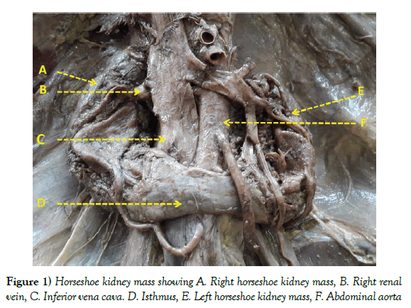
During routine dissection of a male cadaver by the medical students, a horseshoe shaped kidney (HSK) mass was observed (Figure 1). The cadaver had been fixed using 10% of formalin and stored in cadaver tanks before being taken up for dissection. Ethical clearance was obtained. Meticulous dissection of the horseshoe mass was carried out and following were the observations.
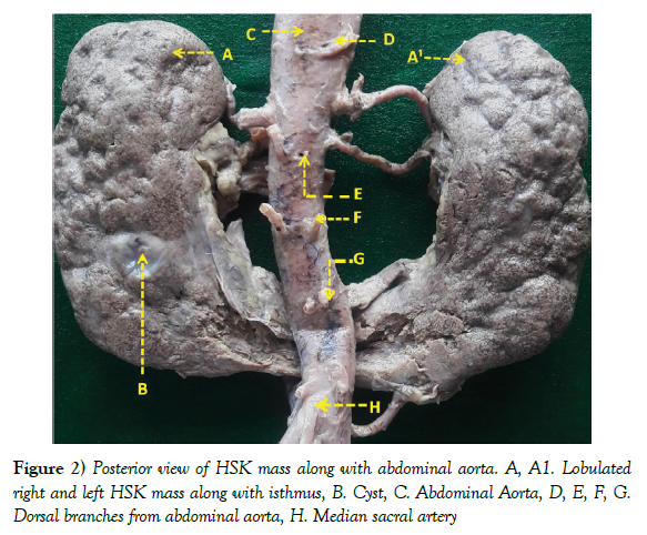
The mass of kidney was observed to be horseshoe shaped. The two parts representing right and left kidneys were joined by parenchymatous mass forming isthmus (Figure 1). The mass was lobulated and also showed a cyst measuring 10 mm x 9 mm on the posterior surface of left kidney (Figure 2). Table 1 gives the measurements of the HSK mass. The HSK mass was placed at a lower level than normal, the upper pole being opposite the L2 vertebra and lower pole being opposite L4 vertebra. The isthmus was placed opposite the L4 vertebra. The HSK mass was lying anterior to aorta and inferior vena cava with the isthmus lying posterior to inferior mesenteric artery.
| Length | Width | Thickness | |
|---|---|---|---|
| Right HSK mass | 100.11 | 45 | 27 |
| Left HSK mass | 113 | 51 | 28 |
| Isthmus | 18 | 36 | 3.53 |
Table 1: Measurements of the Horseshoe shaped mass of both sides and the intervening isthmus (in mms)
Arterial supply
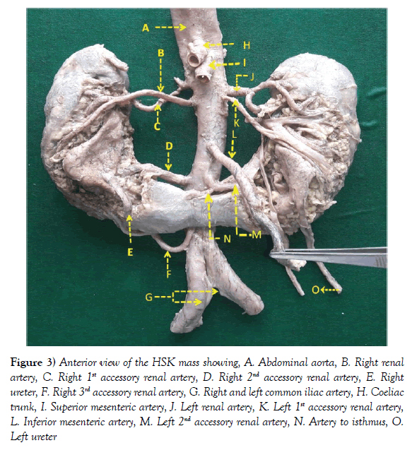
The right and left renal arteries were found to lie at right angles to the abdominal aorta, positioned slightly below the superior mesenteric artery, the left being slightly higher than the right. Right mass of the HSK was also supplied by 3 accessory renal arteries arising from abdominal aorta as follows: (Figure 3)
Figure 3) Anterior view of the HSK mass showing, A. Abdominal aorta, B. Right renal artery, C. Right 1st accessory renal artery, D. Right 2nd accessory renal artery, E. Right ureter, F. Right 3rd accessory renal artery, G. Right and left common iliac artery, H. Coeliac trunk, I. Superior mesenteric artery, J. Left renal artery, K. Left 1st accessory renal artery, L. Inferior mesenteric artery, M. Left 2nd accessory renal artery, N. Artery to isthmus, O. Left ureter
i. 1st right accessory renal artery took origin slightly below the superior mesenteric artery, lying above the right renal artery.
ii. 2nd right accessory renal artery took origin 16.11 mm below the inferior mesenteric artery.
iii. 3rd right accessory renal artery took origin behind the isthmus. Left mass of the HSK was also supplied by 2 accessory renal arteries arising from abdominal aorta as follows: (Figure 3)
i. 1st left accessory renal artery took origin slightly below the left renal artery.
ii. 2nd left accessory renal artery took origin 15.06 mm below the inferior mesenteric artery at the same level as the 2nd accessory renal artery of the right side. This artery also gave a branch supplying the isthmus.
Both the right and left 2nd accessory renal arteries were observed to have a larger diameter compared to the corresponding right and left renal arteries. The coeliac trunk, superior and inferior mesenteric and testicular arteries showed no variations in position.
Venous drainage
The HSK was drained by right and left renal veins which lay anterior to the corresponding arteries. The left renal vein drained into the inferior vena cava after crossing from left to right side across the abdominal aorta. It was seen draining the left suprarenal vein superiorly and the left testicular vein inferiorly. It also received variable number of tributaries from the rest of left side of the HSK mass. A separate vein from the isthmus drained into the left renal vein.
The right renal vein drained directly into the Inferior vena cava (Figure 1). The right testicular vein drained separately into the inferior vena cava.
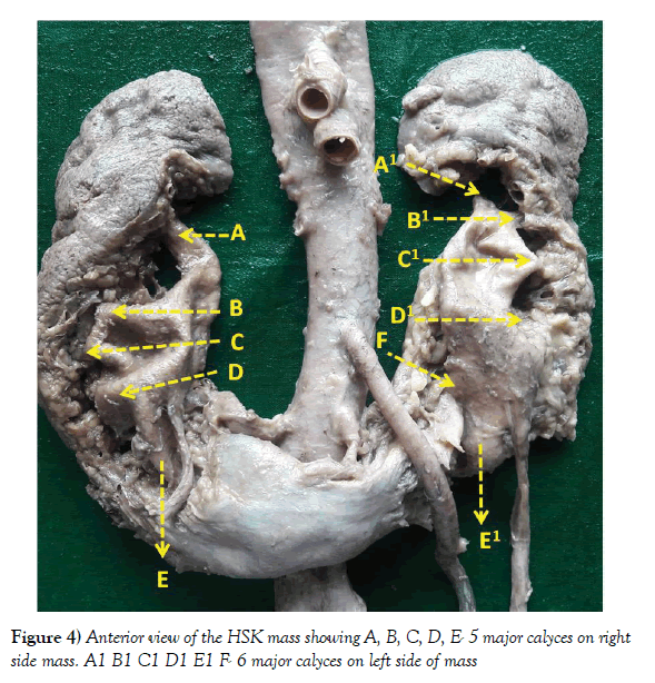
The renal pelves of both sides were directed anteriorly. The ureters ran downwards crossing the isthmus anteriorly (Figure 4). The major calyx part of the calyceal system in the present case was extararenal in position. There were 5 major calyces opening into a single pelvis on right side and 6 on the left side, draining into the single ureters respectively. The lowermost calyces on both sides were found to be draining the isthmus region.
Discussion
The history of observation of HSK goes back to 1523 when it was observed and reported in the isagoge breves. This was reported by Boyden in 1931 [5]. Since then there have been many reports of the horseshoe shaped kidney [2,3,5].
The HSK can be found anywhere along the path of ascent of the kidneys, but its commonest placement is below origin of the inferior mesenteric artery which stops further ascent of the isthmus. The isthmus may also be found between the IVC and the Aorta or posterior to both these blood vessels [6]. In 14% of the cases of HSK the isthmus is made of fibrous tissue, but in majority of the cases it is made of renal parenchyma [4,6].
Embryogenesis: The developing kidneys ascend upwards from the pelvic cavity where the metanephric blastema is formed. Any abnormal flexion or growth of developing spine brings the immature kidneys together for longer period of time than normal, leading to their partial fusion resulting in HSK [7]. As the fused horseshoe shaped mass ascends upwards, its ascent is arrested by the isthmus region failing to clear the inferior mesenteric artery. This also arrests normal rotation of kidneys leaving the renal pelves and ureters oriented anteriorly [8]. Recent theory proposes that, HSK is the result of teratogenic events involving migration of cells that form isthmus [4].
HSK leads to a number of variations in the vascular and excretory systems [6]. The renal arteries arise normally only in 20% of the HSKs reported [9]. The isthmus has been reported to be supplied by 2 arteries originating from the inferior mesenteric artery [2] In present case the isthmus was supplied by the 2nd left accessory renal artery.
There is report of one case where 1 accessory renal artery supplied the right kidney and 2 accessory renal arteries the left kidney [10]. The present case showed more number of accessory renal arteries supplying both right and left HSKs. 4 right renal arteries, 3 left renal arteries and 1 direct branch of aorta supplying the isthmus have also been reported which are findings similar to present study. However in present case the isthmus was supplied by a branch from the 2nd left accessory renal artery [9].
HSKs have been found to be associated with ureteric obstruction, hydronephrosis, infection and urolithiasis. The condition may also have associated conditions such as ureteral duplication, retrocaval ureter, adult polycystic kidney disease etc. [11] Present case also showed presence of cyst on posterior aspect of left HSK mass. The HSK is also associated with congenital anomalies outside the genitourinary system [11].
While transplanting a horseshoe kidney, the kidney size, the vascular anatomy as well as the structure and situation of the calyces are factors to be taken into consideration [12].
The anatomy of the urine collecting calyceal system in the isthmus is important during splitting of the horseshoe kidney, through the isthmus. This may be needed if either of the kidney masses or both are being retrieved for kidney transplant as well in case of any related surgeries [13].
There are also reports of transplanting of the horseshoe kidney mass en bloc [12].
Conclusion
The present article reports a case of HSK mass found during routine dissection of a formalin fixed male cadaver. The mass was lobulated. A cyst was observed on the posterior surface of left HSK mass. The HSK mass was placed at a lower level than normal. The right and left renal arteries were found to lie at right angles to the abdominal aorta, positioned slightly below the superior mesenteric artery. Right mass of the HSK was also supplied by 3 accessory renal arteries arising from abdominal aorta while the left mass of the HSK had 2 accessory renal arteries. The renal pelves of both sides were directed anteriorly and the ureters ran downwards crossing the isthmus. The major calyx part of the calyceal system was extararenal in position. There were 5 major calyces opening into a single pelvis on right side and 6 on the left side, draining into the single ureters respectively. HSKs have been found to be associated with ureteric obstruction, hydronephrosis, infection and urolithiasis. While transplanting a horseshoe kidney, the kidney size, the vascular anatomy as well as the structure and situation of the calyces are factors to be taken into consideration. The anatomy of the urine collecting calyceal system in the isthmus is important during splitting of the horseshoe kidney, through the isthmus. There are also reports of transplanting of the horseshoe kidney mass en bloc.
REFERENCES
- Gray H, Standring S. The Anatomical Basis of Clinical Practice.
- Yoshinaga K, Kodama K, Kanii I, et al. Morphological study of a horseshoe kidney with reference to the vascular system. Anat Sci Int. 2002;77:134-9.
- Basar H, Basar R, Basar MM, et al. The comparison of incidence of horseshoe kidney in autopsy cases versus urologic patient population. OFAJ. 1999;76:137-9.
- Tijerina GO, Uresti J, Urratia VE, et al. Anatomical study of horseshoe kidney. Int J Morphol. 2009;27:491-4.
- Boyden EA. Description of a horseshoe kidney associated with left vena cava and disc shaped suprarenal glands, together with a note on horseshoe kidneys in human embryos. Anat Rec. 1931;51:187-211.
- Roque R, Pina A, Martintintio, et al. Horse shoe kidney transplantation. PJNH. 2007;21:319-24.
- O’Brien J, Buckley O, Doody O, et al. Imaging of horseshoe kidney and their complications. J Med Imag Radio Oncol. 2008;52:216-26.
- Basso LS, Pasqualloto FF, Godoy AEG. Abnormal vascular supply of horseshoe kidney: case report and review of literature. Anatomy. 2011;5:1-4.
- Vaniya VH. Horseshoe kidney with multiple renal arteries and extrarenal calyces: A case report. J Anat Soc. 2004;52:52-4.
- Ephraim Vikram Rao K, Batulla SR. Horseshoe kidney a review article. IJRMS. 2015;3:3004-7.
- Turkvatan A, Oiar T, Lumhur T. Multidetector CT urography of renal fusion anomalies. Diag Interv Radio. 2009;15:127-34.
- Bang JB, Lee JM, Oh CK, et al. Enbloc transplantation of horseshoe korea. ASTR. 2017;92:168-72.
- Stroosma OB, Schurink GW, Smits JM, et al. Transplanting horseshoe kidney:a worldwide survey. J Urol. 2001;166:2039-42.


