A Study of Morphometric Measurements of Scapula and Suprascapular notch
2 Senior Resident, Department of Anatomy, Yenepoya Medical College, Mangalore-575018, Karnataka, India
3 Professor, Department of Anatomy, Yenepoya Medical College, Yenepoya University, Deralakatte, Mangalore -575018, Karnataka, India
Received: 01-Sep-2022, Manuscript No. ijav-22-5365; Editor assigned: 05-Sep-2022, Pre QC No. ijav-22-5365 (PQ) ; Accepted Date: Sep 23, 2022; Reviewed: 19-Sep-2022 QC No. ijav-22-5365; Revised: 23-Sep-2022, Manuscript No. ijav-22-5365 (R); Published: 30-Sep-2022, DOI: 10.37532/1308-4038.15(9).218
This open-access article is distributed under the terms of the Creative Commons Attribution Non-Commercial License (CC BY-NC) (http://creativecommons.org/licenses/by-nc/4.0/), which permits reuse, distribution and reproduction of the article, provided that the original work is properly cited and the reuse is restricted to noncommercial purposes. For commercial reuse, contact reprints@pulsus.com
Abstract
Suprascapular notch (SSN) of the scapular is a depression on the lateral part of its superior border, which is bridged by the superior transverse scapular ligament as it transmits the suprascapular nerve to the supraspinous fossa. Study objectives were to correlate the variations in morphometric features of the human dry scapula and to calculate scapular index and infraspinous index of Southern part of Karnataka, India with an aim to generate reference data for both clinical and research purposes. A cross sectional study design was carried out with one hundred and fifty (150) dry adult human scapulae (70 right and 80 left). The variables of study included the sidedness of the scapular, shape of the suprascapular notch; superior transverse diameter (STD), inferior transverse diameter (ITD) and the maximum depth (MD) of the suprascapular notch .Scapular Index was calculated by scapular breadth X 100/ scapular height and Infraspinous Index was calculated by scapular breadth X100/ Infraspinous height. The metric variables were measured with a Vernier Calliper. The values of STD, ITD and MD showed no statistical difference (P > 0.05).between the right and the left sides. The mean of right scapular index was 74.82 ± 6.052mm which was higher than left scapular index 72.41 ± 5.893mm. There was significant statistical correlation between right and left side. This shows that right scapula is slightly shorter than left scapula. The mean of right infra spinous index was 96.98 ± 9.54 mm which is higher than left infra Spinous index 96.38 ± 10.05 mm.
Keywords
Suprascapular notch; Suprascapular nerve; Scapular morphometry; Scapular index
INTRODUTION
The scapula is a triangular flat bone that lies on the posterolateral aspect of the thorax, overlying the 2nd to 7th ribs [1].The scapula, the clavicle and the manubrium of the sternum, constitute the pectoral/shoulder girdle. The scapula has three borders (Superior, lateral and medial borders), three angles (Lateral, superior and inferior angles), two processes (coracoid and acromion process) and two surfaces (Anterior and posterior surfaces). Its convex posterior surface is unevenly divided by the spine of the scapula into a small supraspinous fossa and a much larger infraspinous fossa while the concave costal surface (anterior surface) has a large subscapular fossa. The triangular body of the scapula is thin and translucent superior and inferior to the scapular spine [2].The suprascapular notch is seen as a depression in the lateral part of the superior border of the scapula, just medial to the base of the coracoids process. It is converted into a foramen by the superior transverse scapular ligament. The suprascapular nerve and vein pass under the ligament while the suprascapular artery passes above it [3-4].A good knowledge of the morphormetric variation of the scapulae is important in clinical investigation as it helps to understand shoulder and back pains [5] as well as the arrangement of the neurovascular structures.
The suprascapular notch is frequently bridged by bone rather than a ligament, converting it into foramen in some animals but its incidence is much less in humans [6].The suprascapular nerve is formed by fibres from C5, C6 and occasionally from C4 [7]. It supplies the supraspinatus and infraspinatus, and sensory branches to rotator cuff muscles and ligamentous structures of the shoulder and acromioclavicular joints [8].
Accordingly, this notch is an important landmark for suprascapular nerve block during arthroscopic shoulder operations [9-10].Variations in the morphology of suprascapular notch have been identified as one of the causes of suprascapular nerve entrapment [11-12]; narrow suprascapular notches have been frequently observed in patients with suprascapular nerve entrapment syndrome [13-15]. Suprascapular nerve neuropathy is a frequent symptomatic presentation in some individuals who have been involved in violent overhead activities, using a particular upper limb. Thus, morphometric variations of the suprascapular notch are very important clinically for understanding possible predisposing factors for compression of the suprascapular nerve in this region. Many researchers had attempted classification of the supraclavicular notch. Perhaps the most popular classification is that produced by Rengachary et al classified this notch into six types, based on its shape.
Good understanding of the anatomical variations of the suprascapular notch is necessary both for investigation and intervention in pathologies around the shoulder regions. Furthermore, it is known that anthropometric data obtained from biological features in one location may not be a standard reference for other locations, given the fact of varying nature of racial, ethnic, genetic and geographical factors across populations. Upon the backdrop of paucity of data in our area, this study was put up with objectives to correlate the variations in morphometric features of the scapula and suprascapular notch and to calculate scapular index and infraspinous index of human dry scapulae of Southern part of Karnataka,India with an aim to generate reference data for both clinical and research purposes in the region.
MATERIALS AND METHODS
A cross sectional study design was carried out with one hundred and fifty (150) dry adult human scapulae (70 right and 80 left). The sex was unknown. The scapulae were obtained from the osteology collections held in the museum of departments of Anatomy. All ethical principles for human research were followed and ethical approval was obtained from the Institutional Ethics Committee of the medical college from where data was collected. The inclusion criteria were the entire human scapula which are completely ossified and with no deformity. Scapula with any deformities, pathologies with broken notches was excluded.
Study Variables: The following variables were studied and recorded: sidedness of the scapular, shape of the suprascapular notch; superior transverse diameter (STD), inferior transverse diameter (ITD) and the maximum depth (MD) of the suprascapular notch (Figure 1). The metric variables were measured in centimeter (cm). The STD was measured as the maximum distance between superior-most edges of suprascapular notch (SSN). The ITD was measured as the maximum distance between the edges of the curved arch at the base of the suprascapular notch (SSN). The MD of suprascapular notch was measured as the maximum vertical distance between deepest points at the base of suprascapular notch to an imaginary line between superior edges of notch. Scapular Index calculated by scapular breadth X 100/scapular height. Scapular index expresses scapular breadth as percentage of scapular height so if scapular index is less means scapula is longer, if scapular index is more that means scapula is shorter.
Infraspinous Index calculated by scapular breadth X100/Infraspinous height. Infraspinous index expresses scapular breadth as percentage of infraspinous height, if infraspinous index is more that means broader scapula, if infraspinous index is less means leaner scapula. Any variation from the normal anatomy of scapula was noted. The measurements above were taken three times and the average was recorded. This is to reduce measurement error. The measurements was taken with a digital Vernier caliper (Vernier Caliper with Fine Adjustment, Yuzuki Company, India), with precision of 0-600 mm/24 Inch.
Method of classification: [16] method of classification of suprascapular notch was used as follows: Type I – the entire superior border of the scapula shows a wide depression from the medial superior angle to the base of coracoid process; Type II – a wide and blunt V-shaped notch; Type III – a symmetrical U-shaped notch; Type IV – a small, V-shaped notch; Type V – similar to type III with the medial part of the ligament ossified; and Type VI – Ligament completely ossified and forming a foramen.
Statistical analysis: Statistical analysis was done by using the Statistical Package for Social Sciences (SPSS) version 20.0 (SPSS Inc., Chicago Illinois, USA). The mean and standard deviation of the values were obtained. Comparison of means for the right and left sides was done with Z test. Frequencies of the various shapes were obtained while chi- square was used to compare for both sides. Difference was deemed statistically significant if the p < 0.05
RESULTS
Table 1 shows the mean and standard deviations of the metric variables. The superior transverse diameter (STD) had the highest value, followed by the maximum depth (MD) while the inferior transverse diameter (ITD) had the least value. This applied to the right and left scapulae separately and when both are combined. The values of STD and ITD were slightly higher in the right than in the left scapular, while the value of the MD was higher on the left side as compared to the right side. However, these differences were not significant (P > 0.05).
Measurements and indices of scapula(mm) |
Scapulae with longer maximal depth (MD>STDv):R | Scapulae with longer superior transverse diameter (STD>MD):L | ||||
|---|---|---|---|---|---|---|
| Mean(mm) | Standard deviation | Min-Max(mm) | Mean(mm) | Standard deviation | Min-Max(mm) | |
| 1. Morphological length | 138.83 | 11.96 | 109-165 | 142.67 | 12.43 | 103-170 |
| 2. Morphological width | 100.11 | 7.75 | 80-114 | 103.33 | 6.51 | 81-116 |
| 3. Projection length of scapular spine | 131.71 | 9.923 | 103-151 | 131.11 | 9.78 | 112-152 |
| 4. Maximal width of scapular spine | 44.05 | 4.00 | 36-53 | 42.68 | 4.1 | 33-53 |
| 5. Length of acromion | 49.79 | 6.86 | 33-60 | 48.96 | 7.19 | 32-84 |
| 6. Maximal length of the coracoid process | 43.27 | 5.05 | 31-53 | 44.55 | 4.75 | 32-57 |
| 7. Length of the glenoid cavity | 35.44 | 4.00 | 28-47 | 35.31 | 4.30 | 20-45 |
| 8. Width of glenoid cavity | 24.53 | 2.89 | 19-31 | 25.14 | 3.44 | 18-40 |
| 9. Width-length index (%) | 62.4 | 4.5 | 59.4-71.5 | 63.8 | 4.6 | 59.3-72.6 |
| 10. Glenoid cavity index (%) | 73.4 | 6.2 | 58.3-84.1 | 74.1 | 6.8 | 59.6-85.3 |
Table 1. Measurements and indices of the scapulae and suprascapular notch.
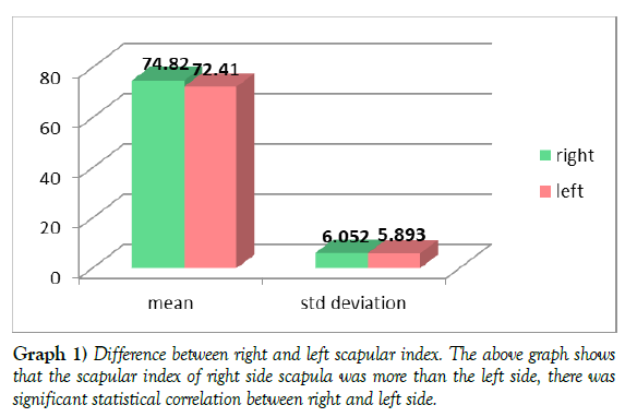
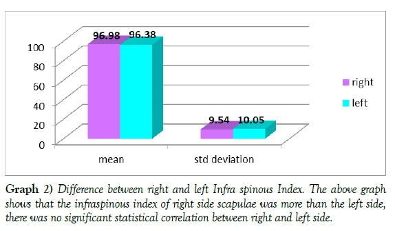
Table 2 (Graph 1-2) shows the mean of right scapular index was 74.82 ± 6.052mm which was higher than left scapular index 72.41 ± 5.893mm. There was significant statistical correlation between right and left side. This shows that right scapula is slightly shorter than left scapula. The mean of right infra spinous index was 96.98 ± 9.54 mm which is higher than left infra Spinous index 96.38 ± 10.05 mm.
Details of measurements |
Right scapular index(mm) | Left scapular index(mm) | Right Infra Spinous Index(mm) |
Left Infra Spinous Index(mm) |
|---|---|---|---|---|
| Numbers | 70 | 80 | 70 | 80 |
| Mean | 74.82 | 72.41 | 96.98 | 96.38 |
| Std. deviation | 6.052 | 5.893 | 9.54 | 10.05 |
Table 2.Difference between scapular index and Infra Spinous Index (right and left).
The distribution of various types of suprascapular notch was significant different. However, the same pattern of frequency was observed in both the right and left scapular. Type III was the most frequent. Type II was the next most frequent, which is followed by type I and V. Type IV was the least frequent while type VI was not observed in the present study.
DISCUSSION
The present study aimed at characterizing the morphometric variables of the suprascapular notch (SSN) of dry human scapulae of the Southern India.
The variables of interest included the superior transverse diameter (STD), the inferior transverse diameter (ITD), the maximal depth (MD) and the shape of the SSN which was used in classifying the SSN into various types based on Rengachary et al classification. The result showed that there is no significant difference in the values of STD, ITD and MD between the right and left SSN. [17] Reported that the right MD was significantly deeper than that of the left, while there is no significant difference in the STD. This suggests that the shapes of the SSN can be affected by laterality, and so does the nerve entrapment syndrome. Jezierski et al noted in their study that suprascapular notch containing both suprascapular nerve and vein was significantly wider and shallower.
The STD in the present study was a little narrower (1.285cm) than that of Manikum et al (2015) in South Africa (1.39cm) while the MD in the present study was deeper (0.997cm) than that in Manikum et al (2015) (0.68cm). The size and dimensions of the SSN has been considered a possible factor for suprascapular nerve entrapment [18]. This is because the SSN is the most likely site for nerve compression all the through the course of the suprascapular nerve [19-20]. SSN is therefore regarded as the most important point in the course of the suprascapular nerve [21]. However, compression of the suprascapular nerve may also occur at the base of the scapular spine, though this is less common [22].
Compression of the suprascapular nerve would lead to suprascapular nerve entrapment syndrome. This disorder is characterized by pain on the posterolateral aspect of the shoulder, weakness of the arm, difficulty in external rotation and abduction, resulting from paresis and atrophy of the infraspinatus and supraspinatus muscles [23].Flower and Garson (1879) measured the scapular index of Europeans and Negroes. European scapular index was 65.91 and that of Negroes was 68.16, which showed that Negroes had shorter scapula than that of Europeans. In present study right scapular index 74.815±6.052 and left scapular index 72.41±5.893 is more than Europeans and Negroid population suggesting that scapulae of South Indian population are shorter than European and Negroid population.
The mean of right infra spinous index was 96.98±9.54 mm which is higher than left infra Spinous index 96.38±10.05 mm. There was no significant statistical correlation between right and left side. This shows that right scapula is broader than left scapula. This when compared with study done by Flower et al (1879), suggesting infraspinous index of European as 87.79 mm and that of Negroes as 93.88 mm. Scapulae of Negroes were longer and broader than that of Europeans? In present study right infra spinous index was 96.98 ± 9.54 mm and left infra spinous index 96.38±10.05 mm is more than Europeans and Negroid population suggesting that scapulae of South Indian population are broader than European and Negroid population. Therefore, the present study observation showed that the scapula of South Indian population are more shorter and broader than European, Turkish and Negroid population, but got lesser values in most of the parameters compared to Lexington population and Canadian population. These differences may be due to the difference in general built and stature of South Indians compared to other population.
In the present study, Type III SSN was the most frequent type of suprascapular notch (71%). This is similar to the findings of Dushyant et al, (2014) (45%), Usha et al, (2014) (52%), and Polguj et al, (2014) (66.9%). However, Sutaria (2013) and Vandana et al (2013) reported type III as the third frequent type. Indeed type III SSN is the most common type as suggested by numerous works in Europe Asia [24] and in Africa. The shape of the SSN is important in the aetiopathogenesis of suprascapular nerve entrapment as it has been hypothesized that ‘V’ shaped notch is more likely to cause nerve entrapment than ‘U’ type [25]. Narrow notch has been has been found in patient with this suprascapular neuropathy [26].
Type VI SSN involves ossification of the superior transverse scapular ligament. It is regarded as one of the predisposing factors for the nerve entrapment since the notch is reduced in size [26]. In the present study, type VI SSN was not found. This may perhaps reflect reduced incidence of suprascapular nerve entrapment syndrome in the study population. However, in the literature, the incidence of complete ossification of the superior transverse scapular ligament was stated to be between 3 to 12.5% (Polguj et al, 2012). This is any way highly variable as a study in Brazilian population recorded prevalence as high as 30.76%.
Surgical removal of the ossified ligament is a treatment option in the management of suprascapular nerve entrapment syndrome in patients with complete ossification of the superior transverse scapular ligament. Furthermore, presence of an ossified superior transverse scapular ligament may pose a problem during decompression of the suprascapular notch if the condition is not fully appreciated [26].
In the present study, classification of SSN was based on Rengachary et al classification of SSN. This is because it is the most popular and verified classification system in the literature. Several newer classification systems have emerged over the years. Simply classified the SSN into two types: U and V shaped SSN classified the SSN into three types: U, V and J shaped SSN. Developed a classification based on the vertical and transverse dimensions of the notch. Polgul et al (2011) developed a classification system for SSN in an attempt to be more specific in classifying the SSN. They incorporated three measurements which include the superior transverse diameter (STD), the middle transverse diameter (MDT) and the maximum depth (MD).
In conclusion, the present study has provided a database on the morphometric variables of the suprascapular notch in the Southern part of India. There was no laterality on the dimensions of the SSN. Type III was the most prevalent type of SSN in the present study while the suprascapular foramen (Type VI) which forms by the ossification of the suprascapular ligament was not observed in the present study. This study will therefore be useful to anatomists, radiologists, neurosurgeons, orthopaedic surgeons and related disciplines for a better understanding, diagnosis and management of+ suprascapular nerve entrapment syndrome.
ACKNOWLEDGEMENT
None.
CONFLICT OF INTEREST
None.
REFERENCES
- Aghera BR, Ahmed S, Javia M, Agarwal GC et al. Morphological and morphometric study of supra scapular notch and incidence of ossified superior transverse scapular ligament in human dry scapulae and its clinical implication. Int J Anat Res. 2017; 5(3.2):4212-4215.
- Bage NN, sriambika K, Murugan M, Nim VK et al. Morphometric study of suprascapular notch as a factor of suprascapular nerve entrapment and dimensions of safe zone to prevent suprascapular nerve injury. Int J Anat Res. 2017; 5(2.3):4015-4019.
- Büyükmumcu M, Seker M, Ozbek O, Akin D et al. Complete ossification of the superior transverse scapular ligament in an turkish male adult. Int J Morphol. 2013; 31(2):590-593.
- Dushyant A, Brijendra S, Geetika AA. Human scapulae; suprascapular notch, morphometry and variations. IJCAP. 2014; 1(1):1-7.
- Flower WH, Garson JG. The scapular index as a race character in man. J Anat Physiol. 1879; 14(1):13-17.
- Gray DJ. Variations in human scapulae. Am J Phys Anthropol. 2013; 29(1):57-72.
- Iqbal K, Iqbal R, khan SG. Anatomical variations in shape of suprascapular notch of scapula. J Morphol Sci. 2010; 27:1-2.
- Jacob PJ, Arun KB. Suprascapular nerve entrapment syndrome. Keral Jour Ortho. 2012; 25(1):21-24.
- Jezierski H, Podgórski M, Stefanczyk L, Kachlik D et al. The influence of suprascapular notch shape on the visualization of structures in the suprascapular notch region: studies based on a new four-stage ultrasonographic protocol. Biomed Res Inter. 2017;1-7.
- Kannan U, Kannan NS, Anbalagan J, Rao S et al. Morphometric study of suprascapular notch in indian dry scapulae with specific reference to the incidence of completely ossified superior transverse scapular ligament. Clin Diagn Res. 2014; 8(3):7-10.
- Manikum C, Rennie C, Naidu ECS, Azu OO et al. A morphological study of the suprascapular notch in a sample of scapulae at the University of Kwazulu natal. Int J Morphol. 2015; 33(4):1365-1370.
- Moore KL, Dalley AF, Agur AMR. Moore clinically oriented anatomy. United States of America. 2014.
- Natsis K, Totlis T, Tsikaras P, Appell HJ et al. Proposal for classification of the suprascapular notch: A study on 423 dried scapulas. Clinical anatomy. 2007; 20:135-139.
- Polguj M, Jdrzejewski KS, Podgórski M, Topo M et al. Correlation between morphometry of the suprascapular notch and anthropometric measurements of the scapula. Folia Morphol. 2011; 70(2):109-15.
- Polguj M, Podgórski M, Jędrzejewski J, Topol M et al. The double suprascapular foramen: unique anatomical variation and the new hypothesis of its formation. Skeletal radiol. 2012; 41:1631-1636.
- Polguj M, Sibiński M, Grzegorzewski A, Grzelak P et al. Variation in morphology of suprascapular notch as a factor of suprascapular nerve entrapment. Int Orthop. 2012; 37(11):2185-2192.
- Polguj M, Sibiński M, Grzegorzewski A, Grzelak P et al. Suprascapular notch asymmetry: a study on 311 patients. Biomed research international. 2014.
- Rakshitha C, Komala N. Suprascapular notch in human scapula: a morphometric study. Int J Anat Res. 2018; 6(1):4840-4843.
- Rakshitha C, Komala N. Suprascapular notch in human scapula: a morphometric study. Int J Anat Res. 2018; 6(1.1): 4840-4843.
- Rengachary SS, Burr D, Lucas S, Hassanein KM et al. Suprascapular entrapment neuropathy: A clinical, anatomical, and comparative study: Part 2: Anatomical study. Clin Neurosurg. 1979; 5:447-451.
- Sutaria L, Nayak T, Patel S, Jadav H et al. Morphology and morphometric analysis of suprascapular notch. Inter J Biomed and Advance Res. 2013; 4(1):35-39.
- Ticker JB, Djurasovic M, Strauch RJ, April EW et al. The incidence of ganglion cysts and other variations in anatomy along the course of the suprascapular nerve. J Shoulder Elbow Surg. 1988; 7:472-478.
- Tubbs RS, Nechtman C, D’antoni AV, Shoja MM et al. Ossification of the suprascapular ligament: a risk factor for suprascapular nerve compression?. Int J Shoulder Surg. 2013; 7:19-22.
- Usha K, Kannan NS, Anbalagan J, Rao S. Morphometric study of suprascapular notch in indian dry scapulae with specific reference to the incidence of completely ossified superior transverse scapular ligament. J Clin Diagn Res. 2014; 8(3):7-10.
- Vaidya VK, Srivastava G, Munsif T, Tewari V et al. The morphological and morphometric study of suprascapular notch and its variations. Era's J Med Res. 2018; 5(1):22-27.
- Vandana R, Sudha P. Morphometric study of suprascapular notch. Natl J Clin Anat. 2013; 2(3):140-144.
Indexed at, Google Scholar, Crossref
Indexed at, Google Scholar, Crossref
Indexed at, Google Scholar, Crossref
Indexed at, Google Scholar, Crossref
Indexed at, Google Scholar, Crossref
Indexed at, Google Scholar, Crossref
Indexed at, Google Scholar, Crossref
Indexed at, Google Scholar, Crossref
Indexed at, Google Scholar, Crossref
Indexed at, Google Scholar, Crossref
Indexed at, Google Scholar, Crossref
Indexed at, Google Scholar, Crossref
Indexed at, Google Scholar, Crossref
Indexed at, Google Scholar, Crossref
Indexed at, Google Scholar, Crossref
Indexed at, Google Scholar, Cross Ref
Indexed at, Google Scholar, Crossref
Indexed at, Google Scholar, Crossref


