A study on variations of accessory coracobrachialis muscle along with variations of biceps brachii muscle
Received: 08-May-2018 Accepted Date: Jun 12, 2018; Published: 25-Jun-2018, DOI: 10.37532/1308-4038.18.11.66
Citation: Maiti D, Bhattacharya S. A study on variations of accessory coracobrachialis muscle along with variations of biceps brachii muscle. Int J Anat Var. 2018;11(2):66-69.
This open-access article is distributed under the terms of the Creative Commons Attribution Non-Commercial License (CC BY-NC) (http://creativecommons.org/licenses/by-nc/4.0/), which permits reuse, distribution and reproduction of the article, provided that the original work is properly cited and the reuse is restricted to noncommercial purposes. For commercial reuse, contact reprints@pulsus.com
Summary
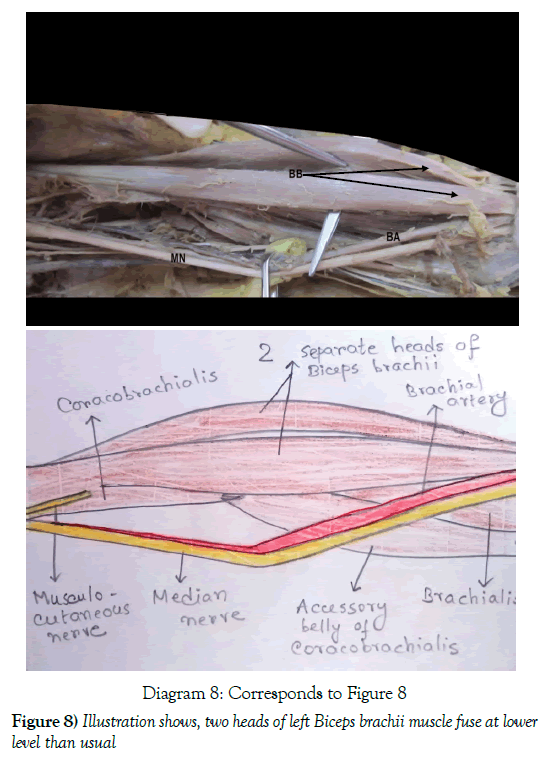
Coracobrachialis is a muscle of arm that shows several variations in its attachment. After dissecting twenty eight arms of embalmed cadavers to study the variations, we found an accessory belly of coracobrachialis muscle which originated from the tendon of coracobrachialis muscle at the middle of the medial side of shaft of humerus and was inserted into medial supracondylar ridge and medial epicondyle of humerus. The coracobrachials muscle originated from the tip of coracoid process with conjoint origin of biceps brachii muscle. We also observed that the two heads of biceps brachii muscle of that arm fused at a lower level than usual, almost at the level of an imaginary line joining the two epicondyles of humerus and formed a very short tendon to get inserted into the radial tuberosity of radius.
Keywords
Coracobrachialis; Accessory belly of coracobrachialis; Biceps brachii; Variations; Medial intermuscular septum; Medial epicondyle of humerus; Ulnar nerve; Muscle tansplant
Introduction
Morphologically, coracobrachialis muscle represents the adductor compartment of arm. But it is not very important in human beings as adductor muscle, due to the upright posture, as gravity plays the role of main adductor [1]. In some mammals, it is tricipital in origin. In human beings, the upper two heads are usually fused while taking origin from the coracoid process and enclose musculocutaneous nerve in between so that it seems that musculocutaneous nerve pierces coracobrachialis muscle. Sometimes, two upper heads are found separately in human beings. The musculocutaneous nerve is situated between two separate heads till insertion [2,3]. The third head is usually suppressed in human beings.
Coracobrachialis muscle is reported to have many variations, like –muscle originated from the coracoid process and the capsule of the shoulder joint. It was inserted into the antebrachial fascia and the medial epicondyle of the humerus [4], presence of coracobrachialis brevis inserting into the capsule of shoulder joint [5]. In one case, the proximal accessory bands of corachobrachialis brevis muscle started from the root of coracoid process and a few aponeurotic fibers from the conoid ligament of clavicle and inserted to the medial intermuscular septum [6]. The presence of two heads of origin for the coracobrachialis muscle, which are situated superficial (anterior) and deep (posterior) to the musculocutaneous nerve. The superficial head arises mainly from most of the medial border of the tendon of the short head of the biceps brachii muscle. The deep head arises from the apex of the coracoid process of the scapula and the adjoining lateral border of the tendon of the short head of the biceps brachii muscle. The musculocutaneous nerve provides a separate branch for each head. In cases where the coracobrachialis muscle was not pierced by the musculocutaneous nerve, the muscle was formed of one head that has an origin analogous to that of the superficial head. One case showed a splitting of the deep head into two bellies shortly after its origin, where the muscle appeared as being formed of three heads. Variations in the insertion were present as an additional aponeurotic insertion above the usual insertion and an aponeurotic extension to the deep fascia on the medial aspect of the arm [7]. The lower head which is usually suppressed in human beings; sometimes present as the ligament of Struthers which is attached between an occasional bony spur called supratrochlear spur from antero-medial surface of lower part of humerus and medial epicondyle of humerus. Median nerve and brachial artery pass deep to the ligament and are vulnerable to be compressed by being entrapped between the ligament and bony surface [1].
Materials and Methods
The embalmed adult cadavers used for gross anatomy dissection courses were utilized for this study. Twenty eight arms were dissected to determine the morphological criteria of the coracobrachialis muscle. We followed the following steps:
1. Careful dissection of the sites of origin and insertion of the muscle.
2. Any variability in the innervation of the muscle and its relationship with the musculocutaneous nerve.
Results
Two interesting findings we noticed in this study:
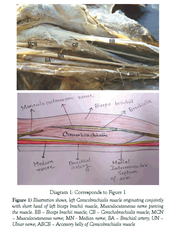
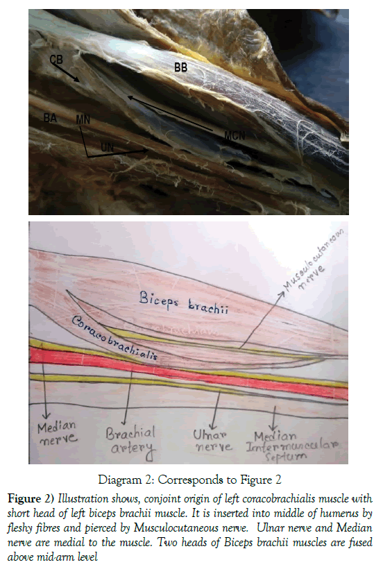
A) Almost in all cases the musculocutaneous nerve pierced the coracobrachialis muscle. Most of the fleshy part of the muscle arose from the tip of coracoid process with conjoint origin of short head of biceps brachii muscle (Figure 1). The muscle was inserted by fleshy fibers at the middle of the medial border of the humerus, between the flexor compartment and the extensor compartment of arm (Figure 2). The two heads of biceps brachii muscle were fused above mid arm level to form a single long tendon (Figure 2).
Diagram 1: Corresponds to Figure 1
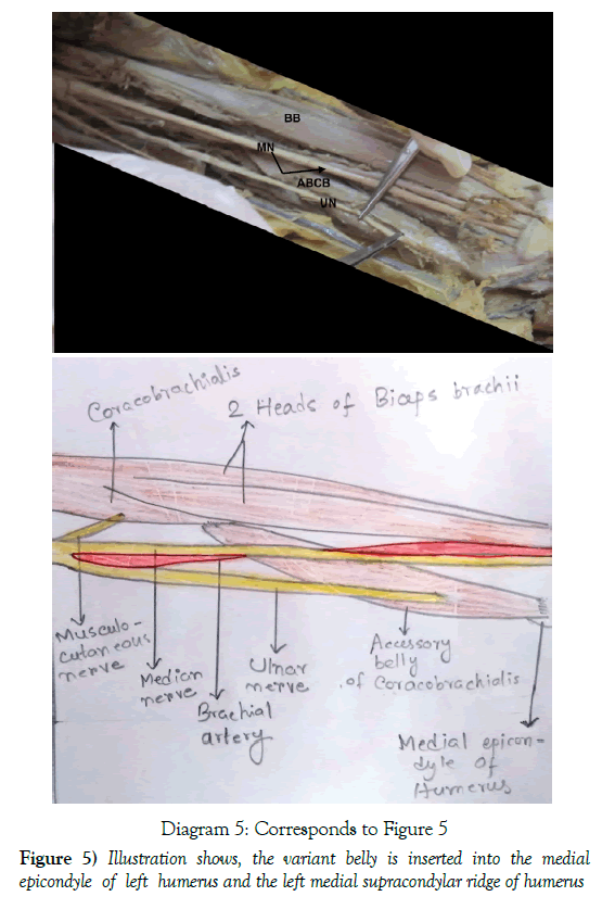
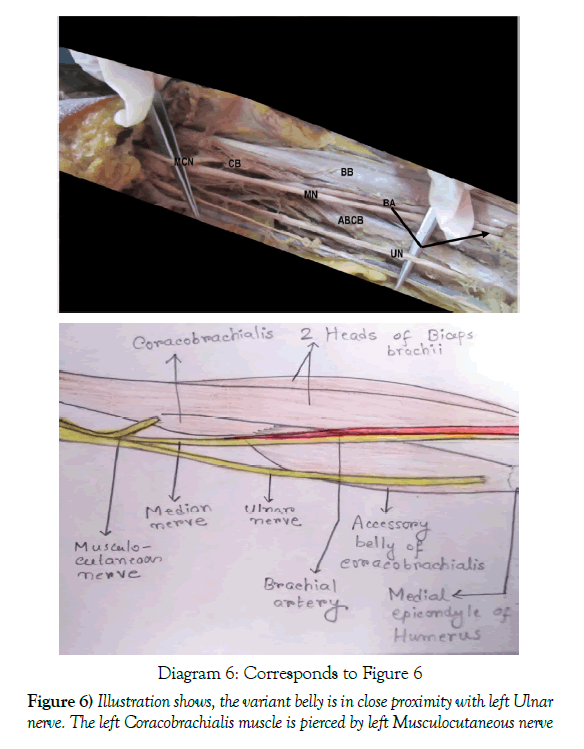
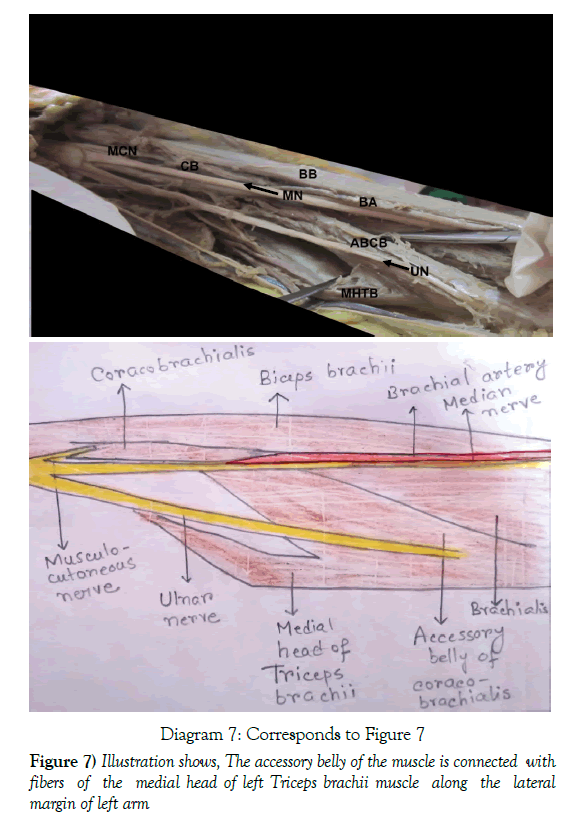
BB – Biceps brachii muscle;
CB – Corachobrachialis muscle;
MCN – Musculocutaneous nerve;
MN – Median nerve;
BA – Brachial artery;
UN – Ulnar nerve;
ABCB – Accessory belly of Coracobrachialis muscle
Figure 1) Illustration shows, left Coracobrachialis muscle originating conjointly with short head of left biceps brachii muscle, Musculocutaneous nerve piercing the muscle.
Diagram 2: Corresponds to Figure 2
Figure 2) Illustration shows, conjoint origin of left coracobrachialis muscle with short head of left biceps brachii muscle. It is inserted into middle of humerus by fleshy fibres and pierced by Musculocutaneous nerve. Ulnar nerve and Median nerve are medial to the muscle. Two heads of Biceps brachii muscles are fused above mid-arm level
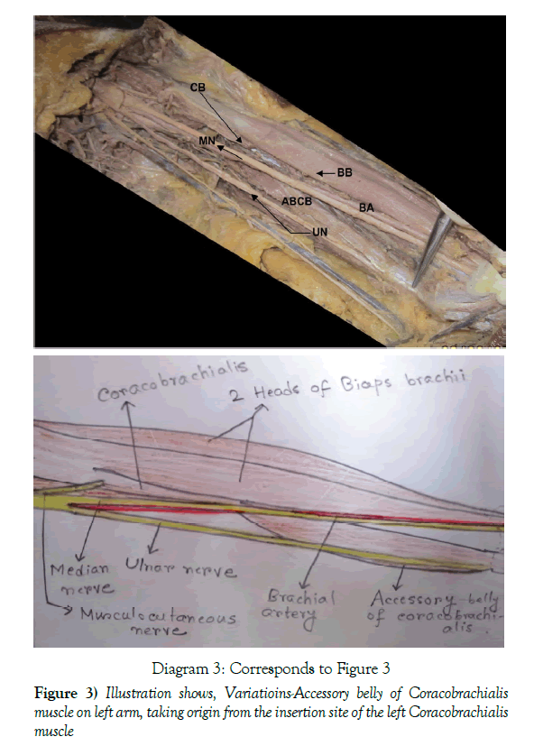
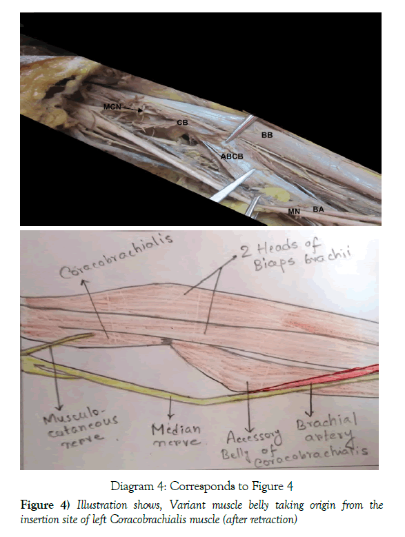
(B) One case showed an accessory belly of coacobrachialis muscle in the left upper limb of a female cadaver. It originated from the fleshy part of the coracobrachialis muscle at its insertion at the middle of medial border of left humerus (Figures 3 and 4) and was inserted into medial epicondyle of humerus and medial supracondylar ridge on the left arm (Figure 5). The coracobrachialis muscle was pierced by musculocutaneous nerve as usual (Figures 4 and 6). The fleshy accessory belly was also fused in part with medial head of triceps brachii along medial border of humerus where the medial intermuscular septum appeared to be deficient (Figure 7). The ulnar nerve was in close relation with the substance of lower part of this belly for a reasonable distance (Figures 5-7), within the fibrous part of the muscle. We also observed that the biceps brachii muscle of this limb was continuous as two separate heads roughly up to a line joining two epicondyles of humerus, and then they joined forming a very short tendon, inserted into the radial tuberosity of radius (Figure 8).
Discussion
The coracobrachialis muscle is more important morphologically than functionally. The coracobrachialis muscle in the arm is morphologically the sole representative of adductor group of muscle in the arm, but such function during the process of evolution became insignificant in man. Three distinct parts of the coracobrachialis muscle are described in amphibians, reptiles, and monotremes:
1) Coracobrachialis brevis (profundus), which is inserted into the humerus superior to tendon of latissimus dorsi, 2) Coracobrachialis medius (proprius), which is inserted into the humerus inferior to tendon of latissimus dorsi, and 3) Coracobrachialis longus (superficialis) or Wood’s muscle, which extends inferiorly on the shaft of humerus bridging the median nerve and brachial artery. Some apes and prosimians have coracobrachialis muscle composed of two parts, which is equivalent to coracobrachialis brevis.
The coracobrachialis in man is formed of one muscular part that probably represent the persistence of coracobrachialis medius of lower animals or the fusion of two heads of that muscle are observed in apes and prosimians, trapping the musculocutaneous nerve between them [8].
The accessory belly of coracobrachialis muscle we observed was unique in origin. It was in association with an important nerve of upper limb - the ulnar nerve which is in close proximity with the fleshy fibers of the muscle and within the fibrous part of the muscle. There was possibility for this belly of getting innervation from ulnar nerve too. But as we did not find any separate branch from ulnar nerve supplying the accessory belly of corachobrachialis muscle in this case, getting neural supply from the ulnar nerve, for this muscle, is only a probability. As the belly was sufficiently fleshy in comparison with the two heads of biceps bachii and as it is connected with the triceps brachii muscle, it might have acted as an assistant muscle of extension of arm. According to some authors the accessory head of the cora- cobrachialis continues with the medial head of triceps to be inserted on the olecranon process of ulna. This additional belly extending towards the ulna through the triceps brachii can be called “coracoulnaris”. The “coracoulnaris” muscle can help in the extension of the elbow and pronation of the forearm [9].
So, any injury of ulnar nerve in axilla may hamper the extension of this elbow if the variant belly was supplied by ulnar nerve. Lateral part of this muscle is not related to any neurovascular structure, so this part may be used as graft in muscle transplantation after mastectomy surgery. It may also lead to confusion in MRI or CT scan evaluation of this part of arm.
The origin of morphological variation of the coracobrachialis may be explained on the basis of the embryogenesis of the muscles of the arm. The intrinsic muscles of the upper limb differentiate in situ from the limb bud mesenchyme of lateral plate mesoderm. At a certain stage of development, the muscle primordia within the different layers of the arm fuse to form a single muscle mass. Thereafter, some muscle primordia disappear through cell death [8]. Failure of muscle primordia to disappear during embryologic development may account for the presence of the accessory belly of coracobrachialis muscle reported in this case.
Therefore, general surgeons, orthopedic surgeons and medical professionals should have more knowledge about this anatomical variation when dealing with some known clinical syndromes like humeral fracture with unusual bone displacement or shoulder pain syndromes.
Conclusively, this work showed an important anatomical variation of the coracobrachialis muscle and its probable innervations which is highly significant for some developmental understanding and surgical approach.
Acknowledgement
We would like to thank Dr. (Professor) Pranab Mukherjee for his proper guidance. We thank Dr. Anirban Sadhu for his kind help in writing this paper. We must thank Dr. (Professor) Alpana Dey, Head of the Department of Anatomy, R.G. Kar Medical College, Kolkata for her support and encouragement. We like to thank all our colleagues for their substantial support.
REFERENCES
- Dutta AK. Essentials of Human Anatomy Superior and Inferior Extremities. Current Books International. 2004;p:59-60.
- Standring S, Borley NR, Collins P. Gray’s Anatomy - The Anatomical Basis of Clinical Practice. Churchill Livingstone Elsevier. 2008;p:824-5.
- Snell RS. Clinical Anatomy for Medical Students. 1992;p;413-6.
- Kopuz C, Icten N, Yildirim M. A rare accessory coracobrachialis muscle: a review of the literature. Surgical and Radiological Anatomy. 2002;24:405-9.
- Alashkham A, Alraddadi A, Soames R. Variation of Coracobrachialis. Axis: The Online J CAHId. 2013;5.
- Chouke KS. Variation of the coracobrachialis muscle. Anat Rec. 1924;27:157-61.
- Mostafa M, EI Naggar. A study on the morphology of the coracobrachialis muscle and its relationship with the musculocutaneous nerve. Folia morphologica. 2001;60:217-24.
- Guha R, Satyanarayana N, Reddy CK. Variant insertion of coracobrachialis muscle morphological significance, embryological basis and clinical importance. J College Med Sci-Nepal. 2010;6:42-6.
- Sawant SP, Shaguphta T. An accessory head of the coracobrachialis muscle - a case report. IJSER.


