A unique case of midline gall bladder with accessory lobe of liver
Received: 10-Aug-2018 Accepted Date: Aug 31, 2018; Published: 10-Sep-2018
Citation: Shukla L. A unique case of midline gall bladder with accessory lobe of liver. Int J Anat Var. Sep 2018;11(3):107-108.
This open-access article is distributed under the terms of the Creative Commons Attribution Non-Commercial License (CC BY-NC) (http://creativecommons.org/licenses/by-nc/4.0/), which permits reuse, distribution and reproduction of the article, provided that the original work is properly cited and the reuse is restricted to noncommercial purposes. For commercial reuse, contact reprints@pulsus.com
Abstract
During routine dissection of abdomen of an adult male cadaver, we found variable position of gall bladder and a small accessory lobe of liver. Gall bladder was occupying middle of fissure for ligamentum teres hepatis and a triangular shaped accessory liver lobe was also present left to porta hepatis near the lower part of fissure for ligamentum venosum. Reporting of such kind of cases are important for radiologists and gastroenterologists for diagnostic as well surgical procedures.
Keywords
Mid line gall bladder; Accessory liver lobe; Ligamentum teres hepatis; Common bile duct; Porta hepatis
Introduction
Variations in position of gallbladder (GB) are not uncommon. GB is being classified into a right sided or left sided depending upon its position to falciform ligament and fissure for ligamentum teres hepatis [1]. Similarly, variations in size, shape, fissures and occurrence of accessory lobes of liver are not uncommon entity. Incidence of accessory lobe of liver (ALL) is ranging from 0.7% to 14.6% [2,3]. The present case is unique due to the presence of midline position of gall bladder as well as presence of an ALL.
Case Report
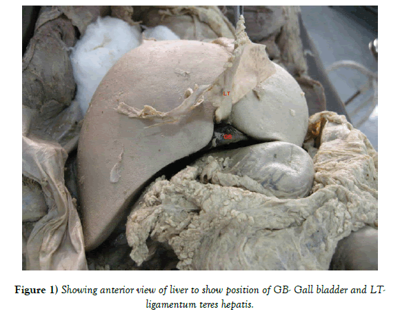
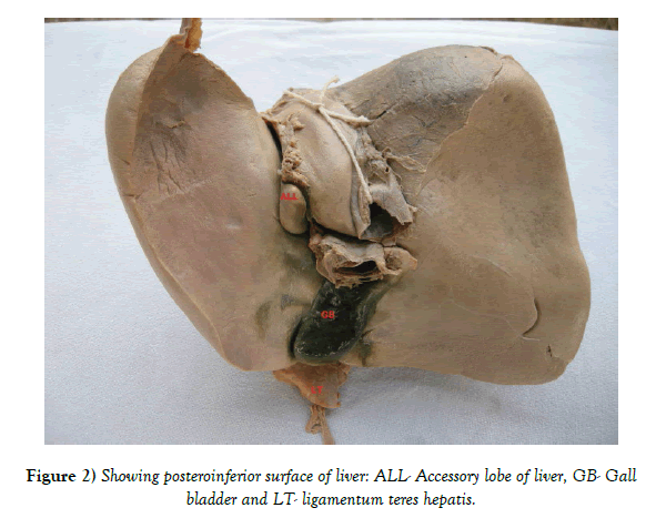
During routine dissection of abdomen of an approximately 50 year old male cadaver, fixed in 10% formalin solution for undergraduate training, GB was found to be occupying the middle of fissure for ligamentum teres hepatis (Figure 1). On exploration; neck of GB and cystic duct were found to cross common bile duct (CBD) from left to right side on its posterior aspect to open into CBD from its right side. A triangular shaped accessory lobe was also observed on the posteroinferior surface of liver. It was present left to porta hepatis near the lower part of fissure for ligamentum venosum (Figures 1 and 2). It was measuring 21 mm (height) x 18 mm (length) x 10 mm (transverse). Arterial supply of liver and gall bladder was by the branches of hepatic artery; additionally left gastric artery was also giving one branch to liver. Extra hepatic biliary duct system was found to be normal. Major organs and vessels of abdominal and thoracic cavities were found to be without any variation.
Discussion
After a thorough search of literature no matching result was found for a midline GB. We suggest modifying Hochstetter’s classification by adding a third type “midline GB” occupying falciform ligament and fissure for ligamentum teres hepatis. Developmentally, there are two explanations for the development of midline or left sided GB depending on the way cystic duct joins the biliary tree. First the normal GB bud may migrate to the left lobe instead of the right lobe. Second, GB may develop from the left hepatic instead of right hepatic duct side [4] Midline position of GB could be due to first explanation as the cystic duct joined the CBD from the right side in the present case.
Developmentally, an ALL is usually the result of embryonic heteroplasia. The accessory liver tissue could be formed due to the displacement of primitive rudiment of the organ [5] or by persistence of the mesoderm septa during proliferation of the hepatic enlarge [6] or by further branching of the foregut diverticulum [7] as in the present case.
ALL is being classified by different workers depending upon various parameters like location, volume, weight, accessory lobe joined to normal hepatic tissue or completely separated, pedunculated or sessile [8]. In the present case, ALL was joining liver completely by liver tissue.
ALL usually remains asymptomatic and accidentally detected during laprotomy or autopsy [9]. However, occasionally it may present with precordial pain, nausea and vomiting. It may be associated with congenital biliary atresia, congenital diaphragmatic defects and angiocavernoma [8].
Knowledge of such kind of variations in liver and GB are important for both the gastroenterologists and radiologists while performing successful procedures like percutaneous cholecystectomy, cholangiography and endoscopic retrograde cholangiopancreatography etc.
REFERENCES
- Hsu SL, Chen TY, Huang TL, et al. Left-sided gall bladder: Its clinical significance and imaging presentations. World J Gasterol. 2007;13:6404-9.
- Anand SS, Chauhan MS. Intrathoracic accessory lobe of the liver. Indian J Nucl Med. 2002;17:44-5.
- Muktyaz H, Nema U, Rakesh G, et al. Morphological variations of liver lobes and its clinical significance in North Indian population. GJMMS. 2013;1:1-5.
- Gross RE. Congenital anomalies of the gall bladder. Arch Surg. 1936;32:131-62.
- Mc Gregor AL. Synopsis of Surgical Anatomy. 1950;418.
- Fraser CG. Accessory lobes of the liver. Ann Surg. 1952;135:127-9.
- Ashby EC. Accessory liver lobe attached to the gall bladder. Br J Surg. 1969;56:311.
- Wang C, Cheng L, Zhang Z, et al. Accessory lobes of the liver: A report of 3 cases and review of the literature. Intractable Rare Dis Res. 2012;1:86-91.
- Pujari BD, Deodhare SG. Symptomatic accessory lobe of liver with a review of the literature. Postgrad Med J. 1976;52:234-6.


