A unique variation in branching pattern of axillary artery
P. S. Chitra* and V. Anandhi
Department of Anatomy, K.A.P.V. Government Medical College, Tiruchirappalli, Tamil Nadu, India
- *Corresponding Author:
- Dr. P. S. Chitra, MS
Assistant Professor, Department of Anatomy, K.A.P.V. Government Medical Coll. Tiruchirappalli - 620062 Tamil Nadu, India
Tel: +91 944 3864424
E-mail: pschitraezhilko@gmail.com
Date of Received: October 10th, 2011
Date of Accepted: August 29th, 2012
Published Online: January 13th, 2013
© Int J Anat Var (IJAV). 2013; 6: 1–3.
[ft_below_content] =>Keywords
axillary artery, subscapular artery, lateral thoracic artery, posterior circumflex humeral artery, variation
Introduction
The axillary artery is the continuation of the subclavian artery. It is divided into three parts by the pectoralis minor muscle. From the 1st part, one branch, superior thoracic artery arises. From the 2nd part two branches, thoraco-acromial and lateral thoracic arteries arise; and from the 3rd part three branches, the subscapular, anterior and posterior circumflex humeral arteries arise.
Various authors have described the variations in the branching pattern of the axillary artery [1–8]. During routine dissection of the upper limb we came across a unique branching pattern, which has not yet been described in literature.
Case Report
During the educational year 2011-2012, within routine educational dissection in the Department of Anatomy, K.A.P.V.Government Medical College, Tiruchirappalli, we found this unique unilateral variation in the right axilla of a 60-year-old female cadaver in the branching pattern of the axillary artery.
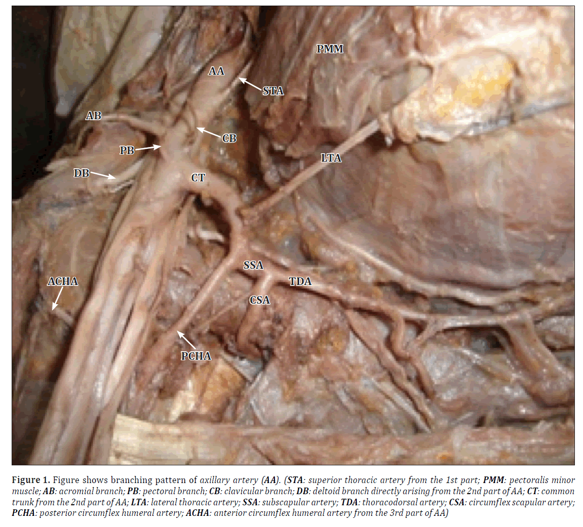
The superior thoracic artery was found to arise from the 1st part of the axillary artery. From the 2nd part, instead of a thoraco-acromial trunk, the four branches of the trunk were found to arise directly from the axillary artery. A common trunk was found to arise from the 2nd part that gave rise to lateral thoracic, posterior circumflex humeral and sub-scapular arteries. The sub-scapular artery gave the circumflex scapular branch and continued as the thoracodorsal artery. The anterior circumflex humeral was found to arise separately from the third part (Figure 1).
Figure 1. Figure shows branching pattern of axillary artery (AA). (STA: superior thoracic artery from the 1st part; PMM: pectoralis minor muscle; AB: acromial branch; PB: pectoral branch; CB: clavicular branch; DB: deltoid branch directly arising from the 2nd part of AA; CT: common trunk from the 2nd part of AA; LTA: lateral thoracic artery; SSA: subscapular artery; TDA: thoracodorsal artery; CSA: circumflex scapular artery; PCHA: posterior circumflex humeral artery; ACHA: anterior circumflex humeral artery from the 3rd part of AA)
Discussion
Variations in the branching pattern of the axillary artery have been described by many authors [1–8].
Saeed et al. have explained a similar variation where a common trunk from the 2nd part of axillary artery gave rise to the lateral thoracic, circumflex humeral, subscapular and thoraco-dorsal arteries [1].
Bhat et al. reported a case in which the common trunk from the 2nd part gave rise to the thoraco-acromial, in addition to the lateral thoracic, posterior circumflex humeral and sub scapular arteries. The anterior circumflex humeral artery was found to arise from the 3rd part [2].
Patnaik et al. described lateral thoracic artery arising from second part of axillary artery in 92% of the limbs and in 6% directly from first part [3].
However, in our present case, the 1st branch showed no variation. The 2nd branch, thoraco-acromial trunk from the 2nd part was absent. Instead, all the four branches of the thoraco-acromial trunk were found arising directly from 2nd part of the axillary artery. A common trunk from the 2nd part gave rise to the lateral thoracic, posterior circumflex humeral and subscapular arteries in addition to few muscular branches. This type of branching pattern has not yet been described in literature.
Arterial variations in the upper limb are due to defects in embryonic development of the vascular plexus of upper limb bud. This may be due to arrest at any stage of development of vessels followed by regression, retention or reappearance, thus leading to variations in the arterial origin and course of major upper limb vessels [8].
Conclusion
The clinical importance of the described axillary variation is of utmost significance for surgeons, cardiologists and vascular specialists. Considering the importance of the axillary artery and the fact that after the popliteal artery it has the second highest rate of puncture and damage in intense movements and its role in bleeding in distal part of limbs (in injuries, surgeries and embolies), knowing the variations of this artery can be significantly helpful in surgeries and clinical procedures like abscess drainage of axilla. Awareness of unusual axillary vasculature is crucial in the use of superficial brachial artery flap in plastic surgery and protection of axillary artery in breast cancer surgery.
References
- Saeed M, Rufai AA, Elsayed SE, Sadiq MS. Variations in subclavian-axillary arterial system. Saudi Med J. 2002; 23: 208–212.
- Bhat KM, Gowda S, Potu BK, Rao MS. A Unique branching pattern of the axillary artery in a South Indian male cadaver. Bratisl Lek Listy. 2008; 109: 587–589.
- Patnaik VVG, Kalsey G, Singla RK. Branching pattern of axillary artery – a morphological study. J Anat Soc India. 2000; 49:127–132.
- Rodriguez-Baeza A, Nehot J, Ferreira B, Reina F, Perez J, Sanudo JR, Roig M. An anatomical study and ontogenetic explanation of 23 cases with variations in the main pattern of the human brachio-antebrachial arteries. J Anat. 1995; 187: 473–479.
- Patnaik VVG, Kalse G, Singla RK. Bifurcation of axillary artery in its 3rd part – a case report. J Anat Soc India. 2001; 50: 166–169.
- Yang HJ, Gil YC, Jung WS, Lee HY. Variations of the superficial brachial artery in Korean cadavers. J Korean Med Sci. 2008; 23: 884–887.
- Tan CB, Tan CK. An unusual course and relations of the human axillary artery. Singapore Med J. 1994; 35: 263–264.
- Hamilton WJ, Mossman HW, eds. Cardiovascular System. In: Human Embryology. 4th Ed., Baltimore, Williams and Wilkins. 1972; 271–290.
P. S. Chitra* and V. Anandhi
Department of Anatomy, K.A.P.V. Government Medical College, Tiruchirappalli, Tamil Nadu, India
- *Corresponding Author:
- Dr. P. S. Chitra, MS
Assistant Professor, Department of Anatomy, K.A.P.V. Government Medical Coll. Tiruchirappalli - 620062 Tamil Nadu, India
Tel: +91 944 3864424
E-mail: pschitraezhilko@gmail.com
Date of Received: October 10th, 2011
Date of Accepted: August 29th, 2012
Published Online: January 13th, 2013
© Int J Anat Var (IJAV). 2013; 6: 1–3.
Abstract
An unusual variation was observed in the right side of a 60-year-old female cadaver during routine dissection. The superior thoracic artery from 1st part of axillary artery was as usual. There was no thoraco-acromial trunk, but the 4 branches of the trunk, deltoid, pectoral, acromial and clavicular were found to arise directly from the 2nd part of axillary artery. A common trunk from the 2nd part was found to give rise to most of the branches of the 2nd and 3rd parts of axillary artery.
-Keywords
axillary artery, subscapular artery, lateral thoracic artery, posterior circumflex humeral artery, variation
Introduction
The axillary artery is the continuation of the subclavian artery. It is divided into three parts by the pectoralis minor muscle. From the 1st part, one branch, superior thoracic artery arises. From the 2nd part two branches, thoraco-acromial and lateral thoracic arteries arise; and from the 3rd part three branches, the subscapular, anterior and posterior circumflex humeral arteries arise.
Various authors have described the variations in the branching pattern of the axillary artery [1–8]. During routine dissection of the upper limb we came across a unique branching pattern, which has not yet been described in literature.
Case Report
During the educational year 2011-2012, within routine educational dissection in the Department of Anatomy, K.A.P.V.Government Medical College, Tiruchirappalli, we found this unique unilateral variation in the right axilla of a 60-year-old female cadaver in the branching pattern of the axillary artery.
The superior thoracic artery was found to arise from the 1st part of the axillary artery. From the 2nd part, instead of a thoraco-acromial trunk, the four branches of the trunk were found to arise directly from the axillary artery. A common trunk was found to arise from the 2nd part that gave rise to lateral thoracic, posterior circumflex humeral and sub-scapular arteries. The sub-scapular artery gave the circumflex scapular branch and continued as the thoracodorsal artery. The anterior circumflex humeral was found to arise separately from the third part (Figure 1).
Figure 1. Figure shows branching pattern of axillary artery (AA). (STA: superior thoracic artery from the 1st part; PMM: pectoralis minor muscle; AB: acromial branch; PB: pectoral branch; CB: clavicular branch; DB: deltoid branch directly arising from the 2nd part of AA; CT: common trunk from the 2nd part of AA; LTA: lateral thoracic artery; SSA: subscapular artery; TDA: thoracodorsal artery; CSA: circumflex scapular artery; PCHA: posterior circumflex humeral artery; ACHA: anterior circumflex humeral artery from the 3rd part of AA)
Discussion
Variations in the branching pattern of the axillary artery have been described by many authors [1–8].
Saeed et al. have explained a similar variation where a common trunk from the 2nd part of axillary artery gave rise to the lateral thoracic, circumflex humeral, subscapular and thoraco-dorsal arteries [1].
Bhat et al. reported a case in which the common trunk from the 2nd part gave rise to the thoraco-acromial, in addition to the lateral thoracic, posterior circumflex humeral and sub scapular arteries. The anterior circumflex humeral artery was found to arise from the 3rd part [2].
Patnaik et al. described lateral thoracic artery arising from second part of axillary artery in 92% of the limbs and in 6% directly from first part [3].
However, in our present case, the 1st branch showed no variation. The 2nd branch, thoraco-acromial trunk from the 2nd part was absent. Instead, all the four branches of the thoraco-acromial trunk were found arising directly from 2nd part of the axillary artery. A common trunk from the 2nd part gave rise to the lateral thoracic, posterior circumflex humeral and subscapular arteries in addition to few muscular branches. This type of branching pattern has not yet been described in literature.
Arterial variations in the upper limb are due to defects in embryonic development of the vascular plexus of upper limb bud. This may be due to arrest at any stage of development of vessels followed by regression, retention or reappearance, thus leading to variations in the arterial origin and course of major upper limb vessels [8].
Conclusion
The clinical importance of the described axillary variation is of utmost significance for surgeons, cardiologists and vascular specialists. Considering the importance of the axillary artery and the fact that after the popliteal artery it has the second highest rate of puncture and damage in intense movements and its role in bleeding in distal part of limbs (in injuries, surgeries and embolies), knowing the variations of this artery can be significantly helpful in surgeries and clinical procedures like abscess drainage of axilla. Awareness of unusual axillary vasculature is crucial in the use of superficial brachial artery flap in plastic surgery and protection of axillary artery in breast cancer surgery.
References
- Saeed M, Rufai AA, Elsayed SE, Sadiq MS. Variations in subclavian-axillary arterial system. Saudi Med J. 2002; 23: 208–212.
- Bhat KM, Gowda S, Potu BK, Rao MS. A Unique branching pattern of the axillary artery in a South Indian male cadaver. Bratisl Lek Listy. 2008; 109: 587–589.
- Patnaik VVG, Kalsey G, Singla RK. Branching pattern of axillary artery – a morphological study. J Anat Soc India. 2000; 49:127–132.
- Rodriguez-Baeza A, Nehot J, Ferreira B, Reina F, Perez J, Sanudo JR, Roig M. An anatomical study and ontogenetic explanation of 23 cases with variations in the main pattern of the human brachio-antebrachial arteries. J Anat. 1995; 187: 473–479.
- Patnaik VVG, Kalse G, Singla RK. Bifurcation of axillary artery in its 3rd part – a case report. J Anat Soc India. 2001; 50: 166–169.
- Yang HJ, Gil YC, Jung WS, Lee HY. Variations of the superficial brachial artery in Korean cadavers. J Korean Med Sci. 2008; 23: 884–887.
- Tan CB, Tan CK. An unusual course and relations of the human axillary artery. Singapore Med J. 1994; 35: 263–264.
- Hamilton WJ, Mossman HW, eds. Cardiovascular System. In: Human Embryology. 4th Ed., Baltimore, Williams and Wilkins. 1972; 271–290.


