A variation in the branching pattern between the musculocutaneous and median nerves in the arm
Received: 06-Sep-2018 Accepted Date: Sep 27, 2018; Published: 04-Oct-2018
Citation: Smit JHT. A variation in the branching pattern between the musculocutaneous- and median nerves in the arm. Int J Anat Var. Dec 2018;11(4):115-116.
This open-access article is distributed under the terms of the Creative Commons Attribution Non-Commercial License (CC BY-NC) (http://creativecommons.org/licenses/by-nc/4.0/), which permits reuse, distribution and reproduction of the article, provided that the original work is properly cited and the reuse is restricted to noncommercial purposes. For commercial reuse, contact reprints@pulsus.com
Abstract
This is a cadaveric case study report of a 47-year-old male who died of Tuberculosis in the Western Cape area of South Africa. In the right axilla a variation in the branching pattern between the median- and musculocutaneous nerves was observed. The innervation to the coracobrachialis muscle came directly from the lateral cord of the brachial plexus. The formation of the musculocutaneous-, median- and ulnar nerves were normal. However, 4.0 cm below this site, the median- and musculocutaneous nerves re-joined into a single nerve. 3.5 cm below the reunion, the combined nerve gave a branch to the biceps brachii muscle. 1 cm below the biceps branch, another branch was given off to the brachialis muscle and then continued into the forearm as the lateral cutaneous nerve. The remaining section of the combined nerve continued with normal distribution, as the median nerve.
Keywords
Musculocutaneous nerve; Median nerve; Variant branches
Introduction
The main terminal branches of the brachial plexus are well documented and described in all anatomical textbooks and atlases [1-3]. A variation in the branching patterns of these nerves are however seldom mentioned, although they are often seen in the dissection room. Knowledge of these “variations” are important for clinical practice, surgical interventions and can be crucial in making the correct diagnosis [4].
Despite the lack of discussions in textbooks, there are variants that have been well documented such as the Martin-Gruber anastomosis between the median- and the ulnar nerves [5]. In this study, 70 cadavers were examined and connections between these two nerves were found in 22.9% of cases.
Guerri-Guttenberg et al. have reported communications between the median and musculocutaneous nerves in 53.6% of cases where 56 foetal and adult cadaver bodies were examined. In two cases the musculocutaneous nerve was completely absent. This led them to a “new” classification for these variations, where they used the entry of the musculocutaneous nerve into the coracobrachialis muscle as their reference point. 84.6% of the connections were proximal, 7.7% were distal and 7.7% had one proximal and one distal connection between these two nerves.
Kumar et al. [6] reported in their study on 50 isolated upper limbs, a 28% incidence where there were connections between the musculocutaneous- and the median nerves. The most frequent variation was a single communicating branch from the musculocutaneous nerve to the median nerve.
Zhang et al. [4] reported a case where there was a total absence of the musculo-cutaneous nerve. A branch of the medial cord innervated the coracobrachialis muscle, while two other branches coming from the median nerve innervated the biceps brachii and brachialis muscles respectively. The median nerve also gave off a lateral antibrachial cutaneous branch to the forearm.
Bhattarai et al. [7] reported an absence of the musculocutaneous nerve ranging between 1.7%-15%. Variations and or absence of the musculocutaneous nerve has also been reported by Fregnani et al. [5]; Pacholczak et al. [8]; Tomar et al. [9]; Chauhan et al. [10] and Oluyemi et al. [11].
Case Report
This study reports on a variation of the branching pattern between the musculocutaneous- and median nerves, which was discovered during the dissection of the right axilla. The cadaver was a male of mixed race from the Western Cape area in South Africa. He was 47-year-old and his cause of death was reported as Tuberculosis.
In the right axilla the following were seen:
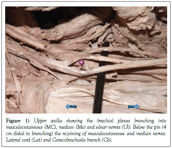
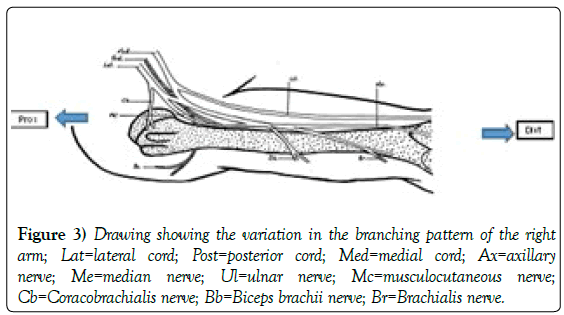
The branch to the coracobrachialis muscle came off the lateral cord of the brachial plexus, proximal to the formation of the musculocutaneous- and median nerves.
The division into musculocutaneous-, median- and ulnar nerves were clearly present anterior to the axillary artery. However, 4 cm distal to this branching, the musculocutaneous nerve re-joined the median nerve (Figure 1).
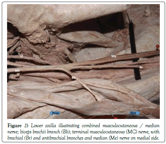
This combined musculocutaneous/median nerve gave off a single branch to the biceps brachii muscle, 3.5 cm distal to the re-joining of the nerves. 2 cm distal to the branch, the nerve splits into two separate branches to innervate the two heads of biceps brachii muscle. 1 cm distal to the main biceps branch, another branch emerged which innervated the brachialis muscle and continued further as the lateral cutaneous nerve of the antibrachium. This terminal branch is the distal musculocutaneous nerve. Under normal circumstances coracobrachialis-, biceps brachii and brachialis muscles will only receive innervation from the musculocutaneous nerve.
The median nerve followed the normal course and distribution as did the ulnar nerve and its branches (Figures 2 and 3).
Discussion
Hollinshead [12] states that nerve fibres that normally run in the lateral section of the median nerve, can join the musculocutaneous nerve instead. These fibres will split away from the musculocutaneous nerve distally, to rejoin the median nerve.
Guerri-Guttenberg et al. [13] in their study of 56 foetal and adult cadavers, reported a total absence of the musculocutaneous nerve in two cases. They also reported a connection between these two nerves in 53.6% of the cases they investigated. Most of these connections were proximal to the point where the musculocutaneous nerve enters the coracobrachialis muscle (84.6%). Only 7.7% of connections in their study were distal to the nerve entry point into the coracobrachialis muscle. In 7.7% of cases reported, there were two connections between these two nerves. One proximal to the entry point and one distal to the entry point.
Kumar et al. [6] in their study of 50 separate upper limb dissections, reported a 28% incidence of connections between the musculocutaneous- and median nerves. Mostly it was only a single connection between these two nerves.
Zhang et al. [4] reported on a single case where there was no musculocutaneous nerve present. Instead, separate branches from the median nerve innervated the coracobrachialis-, biceps brachii- and brachialis muscles.
Variations or total absence of the musculocutaneous nerve have also been reported by Bhattarai et al. [7]; Fregnani et al. [14]; Pacholczak et al. [8]; Tomar et al. [9]; Chauhan et al. [10] and Oluyemi et al. [11] but none of them described a case where the coracobrachialis muscle is innervated from the lateral cord and then the branching pattern between the musculocutaneous and median nerves as described in this case. Although there were similarities to what was described by Zhang et al. [4] of the lower median nerve in the absence of the musculocutaneous nerve, no one described a coracobrachialis branch from the lateral cord and then the branching and re-joining between median- and musculocutaneous nerves, as was seen in this case.
Any variation is clinically important. The coracobrachialis branch coming off the lateral cord can be significant. Surgeons are always looking for the musculocutaneous nerve entering the coracobrachialis as the most superficial and lateral branch of the brachial plexus in order to stay safely away from the other branches. The division and re-anastomosis between the musculocutaneous- and median nerves as described in this case will be of importance if there are any injuries at these levels. Fractures to the proximal humerus or any open wounds to this area with nerve damage will have different outcomes in comparison with a normal brachial plexus distribution.
REFERENCES
- Drake RL. Gray’s Anatomy for Students. Churchill Livingstone Elsevier. 2010.
- Moore KL. Clinical oriented anatomy. Lippincott Williams and Wilkins, Wolters Kluwer business. 2014.
- Agur AMR, Dalley. Grants atlas of anatomy. Lippincott Williams and Wilkins, Wolters Kluwer business. 2009.
- Zhang Y. Absence of musculocutaneous nerve associated with variations of distribution patterns of the median nerve. Inter J Morphol. 2014;32:461-3.
- Rodriguez-Niedenfuhr M. Martin-Gruber anastomosis revisited. J Clin Anatomy. 2002;15:129-34.
- Kumar N. Incidences and clinical implications of communications between musculocutaneous nerve and median nerve in the arm-A cadaveric study. West Indian Med J. 2013;62:744-7.
- Bhattaria C, Poudel P. Unusual variation in musculocutaneous nerves in Nepalese. Kathmandu Uni Med J. 2009;7:408-10.
- Pacholczak R. Absence of musculocutaneous nerve associated with a supernumerary head of biceps brachii: A case report. Surg Radiol Anat. 2011;33:551-4.
- Tomar V, Wadhwa S. Asymmetric bilateral variations in the musculocutaneous and median nerves with high branching of the brachial artery. Acta Medica (Hradec Kralove). 2012;55:189-92.
- Chauhan R, Roy TS. Communication between the median and musculocutaneous nerve-A case report. J Anatomical Soc India. 2002;51: 72-75.
- Oluyemi K. Communication between the median and musculocutaneous nerve and accessory head of biceps brachii: Case report. The Internet J Sur. 2006;12:1-7.
- Hollinshead WH. Anatomy for surgeons. The back and limbs, Harper and Row, Philadelphia. 1982.
- Guerri-Guttenberg RA, Ingolotti M. Classifying musculocutaneous nerve variations. Clinical Anatomy. 2009;22:671-83.
- Fregnani JH. Absence of the musculocutaneous nerve; a rare anatomical variation with possible clinical-surgical implications. Sao Paulo Med J. 2008;126:288-90.


