Aberrant right polar renal artery originating from aortic bifurcation
Yakup Yesilkaya1*, Melih Topcuoglu1, Bozkurt Gulek2 and Barbaros Cil1
1Department of Radiology, Hacettepe University, Faculty of Medicine, Ankara, Turkey
2Department of Radiology, Adana Education and Research Hospital, Adana, Turkey
- *Corresponding Author:
- Yakup Yesilkaya, MD
Hacettepe University, Faculty of Medicine, Department of Radiology Sihhiye Ankara 06100, Turkey
Tel: +90 (312) 3051188
E-mail: dryakup23@hotmail.com
Date of Received: June 8th, 2011
Date of Accepted: December 1st, 2011
Published Online: July 8th, 2012
© Int J Anat Var (IJAV). 2012; 5: 25–26.
[ft_below_content] =>Keywords
aberrant, polar renal artery, angiography
Introduction
Multiple renal arteries are unilateral in approximately 30% of patients and bilateral in approximately 10% [1,2]. Aberrant arteries usually arise from the aorta or iliac arteries anywhere from the level of T11 to the level of L4. In rare cases, they can arise from the lower thoracic aorta or from lumbar or mesenteric arteries [1]. Aberrant renal artery originating from aortic bifurcation as in our case is an extremely rare variation. It is obligatory to pay attention and keep in mind the possibility of accessory renal vessels during renal transplantations and vascular operations although they may be extremely rare as well as the variation in our case; an aberrant polar renal artery originating from aortic bifurcation.
Renal artery variations are common in general population [3,4]. Frequency of aberrant renal arteries varies between 9% to 76% in anatomic and cadaver studies [3,5,6] and it is becoming more important to detect aberrant renal arteries (ARA) due to gradual increase in interventional radiological procedures, urological and vascular operations, and renal transplantation [5–8]. If ARA is missed before these procedures it may lead to multiple complications ranging from hematoma to death. Herein we report a case of aberrant right polar renal artery incidentally found during the evaluation for resistant hypertension.
Case Report
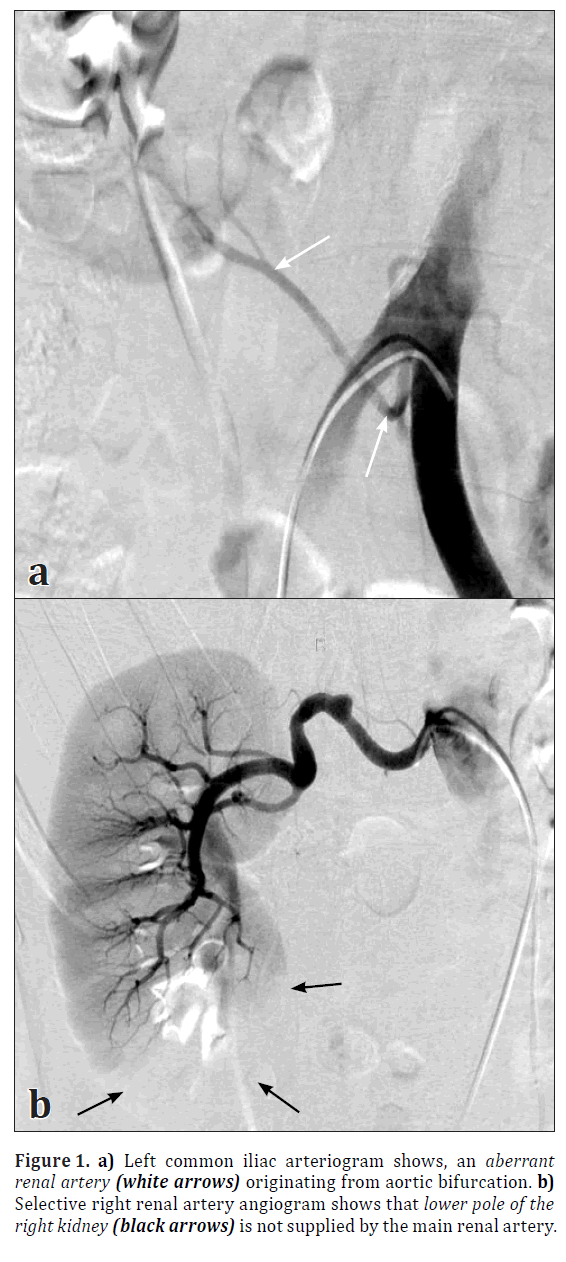
A fifty-year-old woman suffering from hypertension for 20 years was diagnosed with cerebral aneurysms during the investigation for headaches in 2007. Cerebral aneurysms were treated endovascularly with coil embolization. In 2009 patient was referred to our clinic with relapsing headaches and a new cerebral aneurysm was found but endovascular treatment was postponed due to the malign hypertension reaching levels up to 230/160 mm Hg. Renal artery Doppler ultrasound showed bilateral renal artery stenoses indirectly, according to low resistive index data and the prolongation of acceleration times. Then patient underwent bilateral selective renal arteriography, which showed diffuse extensive atherosclerosis of all arterial vessels, and there was no significant renal artery stenosis. However, there was a single renal artery on the left but double renal arteries on the right. The aberrant renal artery was originating from the terminal aorta at the bifurcation point and supplying the lower pole of the right kidney through the renal capsule (Figure 1a, 1b).
Discussion
The renal arteries are a pair of lateral branches of abdominal aorta. They arise from aorta just below the level of origin of the superior mesenteric artery. The accessory renal arteries are also seen frequently [9,10]. Such arteries enter the kidney either above or below the hilum [11]. Aberrant renal arteries constitute the most common and clinically important renal arterial variations and can be seen in up to one-third of the normal population. Multiple renal arteries are unilateral in approximately 30% of patients and bilateral in approximately 10%. Aberrant arteries usually arise from the aorta or iliac arteries anywhere from the level of T11 to the level of L4. In rare cases, they can arise from the lower thoracic aorta or from lumbar or mesenteric arteries. Perihilar branching is another common variant and it is the branching of the main renal arteries into segmental branches at a more proximal level than the renal hilum. Most of the variations of the renal arteries are due to the various developmental positions of the kidneys [12].
In conclusion, the aim of this case report was to emphasize the importance of knowledge of renal vessel variations for surgeons practicing kidney transplants, also for radiologists dealing with interventional vascular procedures. In the literature, variations include double renal artery and polar renal arteries but the important point is the origin of accessory renal vessels. Aberrant polar renal artery originating from aortic bifurcation is an extremely rare renal vascular variation. So it is obligatory to pay attention and keep in mind the possibility of accessory renal vessels during these procedures, although they may be extremely rare as in our case.
References
- Kadir S. Angiography of the kidneys. In: Kadir S, ed. Diagnostic Angiography. Philadelphia, Saunders.1986; 445–495.
- Spring DB, Salvatierra O Jr, Palubinskas AJ, Amend WJ Jr, Vincenti FG, Feduska NJ. Results and significance of angiography in potential kidney donors. Radiology. 1979; 133: 45–47.
- Kadir S. Kidneys. In: Kadir S, ed. Atlas of Normal and Variant Angiographic Anatomy. Philadelphia, W.B. Saunders Company. 1991; 387–429.
- Boijsen E. Renal angiography: Techniques and hazards; anatomic and physiologic considerations. In: Baum S, ed. Abrams’ Angiography. 4th Ed., Philadelphia, Little, Brown and Company. 1997; 1101–1131.
- Khamanarong K, Prachaney P, Utraravichien A, Tong-Un T, Sripaoraya K. Anatomy of renal arterial supply. Clin Anat. 2004; 17: 334–336.
- Satyapal KS, Haffejee AA, Singh B, Ramsaroop L, Robbs JV, Kalideen JM. Additional renal arteries: incidence and morphometry. Surg Radiol Anat. 2001; 23: 33–38.
- Sampaio FJ, Passos MA. Renal arteries: anatomic study for surgical and radiological practice. Surg Radiol Anat. 1992; 14: 113–117.
- Baltacioglu F, Ekinci G, Akpinar IN, Cimsit NÇ, Tuglular S, Akoglu E. Endovascular treatment of renal arter stenosis: technical and clinical results. Diagn Intervent Radiol. 2003; 9: 246–256. (Turkish)
- Singh G, Ng YK, Bay BH. Bilateral accessory renal arteries associated with some anomalies of the ovarian arteries: a case study. Clin Anat. 1998; 11: 417–420.
- Bordei P, Sapte E, Iliescu D. Double renal arteries originating from the aorta. Surg Radiol Anat. 2004; 26: 474–479.
- Bayramoglu A, Demiryurek D, Erbil KM. Bilateral additional renal arteries and an additional right renal vein associated with unrotated kidneys. Saudi Med J. 2003; 24: 535–537.
- Moore KL, Persaud TVN. The Developing Human. 7th Ed., Saunders, An Imprint of Elsevier. 2003; 293.
Yakup Yesilkaya1*, Melih Topcuoglu1, Bozkurt Gulek2 and Barbaros Cil1
1Department of Radiology, Hacettepe University, Faculty of Medicine, Ankara, Turkey
2Department of Radiology, Adana Education and Research Hospital, Adana, Turkey
- *Corresponding Author:
- Yakup Yesilkaya, MD
Hacettepe University, Faculty of Medicine, Department of Radiology Sihhiye Ankara 06100, Turkey
Tel: +90 (312) 3051188
E-mail: dryakup23@hotmail.com
Date of Received: June 8th, 2011
Date of Accepted: December 1st, 2011
Published Online: July 8th, 2012
© Int J Anat Var (IJAV). 2012; 5: 25–26.
Abstract
Variations in the renal vessels and knowledge of the possibility of these variations are becoming more important nowadays due to gradual increase in interventional radiological procedures, urological and vascular operations, and renal transplantation. This report describes a case of aberrant right polar renal artery originating from aortic bifurcation in a fifty-year-old woman, that found incidentally while investigating for hypertension before the endovascular treatment of a cerebral aneurysm.
-Keywords
aberrant, polar renal artery, angiography
Introduction
Multiple renal arteries are unilateral in approximately 30% of patients and bilateral in approximately 10% [1,2]. Aberrant arteries usually arise from the aorta or iliac arteries anywhere from the level of T11 to the level of L4. In rare cases, they can arise from the lower thoracic aorta or from lumbar or mesenteric arteries [1]. Aberrant renal artery originating from aortic bifurcation as in our case is an extremely rare variation. It is obligatory to pay attention and keep in mind the possibility of accessory renal vessels during renal transplantations and vascular operations although they may be extremely rare as well as the variation in our case; an aberrant polar renal artery originating from aortic bifurcation.
Renal artery variations are common in general population [3,4]. Frequency of aberrant renal arteries varies between 9% to 76% in anatomic and cadaver studies [3,5,6] and it is becoming more important to detect aberrant renal arteries (ARA) due to gradual increase in interventional radiological procedures, urological and vascular operations, and renal transplantation [5–8]. If ARA is missed before these procedures it may lead to multiple complications ranging from hematoma to death. Herein we report a case of aberrant right polar renal artery incidentally found during the evaluation for resistant hypertension.
Case Report
A fifty-year-old woman suffering from hypertension for 20 years was diagnosed with cerebral aneurysms during the investigation for headaches in 2007. Cerebral aneurysms were treated endovascularly with coil embolization. In 2009 patient was referred to our clinic with relapsing headaches and a new cerebral aneurysm was found but endovascular treatment was postponed due to the malign hypertension reaching levels up to 230/160 mm Hg. Renal artery Doppler ultrasound showed bilateral renal artery stenoses indirectly, according to low resistive index data and the prolongation of acceleration times. Then patient underwent bilateral selective renal arteriography, which showed diffuse extensive atherosclerosis of all arterial vessels, and there was no significant renal artery stenosis. However, there was a single renal artery on the left but double renal arteries on the right. The aberrant renal artery was originating from the terminal aorta at the bifurcation point and supplying the lower pole of the right kidney through the renal capsule (Figure 1a, 1b).
Discussion
The renal arteries are a pair of lateral branches of abdominal aorta. They arise from aorta just below the level of origin of the superior mesenteric artery. The accessory renal arteries are also seen frequently [9,10]. Such arteries enter the kidney either above or below the hilum [11]. Aberrant renal arteries constitute the most common and clinically important renal arterial variations and can be seen in up to one-third of the normal population. Multiple renal arteries are unilateral in approximately 30% of patients and bilateral in approximately 10%. Aberrant arteries usually arise from the aorta or iliac arteries anywhere from the level of T11 to the level of L4. In rare cases, they can arise from the lower thoracic aorta or from lumbar or mesenteric arteries. Perihilar branching is another common variant and it is the branching of the main renal arteries into segmental branches at a more proximal level than the renal hilum. Most of the variations of the renal arteries are due to the various developmental positions of the kidneys [12].
In conclusion, the aim of this case report was to emphasize the importance of knowledge of renal vessel variations for surgeons practicing kidney transplants, also for radiologists dealing with interventional vascular procedures. In the literature, variations include double renal artery and polar renal arteries but the important point is the origin of accessory renal vessels. Aberrant polar renal artery originating from aortic bifurcation is an extremely rare renal vascular variation. So it is obligatory to pay attention and keep in mind the possibility of accessory renal vessels during these procedures, although they may be extremely rare as in our case.
References
- Kadir S. Angiography of the kidneys. In: Kadir S, ed. Diagnostic Angiography. Philadelphia, Saunders.1986; 445–495.
- Spring DB, Salvatierra O Jr, Palubinskas AJ, Amend WJ Jr, Vincenti FG, Feduska NJ. Results and significance of angiography in potential kidney donors. Radiology. 1979; 133: 45–47.
- Kadir S. Kidneys. In: Kadir S, ed. Atlas of Normal and Variant Angiographic Anatomy. Philadelphia, W.B. Saunders Company. 1991; 387–429.
- Boijsen E. Renal angiography: Techniques and hazards; anatomic and physiologic considerations. In: Baum S, ed. Abrams’ Angiography. 4th Ed., Philadelphia, Little, Brown and Company. 1997; 1101–1131.
- Khamanarong K, Prachaney P, Utraravichien A, Tong-Un T, Sripaoraya K. Anatomy of renal arterial supply. Clin Anat. 2004; 17: 334–336.
- Satyapal KS, Haffejee AA, Singh B, Ramsaroop L, Robbs JV, Kalideen JM. Additional renal arteries: incidence and morphometry. Surg Radiol Anat. 2001; 23: 33–38.
- Sampaio FJ, Passos MA. Renal arteries: anatomic study for surgical and radiological practice. Surg Radiol Anat. 1992; 14: 113–117.
- Baltacioglu F, Ekinci G, Akpinar IN, Cimsit NÇ, Tuglular S, Akoglu E. Endovascular treatment of renal arter stenosis: technical and clinical results. Diagn Intervent Radiol. 2003; 9: 246–256. (Turkish)
- Singh G, Ng YK, Bay BH. Bilateral accessory renal arteries associated with some anomalies of the ovarian arteries: a case study. Clin Anat. 1998; 11: 417–420.
- Bordei P, Sapte E, Iliescu D. Double renal arteries originating from the aorta. Surg Radiol Anat. 2004; 26: 474–479.
- Bayramoglu A, Demiryurek D, Erbil KM. Bilateral additional renal arteries and an additional right renal vein associated with unrotated kidneys. Saudi Med J. 2003; 24: 535–537.
- Moore KL, Persaud TVN. The Developing Human. 7th Ed., Saunders, An Imprint of Elsevier. 2003; 293.


