Abnormal Lumbar Curvature, Via Protective System, Causes Head Bones Displacement Hair Loss and Mental Disorders: The Natural Process of Human Grow-Up
Received: 25-Jun-2020 Accepted Date: Oct 09, 2020; Published: 16-Oct-2020, DOI: 10.37532/1308-4038.20.13.20
This open-access article is distributed under the terms of the Creative Commons Attribution Non-Commercial License (CC BY-NC) (http://creativecommons.org/licenses/by-nc/4.0/), which permits reuse, distribution and reproduction of the article, provided that the original work is properly cited and the reuse is restricted to noncommercial purposes. For commercial reuse, contact reprints@pulsus.com
Abstract
The roots of mental disorders are not known yet, moreover it is believed not to be related to the body at all, but that was a common belief among the ancient scientists and Philosophers; they mostly acknowledged these disorders as body-based symptoms. The study focuses on the idea that mental disorders and hair loss are the results of the natural process of human grow-up happening to everyone but with different symptoms. It verifies the qualifications and possibilities of these results in different anatomic types of humans. A hypothesis specifies the basic method of the article, i. e. if decreasing of the lumbar curvature (l .c.) is obvious and accepted, then consequently the changes in the vertebral column (v. c.) are also acceptable, because of the ligaments which firmly attach the vertebrae together. The v. c., diaphragm, thorax and head bones, working together in a definite and synchronized relation, build a unique protective system (p. s.). The force caused by the b. f. of the l. c. activates the p. s., reaches up the skull and relative to the forces imposed on the skull; displacements of the head bones, through cartilaginous sutures, occur. The process causes: a) the body-based mental disorders afterwards, either through providing asymmetrical pairs, as in the parietal and temporal bones or via imposing pressure on the lobes and b) hair loss as it will be described in the paper.
Keywords
The protective system, Lumbar curvature, Head bones displacement, Skull sutures, Body-based mental disorders
Introduction
Although many great steps are taken to unveil the secrets of the human body, there are still many facts to be discovered. One of these facts is the relation between the body and the mind. Does a body have effects on the mind? If yes, then how?
In the history of psychology, this is a rather new argument. From the ancient eras, there was a belief that mental disorders were the results of supernatural phenomena or being haunted by evils needed to be drawn out by piercing the skull or practicing religious ceremonies by the tribe head, religious men or witches.
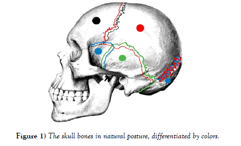
Through the history of science, many scientists tried to make scientific pathways in the body and the mind interaction domain. Plato (427BC-347) Believed that the human body as a balanced system would become imbalance and when it reached the head, caused the great illness or mental illness [1,2]. Avicenna (980-1037) quoted that mental disorders are mere physical illnesses and prescribed herbal medicine. He “in his famous “Floated Man” demonstrated that the soul was a substance [3]. Not that far back Doctor Benjamin Rush (1745-1813) known “the father of American psychiatry” was the first person to certify the mental illness as a disease of the mind [4]. At last, in 1879, in Leipzig Germany, Wilhelm Wundt founded the first laboratory dedicated to psychology as a new field of study [5]. This was the end of the unqualified person ruling. Smashing the ancient totems of ignorance was a great step. Furthermore, one of the prospects of science is searching the relations of body and brain or mind, considering the brain as the organ of mind (Figure 1).
Methods and Materials
1- Fore mentioned Plato’s quotation encouraged the author to search for the imbalance factors of the body. The form of the sphenoid bone which resembles a bird inspired the author to search balance, around the vertical axis dividing the body into two symmetrical halves. Finally, the skeletal system was distinguished to be what Plato meant.2- An experiment determined the progression route of the research, it defines: If the lumbar part is pushed to the dorsal direction, the anterior aspects of the vertebrae move up, the spinous processes move down and thorax is compressed. 3- About 400 Illustrations were drew to determine the natural forms of the v. c., diaphragm, thorax and head bones (mentioned in this section), to find out how they might become imbalanced or unnatural (mentioned in the result section).4- The natural reasons of the anatomical structures were searched and the answer to the question of why this kind of structure came into being, e.g. the reasons of different sutures also guided the author as a method. 5- Aesthetic science criteria were applied in studying human anatomy, e.g. the difference between the natural form of the thorax and the deformed one under backpressure. 6- The findings were closely examined for many years in a great number of volunteer people, mostly their sternal angles and the head bones.7- The variations and their abundance in human anatomy (especially of the crania) as the causes, were evaluated compared to the various types of mental disorders and hair loss, which resulted in justification and supporting the relation.
1 - V. C.
V. c. by itself, ignoring the muscles has natural curvatures because of the shapes of the vertebral bodies. It consists of 7cervical vertebrae, 12 thoracic vertebrae, and 5 lumbar vertebrae. It is built of thin and thick parts, like a pile, the lumbar part (base) bears the whole weight of the body. The size of the vertebral bodies increases downward and thus due to their largeness and the shapes, the minor changes cause great changes in the upper parts.
The v. c. is a curved structure positioned in the space between head and ilium. Suppose the space be12 units (e. g .120 cm.), then the cervical part would be 2(20cm.), thoracic part 6(60 cm.) and the lumbar curve 4(40 cm.). Note that between two fixed points a curve is longer than the straight line.
Thus, when the natural lumbar curvature tends to become a straight line, it would be longer.
2-Diaphragm
Right crus of the diaphragm, broader and longer than the left one, origins from the bodies of LI to LIII and the intervertebral discs but the Left crus origins from the bodies of LI and LII and the intervertebral discs.
3-Thorax
The thorax, consisting of 12 ribs, the first 7 via their cartilaginous ends and 3 through the costal arch frontally attach to the sternum while 11th and 12th are the floating ribs that have free ends [6].
Of the 7 sternocostal joints, the first rib joint is fibrocartilaginous which does not provide any real motion. The sternocostal joints between the ribs 2nd to 7th are all synovial. The second sternocostal joint consists of two separate synovial compartments that are situated between 1) the cartilaginous end of the second rib and the manubrium, 2) the cartilaginous end of the second rib and the body of the sternum to provide motions based on the form of the manubriosternal symphysis joint. This structure shows that the sternal angle could have a hinge-like motion round the second rib, in the other words, the geometric measure the angle between the manubrium and the body of sternum could change [7].
4-Head bones
The head bones are connected via fibrocartilaginous tissue mostly made of collagen and the articulation sites are called sutures. In this section, first, the sutures of the flat cranium bones and their borders are studied and that how the adjacent bones are attached, which one is overlapped or overlapping? All these articulations are classified as a kind of synarthrosis or immovable joint in which the surfaces of the bones are in almost direct contact, not separated by an intervening synovial cavity, but by a series of contact surfaces, processes and indentations of two contiguous bones overlapping one another or interlocked together [8].
The skull bones are shown in different colors. The continuous line shows an overlapping border and the dotted line the overlapped border by the adjacent bone. In the lambdoid suture, the red and blue lines show the overlapping/ overlapped denticulations of the adjacent bones. This kind of suture allows the least or no motion for the occipital against the parietals and the two parietals against each other (Figure 1).
Sutures
The sutures are two kinds: 1-The real suture (sutura Vera) in which the surfaces of the bones are separated by a layer of membrane continuous externally with the pericranium, internally with the dura mater.
There are three kinds of real sutures;
1-1- Sutura dentata, possesses a tooth-like form of the projecting articular processes (between the parietal bones).
1-2- Sutura Serrata, the edges of the two articulating bones are serrated as a fine saw (between two portions of the frontal bone).
1-3- Sutura limbosa, besides the dentated processes, there is a certain degree of beveling of the articular surfaces, so that the bones overlap one another, as the lambdoid suture.
The second kind of the sutures is the false suture (Sutura notha) in which the articulation is formed by roughened surfaces placed in apposition with one another, as the squamous suture, a kind of the false suture, in the temporoparietal suture that is formed by the overlapping of two contiguous bones of broad beveled margins [8].
Now the borders of the cranium bones are studied.
1-Parietal bones
The superior border is dentated to articulate with its fellow of the opposite side, forming the sagittal suture, a sutura dentata. The inferior border is divided into three parts; the anterior is thin and pointed, beveled at the expense of the outer surface, and overlapped by the tip of the great wing of the sphenoid; the middle portion is arched beveled at the expense of the outer surface, and overlapped by the squamous portion of the temporal, sutura squamosa; the posterior portion is thick and serrated for articulation with the mastoid portion of the temporal. The anterior border deeply serrated is beveled at the expense of the outer surface above, and the inner below; it articulates with the frontal bone, forming the coronal suture. The posterior border, deeply denticulated, articulates with the occipital, forming the lambdoid suture, sutura limbosa [8].
2- Frontal Bone
The border of the vertical portion is thick, strongly serrated, beveled at the expense of the internal surface above, where it rests upon the parietal, each side at the expense of the external surface and is overlapped by the lateral parts of the parietal bones.
3- Temporal bone
3-1- The squamous portion
The superior border is thin, beveled at the expense of the internal surface, to overlap the lower border of the parietal bone, forming the squamous suture. The anterior inferior border is thick, serrated, beveled alternately at the expense of the inner and outer surfaces, for articulation with the great wing of the sphenoid.
3-2- The mastoid portion
The superior border of the mastoid portion is rough and serrated, beveled at the expense of the inner surface, for articulation with the parietal inferior border. The posterior border, also uneven and serrated, beveled at the expense of the inner surface, articulates with the inferior border of the occipital bone [8].
4-Occipital bone
The superior border is deeply serrated for articulation with the parietal bone to form the lambdoid suture, a sutura limbosa. The upper half of the inferior border articulates with mastoid portion forming the masto-occipital suture. The lower half articulates with the petrous portion of the temporal, forming the petro-occipital suture [8].
The timetable of ossification
• Birth to 5 years: secondary ossification centers appear in the epiphyses.
• 5 years to 12 years in females,
• 5 to 14 years in males: Ossification is spreading rapidly from the ossification centers and various bones are becoming ossified.
• 17 to 20 years: Bone of upper limbs and scapula becoming completely ossified.
• 18 to 23 years: Bone of the lower limbs becomes completely ossified [7].
• 18 to 25 years: the cartilage between the sphenoid and occipital becomes completely ossified [8].
• 23 to 25 years: The bone of the sternum, clavicles, and vertebrate become completely ossified.
• By 25: nearly all bones are completely ossified [7].
Results
In this section, the results of the p. s. activation are represented in the order of the method section.
1- V. C.
It was supposed before the v. c. as a curved line measured approximately 12.3 units. When the l. c. decreases gradually and becomes a straight line, it is about 0.3 units taller than the vertical line between the up and down points. For example, a v. c. with 123 cm length, each unit of 10 cm, would be inserted in an interval of 120 cm long, so it is about 3 cm longer than the vertical line between head and ilium [7]. Due to the action of the long and short ligaments that cooperate to keep the length of the spinal cord constant, the increase of the v. c. is impossible, so when the lumbar curvature is pushed back gradually, ligaments pull down the anterior aspects of the vertebral column and push up the posterior aspects (a see-saw motion).
2-Diaphragm
The forces of the vertebral bodies would sooner reach the right crus of diaphragm, because it is longer than the left to make the necessary adjustments on the diaphragm. Notice the right dome is also higher than the left. Otherwise, diaphragm with two cruses and domes of different lengths and heights would lose its balance. Note that the cruses of the diaphragm are only attached to the lumbar vertebrae and the diaphragm is dependent only on the lumbar and not thoracic vertebrae.
3-Thorax
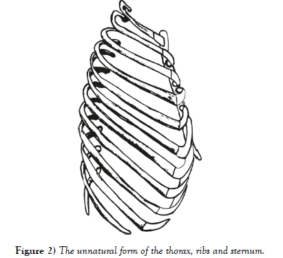
The force of the l. v. imposes pressure on the thoracic vertebrae through its see-saw action i.e. in parallel but opposite directions. When the spinous processes move up and their bodies are pulled down, thus the transverse processes act as a mechanical lever force the ribs down via their heads. Note that rib head is firm, dense, round in cross-section, and its form does not change under the back force. The bodies are, from the second down, spongy, vascular and, trabecular covered with a thin layer of compact bone, resembling a blade that could change from a vertical (natural) position to an oblique (unnatural) one. The thorax under the back force is compressed, the second to tenth vertebrae force the ribs down to adopt the oblique posture made possible through the cartilaginous ends of 2nd to 7th ribs and costal arch.
The first rib is dense, thick and bony and atypically its body lies in a horizontal plane along its ridge (would not change to oblique position), possessing only one articulation facet on its vertebra and a fibrocartilaginous sternocostal joint, not synovial as the other ribs.
The thorax is divided into two chambers:1) The lower, larger volume from the second rib at the top and the tenth rib at the bottom (inferior thoracic aperture). 2) The Upper and the smaller part, which has the first rib at its top (superior thoracic aperture) and the second rib at the bottom. These two chambers are fused at their fusion site i.e. sternal angle (Figure 2).
When there is no back force, the first rib moves upward, it pulls the manubrium upward through its sternocostal joint because it does not work as a movable joint.
4-Head bones displacement
4-1- Occipital displacement
The occipital is the first one of the head bones that engages with the upcoming force. The structure of the occipital bone resembles a vertebra. It possesses a basilar part like a body, two lateral portions as transverse processes, a posterior part that matches the spinous process and condyles which articulate with the superior articular surfaces of the atlas. When the force reaches it up, its posterior part is pushed up and the anterior part of the basilar part is pulled down. It adopts a slope angle like the angle between the adjacent articular surfaces of vertebrae.
4-2- Parietal bones displacement
Tow parietal bones are attached together by the sagittal suture. It is a sutura dentata, in which there are denticulations without overlapping, not as it was in the lambdoid suture, therefore, it can act like a door hinge for the two parietal bones. At the time the occipital bone is elevated, the parietal bones move up so that the lateral sides move inward or get closer to each other and the roof of the sagittal suture becomes palpable under the skin. Here the galea aponeurotica, a very tough tissue attached to the superior temporal line, keeps the parietal bones in their utmost situation, preventing them from moving up too high.
So, the movement of the parietal bones alone without the lambdoid suture, is possible, but when we consider their movement in relation to functioning of the lambdoid suture then two possibilities may take place:
1-No wormian bones: As it was mentioned before, the lambdoid suture was a real suture showing more aspects of overlapping interlocked denticulations, would not allow any movement of the parietal bones. The engagement of interlocked denticulations is so heavy and tight that it would not allow the parietal bones, turn inward round their axis (the sagittal suture). Therefore, they move up firmly attached together keeping their posture fixed to each other. In this case, the tightening of the galea aponeurotica would not happen, because there is no change in the positions of the parietal bones, therefore there is no hair loss.
2-Wormian bones in the lambdoid suture: So, the suture deprives of the strong binding between the occipital and parietal bones; the wormian bones, as a medium, allow the parietal bones turn inward round the sagittal suture. The inferior part of parietal bones gets closer while moving up. In this case, because the tightened galea aponeurotica is tightened, hair losing appears in the individuals.
Note that the fibrous membrane which is continuous externally with precranium and internally with dura mater keeps the bones safe in the displacement course and also the dura mater and the brain.
To trace up the displacement of the temporal bones, a brief description of the squamous suture is mentioned.
Squamous suture
The superior border of the squamous portion of the temporal bone, beveled at the expense of the internal surface, overlaps the inferior border of the parietal, forming the squamous suture, a kind of false suture (sutura notha).
4-3- Temporal bones displacement
The movement of the temporal bones is much different from the foretold bones because the occipital and the parietal bones are along with each other on a horizontal plane, but the rotational movement and inward-outward displacement of the temporal are on a vertical plane that becomes possible via this kind of suture. The roughness, unevenness, and serrations of the articulating surfaces of the posterior border of mastoid with the inferior border of the occipital assure a definite and hazardless movement. The logical study of the temporal movement again starts from two kinds of the lambdoid suture.
1- The lambdoid suture lacks wormian bones; the parietal and occipital bones only move up attached firmly together and the temporal bones are displaced as follow:
• The ultimate posture of the temporal bone is balanced; there is no hair loss (the galea aponeurotica is not tightened) and minor mental disorders caused (the temporal bones are still balanced).
• The ultimate posture of the temporal bone is unbalanced; there is no hair loss (the galea aponeurotica is not tightened), but major mental disorders caused (the temporal bones are in unbalance postures).
2- There are wormian bones situated in the lambdoid suture so that it allows the hinge-like movement of the parietal bones. In this case, two structures may result;
• Balanced spread of wormian bones, the parietals and temporal bones are displaced balanced. Minor mental disorders are caused and also hair loss happens.
• Unbalanced spread of wormian bones; the construction made leads to the unbalanced movement of parietal and temporal bones. Major mental disorders are caused also hair loss happens.
4-4- Frontal bone posture in Parietal bones displacement
Now the article studies the frontal bone articulations with the other bones. Considering the coronal suture, the superior part of the parietals up to the temporal ridge, is limited by frontal, which overlaps them, to move too far upward, but below the temporal ridge, the parietals are limited to move inward, because in the inferior parts they overlap the frontal bones.
Frontal displacement is majorly limited to the form of the falx cerebri which attaches anteriorly to the crista galli; posteriorly it is connected with the upper surface of the cerebellar tentorium. Its superior margin is attached at the midline to the internal surface of the skull, and the internal occipital protuberance. Therefore, the form of falx cerebri is determined by these parts and the whole factors make the frontal bone adopt a sloped posture compared to its natural form.
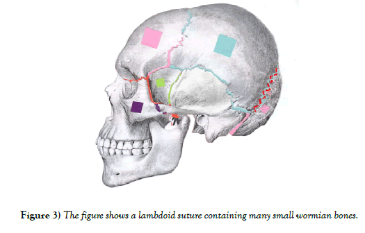
To verify the case closely, in the next figure the bones are differentiated with the colors. When the bones slide over each other, the overlapped borders appear in the same color of each bone. The figure shows a lambdoid suture containing many small wormian bones (Figure 3).
Figure 3: The figure shows a lambdoid suture containing many small wormian bones.
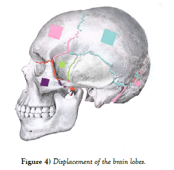
The same view and description but a lambdoid suture void of wormian bones. The same changes take place except in raising the parietals which they are not folded. They are just moved up and impact the temporal bones displace whether balanced or imbalanced, based on the lambdoid suture qualifications (Figure 4).
Figure 4: Displacement of the brain lobes.
Displacement of the brain lobes
Obviously, the positions of the four lobes of the brain are also changed according to the status of the skull bones. The dura mater, sulci and gyri of the brain would make the motions and displacement of the lobes possible. The displacement of the cranial bones has different effects on the functions of the lobes of the brain [16].
Each lobe of the brain is in charge of different functions briefly are mentioned:
1-Occipital lobes
The occipital lobes, the smallest of the four, are responsible for visual processing and interpretation. The cortex is divided into five visual areas; v1 the primary visual cortex, site for the local orientation, spatial-frequency and color properties within small receptive fields, which precepts visual aspects and distributes them distinctively or in correlation with the other visual areas, to the dorsomedial area (DM). The ventral stream (v2 and v4), the dorsal stream visual area MT (V5) and the dorsomedial area are the different parts of the occipital cortex processing the visual aspects, memory information and motor actions in response to the stimuli [9]. The occipital lobe is the endpoint for PGO or the geniculo-occipital waves, beginning from the pons, then moves to lateral geniculate nucleus residing in the thalamus, and finally ends up in the primary visual cortex of the occipital lobe [10].
Although no significant result of studying the brain waves has been earned by the author, but there is a need to consider them in verifying the displaced brain lobes (Figure 5).
2-Parietal lobes
Separated from the frontal lobe by the central sulcus, is divided into two functional regions. The first, anterior parietal lobe contains the primary sensory cortex (SI) involving sensations and perceptions and interpreting the simple somatosensory signals like pain, pressure, touch, vibration, and temperature to form a unique perception known as cognition [11].
The second regions, posterior parietal lobe, with two lobules, are concerned with higher-ranked functions like motor planning action. Processing language and mathematics, proprioception, stereognosis and spatial recognition as well as interpreting visual information are among the functions of the parietal lobules. Many researchers have shown the difference in the right and left parietal lobes functions [12]. In the case of the displacement of parietals and probable relative disorders, the author has not come to major results.
3-Temporal lobes
Only primates have the temporal lobe. Besides its main function as the auditory perception, it coordinates the high-level behaviors as skills, problemsolving, and judgment, planning and reasoning and managing emotions in collaboration with the other lobes. It is also the site for the types of memory, translating and processing all sounds and tones, concerning the knowledge gained and memory [13].
The ability of speech is a very high-level skill that separates humans from other creatures.
Compared to the other lobes the left and right temporal lobes are relatively far away from each other. This is naturally and logically right, because these two lobes, containing the vestibular system, which, are the balance organs for the body and the brain itself and putting them next to each other is useless (Figure 6).
The temporal lobes are also in charge of the proprioceptive system (sense of self) and personality features that are chiefly related to the mental aspects of individuals.
Therefor two unbalanced temporal bones provide two different physical positions of ears, the vestibular systems and lobes. The results of such structures would be mental disorders as anxiety, attention deficit disorder, depression, panic, bipolar, personality disorders and schizophrenia.
These disorders happen because the left and right temporal lobes: 1) Are not synchronized anymore 2) do not know each other 3) have lost their logic relation and cooperation, and 4) are searching for their spatial coordinates. This process, naturally and automatically, forces the brain to solve the problem, hence the mental disorders commence.
Of course, there are many situations as social, family, past, cultural, religious and so on that cause these disorders, but this article is dealing with the bodybased causes.
4- Frontal lobes
The largest lobe with significant functions: Prospective memory, Speech and language, Personality, Decision making and the site for the primary and nonprimary regions for motor cortex [14].
Ossification; The sutures show a great tendency to obliterate as age advances since the sutures are not distinct around 50(17) which denotes the settled unchangeable constructed skeletal process. Ossification time in sutures (almost in the 50s’) and sternal joint (23 to 25) are the most important factor in the process. The first makes the head bones displacements possible and the second let the thorax be compressed around its’ axis [15-17].
Discussion
The following points support the observations cited in the article.
1-Variations occurring in the lumbar part:
• Presence of Lumbosacral Transitional Vertebra (LSTV) or sacralisation of the 5th lumbar vertebra,
• Fusion of 4th and 5th lumbar vertebrae, which both act as retardant factors.
• The numbers of the lumbar vertebrae usually is five, but sometimes four, which may be a helping factor for p. s. start because the lumbar part becomes weaker and sometimes six that acts as retardants because the lumbar part would be stronger.
2-Variations occurring in the skull:
• Wormian bone variations are the most important structures, named after the place they are found in. Due to their large number and different shapes; they are responsible for the different head bones displacements. They are found almost in every suture especially in the lambdoid, or even in a head bone.
• Inca bone, a large wormian bone might be so large that separates the upper half of the occipital bone from the lower half, and so in this case, the mental disorders could be very different.
• Pterion ossicle, which sometimes exists between the sphenoidal angle of the parietal bone and the great wing of the sphenoid bone.
3 -In wormian bones, regular or irregular shapes, symmetrical or unsymmetrical structures, number and size(s) and also the multiplicity of these factors define the wide range of differentiation in individuals and thus mental disorder symptoms.
4- Skull bones displacement and the relevant lobes, certainly changes the volume of the ventricles and cause pressures on the brain glands and the amounts of their secretions.
5- Dentations on the bent part of the sagittal suture are much longer than on the even part because when parietals slide back and up, the gap between them at the bent would be too great and might harm the sagittal sinus.
6-The dentations of the coronal suture are more marked at the sides than at the summit, proving to provide space for the parietal slide back and upward [3].
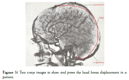
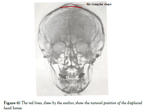
7-Two x-rays images to show and prove the head bones displacement in a patient [18]. (The red lines, drew by the author, show the natural position of the displaced head bones.)
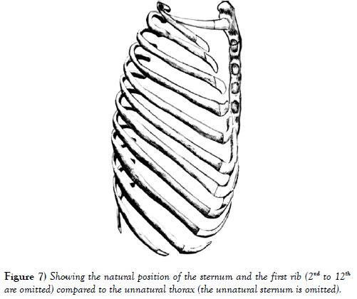
8- (Figure 7) is showing the natural position of the sternum and the first rib (2nd to 12th are omitted) compared to the unnatural thorax (the unnatural sternum is omitted).
9- The thymus gland is located behind the sternal angle and is exposed to the thorax pressures and might function with pressure received from the sternal angle. Note that it is especially large at puberty.
10- Head bones are attached via the sutural ligaments. If the displacement of the head bones is not anticipated, then retention of the cartilaginous joints between them is practically useless after the completion of the ossification (around 50s’).
11- The article focused on the problem regarding both males and females, but future researches would investigate the sex difference more closely.
Conclusion
Nowadays many abnormalities found with the patients, are being considered as mental or neurotic by the doctors because their roots are not known definitely. Studying these symptoms as body-based phenomena, would help the scientists provide the right route to discover and treat mental disorders. Furthermore, the psychologists would find the actual material bases for their treatments.
REFERENCES
- Santoro G, Wood MD, Merlo L, et al. The anatomic location of the soul from the heart, through the brain, to the whole body, and beyond: a journey through Western history, science, and philosophy. Neurosurg. 2009;65:633-43.
- Thirunavukarasu M. A utilitarian concept of manas and mental health. Indian J Psychiatry. 2011;53:99-110.
- Roudgari H. Ibn Sina or Abu Ali Sina (ابن سینا c. 980 –1037) is often known by his Latin name of Avicenna (ævɪˈsɛnə/). JIMC. 2018;1.
- Carlson ET, Simpson MM. The definition of mental illness: benjamin rush (1745-1813). Am J Psychiatry. 1964;121:209-14.
- McLeod SA. Wilhelm Wundt. Retrieved from https://www.simplypsychology.org/wundt.html. 2008.
- Safarini OA, Bordoni B. Anatomy, Thorax, Ribs. Treasure Island (FL): StatPearls2019.
- Drake RL, Vogl AW, Mitchell AWM, et al. Gray’s Atlas of Anatomy. second edition ed. Philadelphia: Churchill Livingstone; 2015.
- Gray H. Anatomy of the Human Body. Philadelphia: lea & febiger. 1918.
- Rehman A, Al Khalili Y. Neuroanatomy, Occipital Lobe. [Updated 2019 Jul 6]. In: StatPearls [Internet]. Treasure Island (FL): StatPearls Publishing; 2019 Jan-. Available from: https://www.ncbi.nlm.nih.gov/books/NBK544320/2019.
- Gott JA, Liley DT, Hobson JA. Towards a Functional Understanding of PGO Waves. Front Hum Neurosci. 2017;11:89.
- Berlucchi G, Vallar G. The history of the neurophysiology and neurology of the parietal lobe. Handb Clin Neurol. 2018;151:3-30.
- Klingner CM, Witte OW. Somatosensory deficits. Handb Clin Neurol. 2018;151:185-206.
- Binder JR. Current Controversies on Wernicke's Area and its Role in Language. Curr Neurol Neurosci. 2017;17(8):58.
- Jawabri K, Sharma S. Physiology, Cerebral Cortex Functions. In: StatPearls [Internet]. Treasure Island (FL): StatPearls Publishing; 2019 Jan-. Available from: https://www.ncbi.nlm.nih.gov/books/NBK538496/.
- Jin SW, Sim KB, Kim SD. Development and Growth of the Normal Cranial Vault : An Embryologic Review. Journal of Korean Neurosurgical Society. 2016;59:192-6.
- Bruner E, Amano H, de la Cuetara JM, et al. The brain and the braincase: a spatial analysis on the Wicke, Lothar, et al. Atlas Der Röntgenanatomie. München: Urban und Schwarzenberg, 1977. midsagittal profile in adult humans. J anat. 2015;227:268-76.
- Sun Z, Lee E, Herring SW. Cranial sutures and bones: growth and fusion in relation to masticatory strain. The anatomical record Part A, Discoveries in molecular, cellular, and evolutionary biology. 2004;276:150-61.
- Wicke, Lothar, et al. Atlas Der Röntgenanatomie. München: Urban und Schwarzenberg. 1977.


