Accessory Belly of the Rectus Femoris Muscle A Case Report
2 Faculty for Anatomical Sciences, Lincoln Memorial University- DeBusk College of Osteopathic Medicine, Harrogate, TN, 37752, USA
Received: 19-May-2022, Manuscript No. ijav-22-4972; Editor assigned: 20-May-2022, Pre QC No. ijav-22-4972 (PQ); Accepted Date: Jun 06, 2022; Reviewed: 03-Jun-2022 QC No. ijav-22-4972; Revised: 06-Jun-2022, Manuscript No. ijav-22-4972 (R); Published: 13-Jun-2022, DOI: 10.37532/1308-4038.15(6).200
This open-access article is distributed under the terms of the Creative Commons Attribution Non-Commercial License (CC BY-NC) (http://creativecommons.org/licenses/by-nc/4.0/), which permits reuse, distribution and reproduction of the article, provided that the original work is properly cited and the reuse is restricted to noncommercial purposes. For commercial reuse, contact reprints@pulsus.com
Abstract
Accounting for variability in human morphology reduces iatrogenic injury and leads to positive clinical outcomes. Variations in the quadriceps femoris muscle group, including accessory muscular components, fascicular orientation, and relationships to the patellar tendon have been the focus of many research studies. Most reports describing variability associated with the rectus femoris muscle have closely examined common but underappreciated accessory muscular slips closer to the hip joint. Here we describe a significant accessory rectus femoris muscle belly in a 103-year-old white male donor discovered during anatomical dissection. The supernumerary muscle belly shared an origin from the ilium with the rest of the rectus femoris muscle and coursed through most of the thigh before blending with the distal vastus lateralis muscle. This finding prompted further investigation and a review of the literature.
Keywords
Rectus femoris muscle; Quadriceps femoris variation; Muscle variation
Introduction
Supernumerary and non-canonical muscle components are commonly encountered in the study of human anatomy but are often underleveraged at the level of clinical application. Many variable or atypical muscle morphologies have risen to the level of clinical consideration. Accessory muscular components of the forearm may interrupt normal contractile activity, resulting in pain [1]. An additional muscle in the lateral compartment of the leg, peroneus quartus, may contribute to swelling and dysfunction at the ankle joint [2]. Pain derived from an extensor digitorum brevis or “le muscle manieux” may be mistaken for a ganglion cyst on the dorsum of the hand [3]. Symptoms arising from these variations are frequently misdiagnosed, which can result in undue pain and superfluous healthcare costs for patients. Knowledge of the potential for variability can also inform other areas in the clinical arena, notably muscular flaps, employed as either local pedicle flaps or free muscle transfers. Utilizing musculature in this way has proven useful in managing amputations, chronic ulceration, and prior graft infections [4-6]. Effectively describing and cataloging natural anatomical variation serves to strengthen clinician awareness and may contribute to alternative treatment options during clinical planning.
CASE REPORT
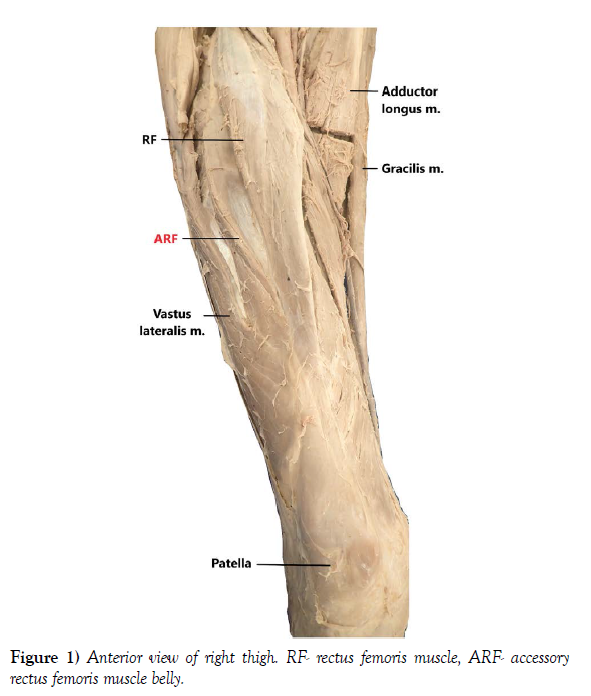
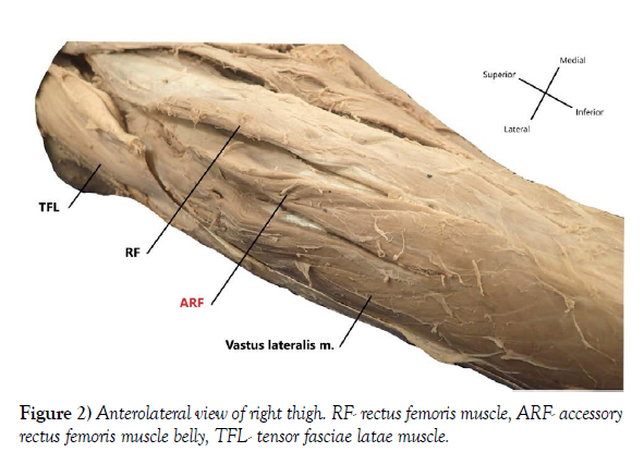
During a routine dissection of a 103-year-old white male formalin-fixed whole-body donor, an additional (third) rectus femoris muscle belly was found fusing distally with the vastus lateralis muscle (Figures 1 and 2). In situ, the additional muscle belly was notably situated between the vastus lateralis muscle and rectus femoris muscle, with most of the mass situated directly proximal to the origin of the patellar tendon. The accessory belly was found to share an origin with the rectus femoris muscle, immediately arising from the anterior inferior iliac spine. The proximal portion of the accessory muscle belly was broad, flat, and tendinous, although the muscle’s width and crosssectional area increased significantly as it blended with the vastus lateralis muscle (Figure 3). The muscle’s trajectory and relationships were carefully dissected until completely revealed.
Discussion
Traditionally, the rectus femoris muscle is described as a bipennate fusiform muscle with two heads, a thick round head arising from the anterior inferior iliac spine and a flat broad head arising from a groove superior to the acetabulum [7]. A review of the literature uncovered several descriptions of the variability discovered in the rectus femoris muscle. Tubbs et al. found the rectus femoris muscle to have a third head in 80 of 96 lower extremities (83%); however, this finding on average was limited to a small 2-4 cm head attached to the iliofemoral ligament and tendon of gluteus minimus muscle [8]. One report describes two unilaterally foreshortened rectus femoris muscles, prematurely narrowing to a thin tendon before joining the confluence of the patellar tendon [9]. Another case report identified a bilateral accessory head emerging from the intertrochanteric line before blending with the rectus femoris muscle in the distal thigh [10]. In a study of 10 cadaveric specimens, Becker et al. reported that 50% had fascicles of the vastus lateralis muscle inserting into the distal rectus femoris tendon, a morphology also described by Macalister in 1875 [11,12]. Similarly, in a Japanese sample of 50 lower extremities (25 male cadavers), Mori found that the “terminal” rectus femoris tendon was fused with the vastus lateralis muscle in 8% of cases and with the fascia of rectus femoris in 12% of cases [13]. Gruber described a rare slip arising from the periphery of the acetabulum and blending with the vastus lateralis muscle [14].
Results of the literature review demonstrated wide variability in rectus femoris muscle morphology, particularly compared to other muscle groups/ compartments. Unanticipated rectus femoris muscle morphology may negatively affect clinical interventions, particularly in cases where muscular flaps may be utilized. Also, significant asymmetrical muscular contributions to the quadriceps femoris group’s tendon may result in patellar instability, interfering with recovery following total knee arthroplasties [15]. Future directions for this research should include larger, more inclusive prevalence studies to establish expectations for variability in form, including interdemographic differences. Additionally, studies investigating variability detectable in medical imaging may encourage clinical integration. Bringing sufficient awareness to anomalous muscular forms may empower clinicians with the ability to better plan procedures, resulting in improved patient outcomes.
Acknowledgements
The authors would like to thank the body donors for willingly offering their remains for the education of future healthcare practitioners and the advancement of scientific knowledge. The authors would also like to extend our gratitude to the Edward via College of Osteopathic Medicine and the DeBusk College of Osteopathic Medicine at Lincoln Memorial University for supporting anatomical research.
REFERENCES
- Ryu JY, Watson K. SSMB Syndrome. Symptomatic Supernumerary Muscle Belly Syndrome. Clin Orthop Relat Res. 1987; 216, 195-202.
- Martinelli B, Bernobi S. Peroneus Quartus Muscle and Ankle Pain. J Foot Ankle Surg. 2002; 8(3), 223-25.
- Coudert X, Deghrar A, Lavarde G et al. Supernumerary Muscle on the Dorsal Surface of the Hand. A Case Report. Ann Chir Main Memb Super. 1993; 12(3), 230-33.
- Shenaq SM, Krouskop T, Stal S et al. Salvage of Amputation Stumps by Secondary Reconstruction Utilizing Microsurgical Free-Tissue Transfer. Plast Reconstr Surg. 1987; 79(6), 861-70.
- Dunn RM, Fudem GM, Walton RL et al. Free Flap Valvular Transplantation for Refractory Venous Ulceration. J Vasc Surg. 1994; 19(3), 525-31.
- Meland NB, Arnold PG, Pairolero PC et al. Muscle-flap Coverage for Infected Peripheral Vascular Prostheses. Plast Reconstr Surg. 1994; 93(5), 1005-11.
- Standring S. Gray’s Anatomy: The Anatomical Basis of Clinical Practice 14th ed. Churchill Livingstone El Sevier. 2008; 1373.
- Tubbs RS, Stetler Jr W, Savage AJ et al. Does a Third Head of the Rectus Femoris Muscle Exist? Folia Morphol. 2006; 65(4), 377-80.
- Stringer MD, Kano M, Fausett C, Samalia L. Unilateral Short Rectus Femoris Muscle Belly. Int J Anat Var. 2012; 5, 56-58.
- Tubbs RS, Salter G, Oakes JW et al. Femoral Head of the Rectus Femoris Muscle. Clin Anat. 2004; 17, 276-78.
- Becker I, Baxer GD, Woodley SJ et al. The Vastus Lateralis Muscle: An Anatomical Investigation. Clin Anat. 2010; 23, 575-85.
- Macalister A. Observations on Muscular Anomalies in the Human Anatomy. Third series with a Catalogue of the Principal Muscular Variations Hitherto Published. Trans Roy Irish Acad Sci. 1875; 25, 1-130.
- Mori M. Statistics on the Musculature of the Japanese. Okajimas Fol Anat Jap. 1964; 40, 282-3.
- Gruber W. Ein Musculus Rectus Femoris Accessories. Arch Path Anat Physiol Klin Med. 1888; 114, 367-8.
- Waligora AC, Johanson, NA, Hirsch BE et al. Clinical Anatomy of the Quadriceps Femoris and Extensor Apparatus of the Knee. Clin Orthop Relat Res. 2009; 467(12), 3297-306.
Indexed at, Google Scholar, Crossref
Indexed at, Google Scholar, Crossref
Indexed at, Google Scholar, Crossref
Indexed at, Google Scholar, Crossref
Indexed at, Google Scholar, Crossref
Indexed at, Google Scholar, Crossref
Indexed at, Google Scholar, Crossref
Indexed at, Google Scholar, Crossref
Indexed at, Google Scholar, Crossref
Indexed at, Google Scholar, Crossref


