Accessory renal artery supplying suprarenal gland and gonad - its clinical importance and embryological basis
Received: 01-Mar-2021 Accepted Date: Mar 22, 2021; Published: 29-Mar-2021, DOI: 10.37532/1308-4038.14(3).186-187
Citation: Jana R, Arya M. Accessory renal artery supplying suprarenal gland and gonad – its clinical importance and embryological basis. Int J Anat Var. 2021;14(3):72-73.
This open-access article is distributed under the terms of the Creative Commons Attribution Non-Commercial License (CC BY-NC) (http://creativecommons.org/licenses/by-nc/4.0/), which permits reuse, distribution and reproduction of the article, provided that the original work is properly cited and the reuse is restricted to noncommercial purposes. For commercial reuse, contact reprints@pulsus.com
Abstract
Renal transplantation is quiet impressive number as it is a treatment of choice for chronic kidney diseases so knowledge of variations in blood supply of kidney, suprarenal gland and gonad during surgical intervention, renal vascular imaging and understanding of medical problem related with kidney diseases. During routine cadaveric study we came across with multiple variations in blood supply to the kidney, suprarenal gland and gonad in a 65 years old male formaldehyde fixed embalmed body. The right kidney was supplied by an accessory renal artery along with main renal artery. Accessory renal artery, which was also supplying supra renal gland and gonad through inferior suprarenal and gonadal artery respectively. Variant blood supply to suprarenal gland, kidney and gonad is important during surgery, interventional radiography and anatomical point of view because it can affect the orientation of the surgeon during laparoscopic adrenalectomy, kidney transplantation, renal vascular imaging to prevent unwanted complications.
Keywords
Accessory renal artery, Hydronephrosis, Suprarenal gland, Inferior suprarenal artery, Gonadal artery
Introduction
Knowledge of abnormal blood supply to kidney, suprarenal gland and gonad is important as day by day number of surgeries is increasing and new methods of surgery are involved to treat the kidney diseases and suprarenal endoscopic surgeries as both suprarenal glands are highly vascular organs and extensively supplied by three sets of blood vessels i.e, superior, middle and inferior suprarenal arteries from inferior phrenic, abdominal aorta and renal arteries respectively. In seventy percent individual kidney is supplied by single renal artery and thirty percent cases is supplied by accessory renal artery [1]. Renal transplantation is the treatment of choice for advanced chronic renal failure. Chronic kidney disease (CKD) encompasses a spectrum of complex pathophysiological processes associated with abnormal kidney function, and a progressive decline in glomerular filtration rate (GFR) [2] and recently an impressive no of renal transplantation is going very high, due to which, surgeons, medical practitioner , radiologist and anatomists should be aware of the variations of blood supply of the organ to understand the etiopathogenesis of hydronephrosis, hypertension, unexplained infertility and to prevent unexpected operative & post-operative bleedings due to renal surgery.
Case Report
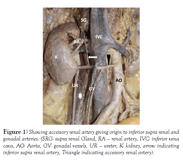
During dissection of posterior abdominal wall we observed right kidney is supplied by right renal artery and accessory renal artery. The accessory renal artery was originated from ventral surface of the abdominal aorta at the level of body of second lumbar vertebra. In the present case it was one more ventral branches of abdominal aorta, after taking origin it was passing to the right, over the inferior vein cava (IVC) and got sandwiched between Right gonadal vein and IVC (Figure 1). Subsequently it was crossing over pelvis of right ureter and reached to the lower end of the hilum of right kidney. The accessory renal artery supplied the lower pole of the right kidney and right suprarenal gland. The inferior supra renal artery was arising at the midway between the right gonadal vein and ureter and ascended vertically over the right renal vein and artery. The right gonadal artery also originated from accessory renal artery in front of IVC and descended down medial to the gonadal vein (Figure 1). Right gonadal vein ascended lateral to gonadal artery and was draining into right renal vein after crossing over accessory renal artery (Figure 1). When we dissected the suprarenal gland to find its vascular supply it was observed that middle suprarenal artery was branch of proximal part of the right main renal artery (Figure 1). And superior supra renal artery was originated as usual from inferior phrenic nerve. The venous drainage pattern was normal.
Figure 1) Showing accessory renal artery giving origin to inferior supra renal and gonadal arteries. (SRG- supra renal Gland, RA – renal artery, IVC- inferior vena cava, AO- Aorta, GV- gonadal vessels, UR – ureter, K- kidney, arrow indicating inferior supra renal artery, Triangle indicating accessory renal artery).
Discussion
Approximately in seventy percent of individuals the kidney is supplied by a single renal artery and it varies in level of origin, obliquity in its extrarenal course each renal artery gives off one or more inferior suprarenal arteries, a branch to the ureter. Accessory renal arteries are common in approximately thirty percent of individuals, and usually arise from the aorta above or below the main renal artery and follow it to the renal hilum. They are regarded as persistent embryonic lateral splanchnic arteries. Accessory vessels to the inferior pole cross anterior to the ureter and maybe obstructing the ureter, thus causing hydronephrosis. Rarely, accessory renal arteries may arise from the coeliac or superior mesenteric arteries near the aortic bifurcation or from the common iliac arteries reported by [1]. The suprarenal glands, gonads and kidneys are vascularized by lateral branches of the descending aorta. The suprarenal gland formed in the posterior abdominal wall between the sixth and twelfth thoracic segment and vascularized by a pair of upper lumbar lateral aortic branches reported by [3]. And also receives blood supply from renal and inferior phrenic artery.
Ranade AV. et al, (2007) revealed the arching pattern of left gonadal artery over left renal vein with two renal arteries and has mentioned that aberrant origin and abnormal course may cause compression of renal vein can lead to renal hypertension and varicocele. Variation in vasculature of kidney, supra renal gland and gonad is very important to the urologist, radiologist (for renal angiography) and can be a possible causative factor of idiopathic varicoceleor orthostatic albuminuria [4].
A study was conducted by Adebisi SS and Singh SP (2000) reported sixty five right and fifty eight left testicular arteries and ten right and eleven left ovarian arteries in eighty two cadavers (sixty eight males and fourteen females) and observed that twenty one right and nineteen left testicular arteries arched over renal vein. In five cases both right and left testicular artery arose from single stem, seven left testicular arteries sprung over renal vein and only three left ovarian arteries arched over renal vein. None of them had originated from accessory renal artery [5]. Aristotle S, et al. (2013) revealed in their study with thirty gross kidneys, out of which two cases, accessory renal artery gave origin to inferior supra renal gland but no artery from accessory renal artery supplied the gonad [6]. Das S.(2008) observed a left renal artery originated from aorta at the level of second lumbar vertebra and divided into two branches and upper one supplying the suprarenal gland and lower one supplying to the left kidney [7].
Conclusion
After extensive literature search no case was found to show accessory renal artery supplying the two important glands of the body; the supra renal artery and gonad. In the present study accessory renal artery a lateral branch of abdominal aorta at the second lumbar vertebral level was compressed by terminal part of right gonadal vein and near hilum was crossing over the pelvis of right ureter can be a cause of unexplained case of hydronephrosis. Most of the cases the gonadal vessels originating from aorta and draining into inferior vena cava (IVC) making an acute angle but in the present case right gonadal vein is draining at right angle to right renal vein instead of into inferior vena cava (IVC) so it may be the cause of varicocele in right gonad and may be one of the causes of unexplained infertility. So knowledge of aberrant blood supply to the kidney, supra renal gland and gonad is important during renal transplant, reno-vascular aneurysm repair, interventional radiologic procedures and urological operations to prevent and control unwanted complications.
Acknowledgements
Authors would like to thank Dr. N. A. Priyadharshini and D. Narayanan for helping during dissection.
REFERENCES
- Standring S. Gray's Anatomy the Anatomical Basis of Clinical Practice. 40th Edition, London, UK; Churchill Livingstone Elsevier. 2008;74:1231-1232.
- Fauci AS. Harrison’s Principles of Internal Medicine. 17th Edition. New York, McGraw hill. 2008;1776-1781.
- Schoenwolf GC. Larsen’s Human Embryology. 4th Edition. London, UK; Churchill Livingstone Elsevier. 2009;410-411.
- Ranade AV, Rai R, Prahbu LV, et al. Arched left gonadal artery over the left renal vein associated with double left renal artery. Singapore Med J. 2007; 48:332-334.
- Adebisi SS, Singh SP. Anomalous Gonadal Arteries in Relation to the Renal Vein: A Preliminary Study in Nigerians. Niger J Surg Res. 2000;2:3-4.
- Aristotle S, Sundarapandian, FeliciaC. Anatomical Study of Variations in the Blood Supply of Kidneys. J Clini Diag Res. 2013;7:1555-1557.
- Das S. Anomalous renal arteries and its clinical implications. Bratisl Lek Listy. 2008;109:182-184.


