Accessory Subscapularis Muscle: A Case Report
Received: 24-Mar-2023, Manuscript No. ijav-23-6269; Editor assigned: 27-Mar-2023, Pre QC No. ijav-23-6269 (PQ); Accepted Date: Apr 14, 2023; Reviewed: 10-Apr-2023 QC No. ijav-23-6269; Revised: 14-Apr-2023, Manuscript No. ijav-23-6269 (R); Published: 21-Apr-2023, DOI: 10.37532/1308-4038.16(4).251
Citation: Kite T, Carpenetti TL. Accessory Subscapularis Muscle: A Case Report. Int J Anat Var. 2023;16(4):276-277.
This open-access article is distributed under the terms of the Creative Commons Attribution Non-Commercial License (CC BY-NC) (http://creativecommons.org/licenses/by-nc/4.0/), which permits reuse, distribution and reproduction of the article, provided that the original work is properly cited and the reuse is restricted to noncommercial purposes. For commercial reuse, contact reprints@pulsus.com
Abstract
Variations of the subscapularis muscle have been sparsely reported in the literature. A classification scheme is lacking and may be a contributing factor. This variation may facilitate clinical syndromes presenting with sensory and motor dysfunction of the axillary nerve distribution, and vascular dysfunction of the posterior circumflex humeral artery. Furthermore, variations of this muscle may complicate surgical approaches to the glenohumeral joint. Routine dissection of the axilla was performed during which muscle fibers arising from the anterior aspect of the subscapularis muscle were found to give rise to an independent muscle band. The muscle band was attached by an independent tendinous insertion on the lesser tubercule of the humeral head. The muscle variation formed a slip around the axillary nerve and was noted bilaterally.
Keywords
Accessory subscapularis muscle, Quadrangular space syndrome, Axillary nerve, Subscapular muscle, Cadaver case report
INTRODUCTION
The subscapularis muscle is situated within the costal surface of the scapula and is a major constituent of the posterior axillary wall. The subscapularis muscle functions primarily in internal rotation and adduction of the arm. Muscle variants are thought to be exceedingly rare, with few reported cases [6]. Even more rare are reports of variations bilaterally. However, in a recent meta-analysis of 2,166 upper limbs, 1,079 were noted to contain the presence of an accessory subscapularis muscle [6]. Prior to this report, this variant had only been reported in the literature on a few occasions. The naming of subscapularis muscle variants is inconsistent in the literature. They have been described in the literature as follows: SCC minor, subscapulocapsularis muscle, scapulohumeral muscle, axillary slip of the subscapularis muscle [6], infraglenoidalis muscle [10]. The discrepant nature of such terminology may lead to misinterpretation of muscle variants. Not only is this variation of anatomical significance, but it may also underly significant clinical findings. Accessory variations of the muscle have been reported to involve the axillary nerve [1-9]. Axillary nerve involvement is clinically relevant, and complications are described in the literature as quadrangular space syndrome (QSS). Individuals who possess this variant may be predisposed to pain, paresthesia, and hyperesthesia over the distribution of the axillary nerve [1]. In extreme cases, overt motor dysfunction may occur, affecting abduction beyond 15 degrees and contributing to glenohumeral joint instability. In addition to the axillary slip variant, the accessory subscapularis muscle (ASM) has been described as varying with respect to the number and location of tendinous insertions on the superior humerus [9], and presence of accessory muscle morphology [1]. The ASM may be more common than reported, playing a more fundamental role in neuropathic pain syndromes of the upper extremity. Here we report a case of bilateral ASM arising from the lateral border of the subscapularis muscle involving the axillary nerve, most closely mimicking the “axillary slip” classification. The pattern reported is most consistent with the findings of Bresich and Krause [1,5]. While the ASM may be more common than thought, the bilateral nature described in this report demonstrates an exceedingly rare type of variation. This report not only enhances existing anatomical knowledge but could prove valuable for medical professionals who utilize this regional anatomy for the workup, diagnosis, and treatment of sensory and motor syndromes affecting the upper extremity.
CASE REPORT
Identification of an ASM was noted after routine dissection of the axilla and proximal humerus. The dissection was completed by first-year medical students according to Grant’s Dissector [2]. The donor was an 88-year-old Caucasian male without obvious trauma to the axilla or proximal humerus.
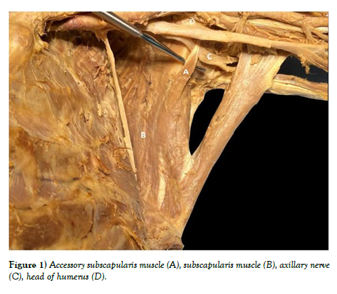
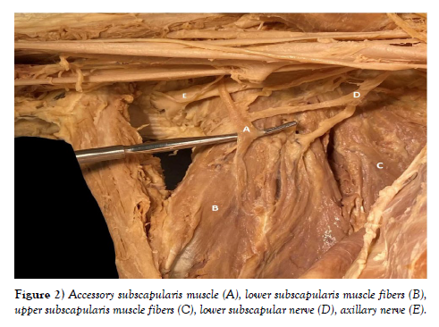
Gross dissection of the left axilla revealed normal anatomy of the radial and axillary nerves, with the radial nerve entering the triangular interval and the axillary nerve entering the quadrangular space. Brachial plexus morphology was normal on the right upper limb with respect to the posterior cord giving rise to upper and lower subscapular nerve branches visualized in Figure 1. However, due to limitations of the dissection, upper and lower subscapular nerves were unable to be visualized in the left upper extremity (Figure 2). Coexisting variation in the brachial plexus has been noted previously with subscapularis muscle variants [1]. The subscapularis muscle was attached to the anterior costal surface of the scapula with a tendinous insertion on the lesser tubercle of the humeral head. The accessory muscle fibers arose from the anterior aspect of the subscapularis muscle giving rise to an independent muscle band and tendinous insertion. This separate muscle belly created a slip over the axillary nerve prior to its branching in the quadrangular space. On the right, the width of the muscle band measured 0.9 mm, the width at the myotendinous junction 0.3 mm, and the length from origin at the lateral scapula to the lesser tubercle was 3.8 mm (Figure 1). From the lateral border of the scapula (the approximate location of its origin) the left sided accessory muscle measured 4.9 mm to its insertion on the lesser tubercle. The width measured 1.2 mm at its origin and 0.7 mm at the myotendinous junction (Figure 2).
DISCUSSION
It was previously believed that the subscapularis muscle was devoid of variation [8]. Our findings, along with the data presented by others, refute this claim. In fact, the ASM may be more prevalent than previously thought. The origins of the muscle fibers are many, generally thought to arise from two or three separate muscular attachments on the subscapular fossa [6]. Innervation is traditionally described as being derived from two branches of the posterior cord of the brachial plexus. The upper and lower subscapular nerves are thought to provide innervation to most of the muscle fibers, however considerable variation of these branches has been reported. A proposed accessory subscapular nerve has also been described infrequently; however, we did not note its presence in our donor [6]. Due to dissection limitations, a confident assessment of the presence of an accessory subscapularis nerve was not possible. Additionally, relationships between variability of the subscapularis muscle and developmental anomalies of the brachial plexus have been noted [1,7]. While our report did not produce these findings, it may be clinically relevant to describe the nature of subscapularis muscle variants and association with variability in the brachial plexus, specifically with respect to the posterior cord.
The subscapularis is the most powerful muscle of the rotator cuff. The anatomical nature and functional demands of the rotator cuff make this one of the most injured structures in the body [5]. As such, it is pertinent to have accurate descriptions of the regional anatomy including morphological variants. Arthroscopic and open repairs of the rotator cuff require precise knowledge of tendinous insertions. Additionally, breast tissue resection and axillary lymph node dissection procedures must account for the variable nature of the axilla [3]. Repair of myotendinous ruptures may not be adequately addressed if the possibility of anatomical variation is not considered. Likewise, the stability of the shoulder joint is a function of its myotendinous sling. Reconstruction in the setting of a glenohumeral joint replacement relies on addressing all possible tendinous attachments. Furthermore, the pattern of variation noted in our donor is one which is functionally associated with a predisposition to Quadrangular Space Syndrome. Breisch and Pires both described similar variants, where an accessory muscle band arises from the anterior aspect of the subscapularis muscle with an independent tendinous insertion onto the lesser tubercle of the humerus [1,8]. More recent investigations report that this so called “axillary arch” may be the most common variable pattern of the subscapularis muscle [6]. The significance of the axillary arch is the entrapment of the axillary nerve as it courses towards its normal anatomic distribution. This primarily affects the deltoid and teres minor muscles, resulting in diminished abduction and external rotation of the arm. Another possibility is the compression of the posterior circumflex humeral artery. Given the nature of its course paralleling the axillary nerve, compression leading to vaso-occlusion, aneurysmal formation, and reduced circulation may occur [3].
A definitive classification scheme for subscapularis variants does not currently exist. Kameda attempted to create a three-tier classification system with respect to innervation and relationship to the brachial plexus [4]. However, this classification scheme does not account for the variable nature of tendinous insertions such as those described in Zielinska’s report [9]. Furthermore, Breisch and Krause reported an exceptionally rare bilateral axillary arch type variant with respect to the brachial plexus, like our findings [1,5]. The existence of multiple muscle bands has also been reported by other investigators [9,10]. Zielinska’s case was extremely unique in that four tendinous insertions were noted, with three attaching to the coracoid process and one to the lesser tubercle. A separate report by this group attempted a classification system based on such tendinous insertions [9]. To establish a universal classification system, these variations, each these factors must be considered. Another point of confusion may be the existence of an axillary arch muscle, reported as Langer’s muscle which arises from muscle fibers of the latissimus dorsi [9]. Care must be taken when dissecting and classifying these variations to avoid misinterpretation of the muscular origin.
We speculate that the phenomenon of axillary nerve compression by an accessory subscapularis muscle may be underdiagnosed as an etiology of quadrangular space syndrome. However, physical signs and symptoms can be confused with that of thoracic outlet syndrome (TOS), and for that reason this condition has been dubbed a TOS mimicker [3]. Only through the collection of more data can we achieve a more accurate estimation of its prevalence in the population.
Furthermore, through our literature review process we have found that the subscapularis muscle is in fact a structure of variable nature. We believe that the heterogenous nature of the variations themselves, along with an inconsistent nomenclature, has complicated our understanding of the prevalence of these variants in the literature. Future work should center on defining a classification scheme and compiling the results for other researchers.
ACKNOWLEDGEMENT
The authors sincerely thank those who donated their bodies to science so that anatomical research could be performed. Results from such research can potentially increase mankind’s overall knowledge that can then improve patient care. Therefore, these donors and their families deserve our highest gratitude. Additionally, we would like to thank the Virginia State Anatomical Program for their facilitation in procuring cadaveric donors for our curriculum.
REFERENCES
- Breisch EA. A rare human variation: The relationship of the axillary and inferior subscapular nerves to an accessory subscapularis muscle. Anat Rec. 1986 Nov; 216(3):440-442.
- Detton A. Grant’s dissector: Sixteenth Edition. WoltersKluwer. 2017; 23-42.
- Hangge PT, Breen I, Albadawi H, Knuttinen MG, Naidu SG, et al. Quadrilateral space syndrome: diagnosis and clinical management. J Clin Med Res. 2018; 7(4):102-107.
- Kameda Y. An anomalous muscle (accessory subscapularis teres latissimus muscle) in the axilla penetrating the brachial plexus in man. Acta Anat (basel) 1976; 96:513-533.
- Krause DA, Youdas JW. Bilateral presence of a variant subscapularis muscle. Int J Anat Var. 2017; 10(4):79-80.
- Mann MR, Plutecki D, Janda P, Pękala J, Malinowski K, et al. The subscapularis muscle‐a meta‐analysis of its variations, prevalence, and anatomy. Clin Anat. 2023; 36(3):527-541.
- Pillay M, Jacob SM. Bilateral presence of axillary arch muscle passing through the posterior cord of the brachial plexus. Int. J. Morphol., 27(4):1047-1050, 2009.
- Pires LAS, Souza CFC, Teixeira AR, Leite TFO, Babinski MA, et al. Accessory subscapularis muscle–A forgotten variation?. Morphologie. 2017; 101(333):101-104.
- Zielinska N, Tubbs RS, Podgórski M, Karauda P, Polguj M, et al. The subscapularis tendon: a proposed classification system. Ann Anat. 2021; 233:151-615.
- Zielinska N, Tubbs RS, Konschake M, Olewnik Ł. Unknown variant of the accessory subscapularis muscle?. Anatomical Science International, 97(1), 138-142.
Indexed at, Google Scholar, Crossref
Indexed at, Google Scholar, Crossref
Indexed at, Google Scholar, Crossref
Indexed at, Google Scholar, Crossref
Indexed at, Google Scholar, Crossref
Indexed at, Google Scholar, Crossref
Indexed at, Google Scholar, Crossref


