Agenesis of common iliac vein encroaching development of inferior vena cava
Padamjeet Panchal* and Harish Chaturvedi
Department of Anatomy, Veer Chandra Singh Garhwali Government Medical Sciences and Research Institute, Srinagar, Uttarakhand, India
- *Corresponding Author:
- Dr. Padamjeet Panchal
Department of Anatomy, Veer Chandra Singh Garhwali Govt. Med. Sci. & Research Inst., Srinagar (Pauri Garhwal) Uttarakhand, 246174, India
Tel: +91 (963) 9897460
E-mail: padampanchal@gmail.com
Date of Received: May 29th, 2012
Date of Accepted: March 25th, 2013
Published Online: June 1st, 2014
© Int J Anat Var (IJAV). 2014; 7: 21–23.
[ft_below_content] =>Keywords
deep vein thrombosis, external iliac vein, internal iliac vein, laparoscopic surgery, magnetic resonance angiography
Introduction
The inferior vena cava (IVC) conveys blood to the right atrium from all structures below the diaphragm. The inferior vena cava is constituted by the junction of two common iliac veins (CIV) anterior to the fifth lumbar vertebral body slightly on its right. The common iliac veins are formed by confluence of the external iliac and internal iliac veins. The external iliac veins (EIV) are the upward continuation of the femoral veins whereas internal iliac veins (IIV) are the union of various tributaries those correspond to the branches of internal iliac artery [1].
During 6th to 8th week of embryonic period, genesis of the IVC adopts complex process of the development, regression and anastomosis of 3 sets of paired veins: the postcardinal, subcardinal, and supracardinal veins [2]. Subsequently, IVC is converted to a unilateral right-sided system consisting of the postrenal, renal, prerenal, and hepatic segments from caudal to cranial [3,4]. During fetal life the venous drainage of the lower limb buds and pelvis is initially carried out by the right and left postcardinal veins, which runs dorsal to the mesonephric ridges. The early postcardinal veins communicate across the midline via an inter-postcardinal anastomosis. This remains as an oblique transverse channel between the iliac veins, and becomes the major part of the definitive left CIV. The IVC is formed from below by the convergence of two CIV and the right postcardinal vein [1].
The spectrum of anatomical variations in the formation and course of IVC has been well described [5,6]. But the variant infrarenal IVC is believed to have a prevalence of less than 2% in the normal population and with complete absence of the IVC occurring in 0.3% of healthy subjects [6,7]. The most common major venous variants are transposition of the IVC, duplication of IVC, circumaortic renal collar and retroaortic renal vein [7].
Case Report
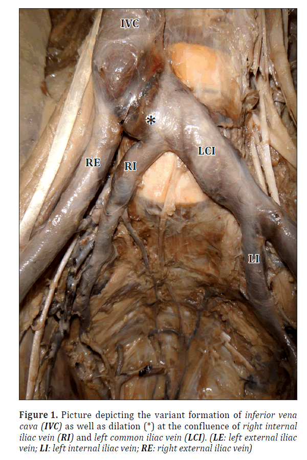
During a routine dissection of the retroperitoneal region of an adult male cadaver fixed in 10% formalin, we came across a variant drainage pattern of formation of inferior vena cava and unilateral absence of common iliac vein. Dissection was carried out using Cunningham’s Manual of Practical Anatomy. We have observed a rare formation of IVC by two channels, right EIV and left CIV. The right CIV was found to be absent due to affluence of right external iliac and internal iliac veins. The latter was rushing cranially and medially, crossing the median plane and draining into the left CIV. A significant dilation was exhibited at the junction of right iliac vein with left CIV (Figure 1). The right EIV was crossed anteriorly by right external iliac artery and ureter.
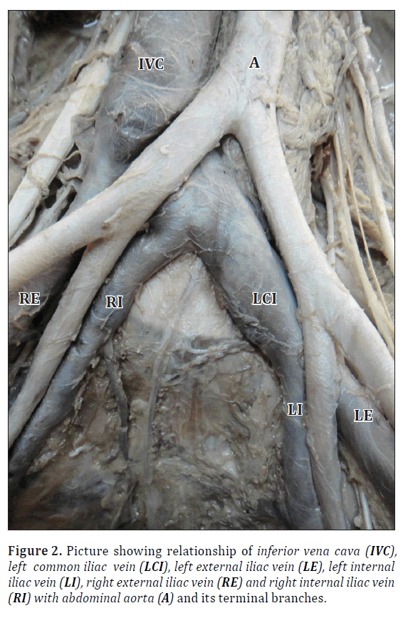
On contrary, the left CIV is formed by the confluence of left external and internal iliac veins. The CIV was running cranially and medially to join right EIV after receiving right IIV as its tributary, thereafter it crossed median plane posterior to the right common iliac artery (Figure 2). The left EIV was crossed anteriorly by left internal iliac artery and ureter.
The origin of internal iliac veins was formed bilaterally from contribution of an anterior and posterior venous tributaries which received drainage from visceral (vesical, prostatic and rectal) and parietal (gluteal and lateral sacral) veins, respectively. The median sacral vein drained into the left internal iliac vein. The right and left lateral sacral veins drained into the corresponding internal iliac veins. Other veins draining into the IVC did not show any variation in their drainage and course. The branching pattern of the abdominal aorta was as usual.
Discussion
Oto et al. and Cardinot et al. reported a case of right IIV draining into the left CIV. Our results are consistent with these reports [9,10]. Singh and Biswas conveyed that the regression of part of the right post cardinal vein and the oblique transverse anastomosis lead to bilateral agenesis of CIV [11]. Present case revealed the agenesis of right CIV only. This may be due to the segmental deterioration of right post cardinal vein caudal to the oblique transverse anastomosis after sprouting out of external iliac and internal iliac veins. Therefore, common iliac vein was not formed on right side resulting shifting of right IIV to left CIV owing to its remodeling. Alicioglu et al. supported the theory proposed by Milner (1980) of perinatal thrombosis as a causative factor that may trigger variation in formation of IVC [5].
Dissemination of such uncommon variation in the formation and course of IVC is helpful not only in preoperative planning of open retroperitoneal and laparoscopic abdominal surgeries but can also prevent any intraoperative inadvertent injuries to anomalous vein [13]. Furthermore, detailed knowledge of variations of veins is crucial in staging of abdominal neoplasm, in radiological interpretation of CT venography as well as therapeutic intervention like placing IVC filters, testicular vein embolization and sampling of renal and adrenal veins [14].
Ruggeri et al. considered IVC variants as a sufficient cause for development of deep vein thrombosis; they also hypothesized correlation between an IVC variants and thrombophilia in the pathogenesis of deep vein thrombosis [15]. Further investigation is needed to validate this hypothesis.
The interventional radiological procedures like CT venography, MR angiography are very sensitive in diagnosing the venous thrombosis. The rare variant of the IVC are not always recognized during the preoperative assessment of patient. This may increase the risk of intraoperative injury to the variants of retroperitoneal and pelvic venous channels, particularly during laparoscopic surgeries. Although the anatomical variants of pelvic and retroperitoneal vessels do not cause any functional damage but they can be relevant during surgery.
Acknowledgements
Our sincere thanks to Dr. Amal Rani Das, Head of Anatomy and all other staff members of Department, for their support us to carry out this work.
References
- Standring S, ed. Gray’s Anatomy. 40th Ed., London, Churchill Livingstone. 2005; 1075, 1219.
- Babaian RJ, Johnson DE. Major venous anomalies complicating retroperitoneal surgery. South Med J. 1979; 72: 1254–1258.
- Chuang VP, Mena CE, Hoskins PA. Congenital anomalies of the inferior vena cava: Review of embryogenesis and presentation of a simplified classification. Br J Radiol. 1974; 47: 206–213.
- Kellman GM, Alpern MB, Sandler MA, Craig BM. Computed tomography of vena caval anomalies with embryologic correlation. Radiographics. 1988; 8: 533–556.
- Alicioglu B, Kaplan M, Ege T. Absence of infrarenal inferior vena cava is not a congenital abnormality. Bratisl Lek Listy. 2009; 110: 304–306.
- Bapat G, Daly K, Razzaq R, Onwudike M. Unusual presentations of congenital absence of the inferior vena cava–Case Reports. Int J Angiol. 2003; 12: 48–50.
- Zhou W, Rosenberg W, Lumsden A, Li J. Successful surgical management of pelvic congestion and lower extremity swelling owing to absence of infrarenal inferior vena cava. Vascular. 2005; 13: 358–361.
- Meyer DR, Huggle H, Andresen R, Huppe T, Friedrich M. Segmental variants of the inferior vena cava – manifestations with embryological correlation in differentiation from secondary vena cava occlusion. Rontgenpraxis. 2001; 54: 101–113. (German)
- Oto A, Akpınar E, Surucu HS, Denk CC, Celik HH. Right internal iliac vein joining the left common iliac vein: case report demonstrated by CT angiography. Surg Radiol Anat. 2003; 25: 339–341.
- Cardinot T, Aragao AH, Babinski MA, Favorito LA. Rare variation in course and affluence of internal iliac vein due to its anatomical and surgical significance. Surg Radiol Anat. 2006; 28: 422–425.
- Biswas S, Singh R. Absence of the common iliac veins. Clin Anat. 2007; 20: 990–991.
- Obernosterer A, Aschauer M, Schnedl W, Lipp RW. Anomalies of the inferior vena cava in patients with iliac venous thrombosis. Ann Intern Med. 2002; 136: 37–41.
- Mathews R, Smith PA, Fishman EK, Marshall FF. Anomalies of the inferior vena cava and renal veins: embryologic and surgical considerations. Urology. 1999; 53: 873–880.
- Trigaux JP, Vandroogenbroek S, De Wispelaere JF, Lacrosse M, Jamart J. Congenital anomalies of the inferior vena cava and left renal vein: evaluation with spiral CT. J Vasc Interv Radiol. 1998; 9: 339–345.
- Ruggeri M, Tosetto A, Castaman G, Rodeghiero F. Congenital absence of the inferior vena cava: Rare risk factor for idiopathic deep- vein thrombosis. Lancet. 2001; 357: 441.
Padamjeet Panchal* and Harish Chaturvedi
Department of Anatomy, Veer Chandra Singh Garhwali Government Medical Sciences and Research Institute, Srinagar, Uttarakhand, India
- *Corresponding Author:
- Dr. Padamjeet Panchal
Department of Anatomy, Veer Chandra Singh Garhwali Govt. Med. Sci. & Research Inst., Srinagar (Pauri Garhwal) Uttarakhand, 246174, India
Tel: +91 (963) 9897460
E-mail: padampanchal@gmail.com
Date of Received: May 29th, 2012
Date of Accepted: March 25th, 2013
Published Online: June 1st, 2014
© Int J Anat Var (IJAV). 2014; 7: 21–23.
Abstract
Anatomical variations of inferior vena cava and its tributaries are not uncommon during abdominal and pelvic surgeries. Among the variations of inferior vena cava the infrarenal type is rare. During the routine dissection of a male cadaver, such rare variation in formation of inferior vena cava was found associated with agenesis of common iliac vein. Although they may not be of functional importance but knowledge of such variations is essential for surgical point of view. Some variations are common and some are rare. It becomes essential to find out their structural and functional importance in the body. Unfortunately, they are missed during preoperative assessment of patients even in the era of advanced radiological interventions unless they are hazardous or altered any function. Therefore, developmental origin of these variants gets equal attention.
-Keywords
deep vein thrombosis, external iliac vein, internal iliac vein, laparoscopic surgery, magnetic resonance angiography
Introduction
The inferior vena cava (IVC) conveys blood to the right atrium from all structures below the diaphragm. The inferior vena cava is constituted by the junction of two common iliac veins (CIV) anterior to the fifth lumbar vertebral body slightly on its right. The common iliac veins are formed by confluence of the external iliac and internal iliac veins. The external iliac veins (EIV) are the upward continuation of the femoral veins whereas internal iliac veins (IIV) are the union of various tributaries those correspond to the branches of internal iliac artery [1].
During 6th to 8th week of embryonic period, genesis of the IVC adopts complex process of the development, regression and anastomosis of 3 sets of paired veins: the postcardinal, subcardinal, and supracardinal veins [2]. Subsequently, IVC is converted to a unilateral right-sided system consisting of the postrenal, renal, prerenal, and hepatic segments from caudal to cranial [3,4]. During fetal life the venous drainage of the lower limb buds and pelvis is initially carried out by the right and left postcardinal veins, which runs dorsal to the mesonephric ridges. The early postcardinal veins communicate across the midline via an inter-postcardinal anastomosis. This remains as an oblique transverse channel between the iliac veins, and becomes the major part of the definitive left CIV. The IVC is formed from below by the convergence of two CIV and the right postcardinal vein [1].
The spectrum of anatomical variations in the formation and course of IVC has been well described [5,6]. But the variant infrarenal IVC is believed to have a prevalence of less than 2% in the normal population and with complete absence of the IVC occurring in 0.3% of healthy subjects [6,7]. The most common major venous variants are transposition of the IVC, duplication of IVC, circumaortic renal collar and retroaortic renal vein [7].
Case Report
During a routine dissection of the retroperitoneal region of an adult male cadaver fixed in 10% formalin, we came across a variant drainage pattern of formation of inferior vena cava and unilateral absence of common iliac vein. Dissection was carried out using Cunningham’s Manual of Practical Anatomy. We have observed a rare formation of IVC by two channels, right EIV and left CIV. The right CIV was found to be absent due to affluence of right external iliac and internal iliac veins. The latter was rushing cranially and medially, crossing the median plane and draining into the left CIV. A significant dilation was exhibited at the junction of right iliac vein with left CIV (Figure 1). The right EIV was crossed anteriorly by right external iliac artery and ureter.
On contrary, the left CIV is formed by the confluence of left external and internal iliac veins. The CIV was running cranially and medially to join right EIV after receiving right IIV as its tributary, thereafter it crossed median plane posterior to the right common iliac artery (Figure 2). The left EIV was crossed anteriorly by left internal iliac artery and ureter.
The origin of internal iliac veins was formed bilaterally from contribution of an anterior and posterior venous tributaries which received drainage from visceral (vesical, prostatic and rectal) and parietal (gluteal and lateral sacral) veins, respectively. The median sacral vein drained into the left internal iliac vein. The right and left lateral sacral veins drained into the corresponding internal iliac veins. Other veins draining into the IVC did not show any variation in their drainage and course. The branching pattern of the abdominal aorta was as usual.
Discussion
Oto et al. and Cardinot et al. reported a case of right IIV draining into the left CIV. Our results are consistent with these reports [9,10]. Singh and Biswas conveyed that the regression of part of the right post cardinal vein and the oblique transverse anastomosis lead to bilateral agenesis of CIV [11]. Present case revealed the agenesis of right CIV only. This may be due to the segmental deterioration of right post cardinal vein caudal to the oblique transverse anastomosis after sprouting out of external iliac and internal iliac veins. Therefore, common iliac vein was not formed on right side resulting shifting of right IIV to left CIV owing to its remodeling. Alicioglu et al. supported the theory proposed by Milner (1980) of perinatal thrombosis as a causative factor that may trigger variation in formation of IVC [5].
Dissemination of such uncommon variation in the formation and course of IVC is helpful not only in preoperative planning of open retroperitoneal and laparoscopic abdominal surgeries but can also prevent any intraoperative inadvertent injuries to anomalous vein [13]. Furthermore, detailed knowledge of variations of veins is crucial in staging of abdominal neoplasm, in radiological interpretation of CT venography as well as therapeutic intervention like placing IVC filters, testicular vein embolization and sampling of renal and adrenal veins [14].
Ruggeri et al. considered IVC variants as a sufficient cause for development of deep vein thrombosis; they also hypothesized correlation between an IVC variants and thrombophilia in the pathogenesis of deep vein thrombosis [15]. Further investigation is needed to validate this hypothesis.
The interventional radiological procedures like CT venography, MR angiography are very sensitive in diagnosing the venous thrombosis. The rare variant of the IVC are not always recognized during the preoperative assessment of patient. This may increase the risk of intraoperative injury to the variants of retroperitoneal and pelvic venous channels, particularly during laparoscopic surgeries. Although the anatomical variants of pelvic and retroperitoneal vessels do not cause any functional damage but they can be relevant during surgery.
Acknowledgements
Our sincere thanks to Dr. Amal Rani Das, Head of Anatomy and all other staff members of Department, for their support us to carry out this work.
References
- Standring S, ed. Gray’s Anatomy. 40th Ed., London, Churchill Livingstone. 2005; 1075, 1219.
- Babaian RJ, Johnson DE. Major venous anomalies complicating retroperitoneal surgery. South Med J. 1979; 72: 1254–1258.
- Chuang VP, Mena CE, Hoskins PA. Congenital anomalies of the inferior vena cava: Review of embryogenesis and presentation of a simplified classification. Br J Radiol. 1974; 47: 206–213.
- Kellman GM, Alpern MB, Sandler MA, Craig BM. Computed tomography of vena caval anomalies with embryologic correlation. Radiographics. 1988; 8: 533–556.
- Alicioglu B, Kaplan M, Ege T. Absence of infrarenal inferior vena cava is not a congenital abnormality. Bratisl Lek Listy. 2009; 110: 304–306.
- Bapat G, Daly K, Razzaq R, Onwudike M. Unusual presentations of congenital absence of the inferior vena cava–Case Reports. Int J Angiol. 2003; 12: 48–50.
- Zhou W, Rosenberg W, Lumsden A, Li J. Successful surgical management of pelvic congestion and lower extremity swelling owing to absence of infrarenal inferior vena cava. Vascular. 2005; 13: 358–361.
- Meyer DR, Huggle H, Andresen R, Huppe T, Friedrich M. Segmental variants of the inferior vena cava – manifestations with embryological correlation in differentiation from secondary vena cava occlusion. Rontgenpraxis. 2001; 54: 101–113. (German)
- Oto A, Akpınar E, Surucu HS, Denk CC, Celik HH. Right internal iliac vein joining the left common iliac vein: case report demonstrated by CT angiography. Surg Radiol Anat. 2003; 25: 339–341.
- Cardinot T, Aragao AH, Babinski MA, Favorito LA. Rare variation in course and affluence of internal iliac vein due to its anatomical and surgical significance. Surg Radiol Anat. 2006; 28: 422–425.
- Biswas S, Singh R. Absence of the common iliac veins. Clin Anat. 2007; 20: 990–991.
- Obernosterer A, Aschauer M, Schnedl W, Lipp RW. Anomalies of the inferior vena cava in patients with iliac venous thrombosis. Ann Intern Med. 2002; 136: 37–41.
- Mathews R, Smith PA, Fishman EK, Marshall FF. Anomalies of the inferior vena cava and renal veins: embryologic and surgical considerations. Urology. 1999; 53: 873–880.
- Trigaux JP, Vandroogenbroek S, De Wispelaere JF, Lacrosse M, Jamart J. Congenital anomalies of the inferior vena cava and left renal vein: evaluation with spiral CT. J Vasc Interv Radiol. 1998; 9: 339–345.
- Ruggeri M, Tosetto A, Castaman G, Rodeghiero F. Congenital absence of the inferior vena cava: Rare risk factor for idiopathic deep- vein thrombosis. Lancet. 2001; 357: 441.


