Agenesis of thyroid isthmus associated with a rare variation of origin of superior laryngeal and cricothyroid arteries: Case report and review of the literature
2 Department of Surgery, B of UHC Point-G, Bamako, Mali
3 Department of Otorhinolaryngology, UHC Gabriel Touré, Bamako, Mali
Received: 29-Apr-2020 Accepted Date: Aug 11, 2020; Published: 18-Aug-2020, DOI: 10.37532/1308-4038.20.13.12
This open-access article is distributed under the terms of the Creative Commons Attribution Non-Commercial License (CC BY-NC) (http://creativecommons.org/licenses/by-nc/4.0/), which permits reuse, distribution and reproduction of the article, provided that the original work is properly cited and the reuse is restricted to noncommercial purposes. For commercial reuse, contact reprints@pulsus.com
Abstract
Agenesis of the thyroid isthmus was observed in an adult male cadaver during a routine dissection at the anatomy laboratory of the Faculty of Medicine and Odontostomatology (FMOS) in Bamako. The two thyroid lobes were completely separated from each other. The left lobe presented the pyramidal lobe. The supra and infra-isthmic arches were absent, the upper right and left thyroid arteries did not anastomose, as did the lower right and left thyroid arteries. On the right side, the superior laryngeal and crico-thyroid arteries were born by a common trunk which came from the external carotid artery by a common trunk with the lingual and superior thyroid arteries.
Keywords
Agenesis of the thyroid isthmus; Superior laryngeal artery; Crico-thyroid artery; External carotid artery.
Introduction
Located in the anterolateral part of the visceral compartment of the neck in front of the laryngo-tracheal axis, the thyroid gland is the largest of the endocrine glands. It is made up of two lateral lobes joined on the midline by a transverse parenchyma bridge which constitutes the thyroid isthmus [1].
A wide spectrum of abnormalities and anatomical variations can occur in the thyroid gland. Common abnormalities include the thyroglossal duct cyst and the persistent pyramidal lobe and rare abnormalities such as agenesis, hemiagenesis of the thyroid gland, aberrant thyroid glands can also be observed [2].
The superior laryngeal and crico-thyroid arteries originate from the superior thyroid artery [1]. Although variations in the origin of the superior laryngeal artery (SLA) are not very common, variations when present can acquire great importance in larynx surgical procedures such as partial laryngectomy, reconstructive surgery and laryngeal transplantation [3,4]. Knowledge of SLA variations can also be useful to clinicians for super-elective intra-arterial injection chemotherapy for larynx and hypopharynx cancers, as well as for successful radical neck dissection to minimize postoperative complications in bloodless surgery [5,6].
We report a case of thyroid isthmus agenesis associated with a variation in origin of the superior laryngeal and crico-thyroid arteries.
Results
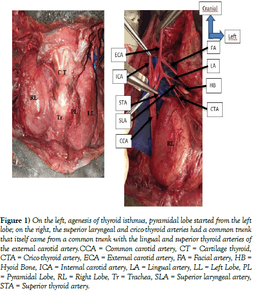
During a routine dissection in an adult male cadaver, at the anatomy laboratory of the Faculty of Medicine and Odontostomatology of Bamako, we observed agenesis of the thyroid isthmus, the two thyroid lobes were completely separate, independent of each other (Figure 1 left). There was no anastomosis between the superior thyroid arteries and between the lower thyroid arteries. The right lobe was 74 mm long and the left lobe was 71 mm long. The latter presented the pyramidal lobe. In this same corpse, on the right side, we observed that the superior laryngeal and crico-thyroid arteries were born from a common trunk which came from the external carotid artery by a common trunk with the lingual and superior thyroid arteries (Figure 1 right). The crico-thyroid artery gave descending branches for the anterior aspect of the right thyroid lobe.
Figure 1: On the left, agenesis of thyroid isthmus, pyramidal lobe started from the left lobe; on the right, the superior laryngeal and crico-thyroid arteries had a common trunk that itself came from a common trunk with the lingual and superior thyroid arteries of the external carotid artery.CCA = Common carotid artery, CT = Cartilage thyroid, CTA = Crico-thyroid artery, ECA = External carotid artery, FA = Facial artery, HB = Hyoid Bone, ICA = Internal carotid artery, LA = Lingual artery, LL = Left Lobe, PL = Pyramidal Lobe, RL = Right Lobe, Tr = Trachea, SLA = Superior laryngeal artery, STA = Superior thyroid artery.
Discussion
Agenesis of the thyroid isthmus is the complete and congenital absence of the thyroid isthmus as defined by Pastor et al. [7]. In their study, they reported isthmus agenesis of the thyroid gland with enlarged lobes in a Caucasian corpse. According to Gruber (cited by Testus and Latarjet), the incidence of agenesis of the isthmus is around 5% [8]. Marshall has documented variations in the gross structure of the thyroid gland in 60 children, ranging in age from a few weeks to 10 years, and the absence of an isthmus would be 10% in this group [9]. Ranade et al reported the absence of an isthmus in 35 of 105 cases (33%), including 8 female corpses [10]. According to the study by Braun et al., the isthmus was missing in 4 cases out of the 58 corpses studied [11].
Won and Chung reported that in 3% of the cases studied, the isthmus was absent and the lateral lobes of the thyroid were separated [12]. The incidence among North West Indians is reported to be 7.9% in the raw samples [13]. According to Dixit et al, in their study, the incidence was slightly higher at 14.6% [14]. In our case, agenesis of the thyroid isthmus was associated with the presence of a pyramidal lobe which was linked to the left thyroid lobe.
The absence of an isthmus is quite rare in humans [15]. Agenesis of the isthmus can be explained as an abnormality in embryological development. The adult thyroid gland has two types of endocrine cells, follicular and parafollicular cells or “C” cells, which are derived from two different families of embryological cells. The follicular cells come from the endodermal cells of the primitive pharynx and the parafollicular cells from the neural crest [16]. The thyroid gland begins to develop as a median thickening of the endoderm on the floor of the pharynx between the first and second pharyngeal pockets. This area later invagers to form the median diverticulum, which appears in the second half of the fourth week. This thyroid diverticulum develops in allometric proliferation, becoming a solid cellular cord called the thyroglossal canal. The duct develops caudally and forks to give rise to thyroid lobes and the isthmus. In parallel with its caudal growth, the cephalic end of the thyroglossal canal degenerates [17].
A high division of the thyroglossal canal can generate two independent thyroid lobes in the absence of an isthmus. The absence of the isthmus may be associated with other types of dysorganogenesis, such as the absence of a lobe or the presence of ectopic thyroid tissue [18].
Clinically, the diagnosis of agenesis of the isthmus can be made by scintigraphy, which can also be performed with an overload of TSH. The diagnosis can also be made using ultrasound, computed tomography (CT), magnetic resonance imaging (MRI) or during surgery. In asymptomatic patients with nodular goiters, fine needle aspiration biopsies and possibly immunohistochemistry tests are useful to support medical decision, but when agenesis is present, the importance of preoperative differentiation between Benign and malignant lesions is critical, given the surgical procedure and the possibility of impaired thyroid function [19]. When an image of the absence of an isthmus is observed, a differential diagnosis against the autonomous thyroid nodule, thyroiditis, primary carcinoma, neoplastic metastases and infiltrative diseases such as amyloidosis [7].
Concerning SLA, its most common origin is the upper thyroid artery, the range reported being from 68% to 92.7% [5,20-25]. According to Deavadas et all [24], in 5% of cases, the SLA came from the external carotid artery. This is more or less in agreement with the studies of Terayama et al. [5], Lang et al. [22], and Ozgur et al. [25], but differ from those of Rusu et al. [20] who reported a 32% higher incidence. There are also case reports of SLA from the external carotid artery. Muralimanju et al. [26] reported a case of left SLA originating from the external carotid artery. The SLA in the same case was born from the common carotid artery 2 cm before its bifurcation. There are rare reports of SLA from common trunks such as the linguofacial trunks [6] and that of a common trunk from the external carotid artery giving rise to SLA and lower thyroid arteries [27]. Motwani and Jhajhria [28] noticed a rare common trunk coming from the anterior surface of the right external carotid artery just above the carotid bifurcation. The common core was divided into five branches, that is to say infra-hyoid, superior laryngeal, superior thyroid, cricothyroid and sternocleidomastoid. In our case, the superior laryngeal and crico-thyroid arteries had a common trunk which came from the external carotid artery by a common trunk with the lingual and superior thyroid arteries. Such a variation has not been reported in the literature.
The varied origin of the superior thyroid artery and the ALS of the carotid arterial system considerably increases the possibility of their misidentification during surgery [21], particularly given that variations are not uncommon. Therefore, detailed knowledge of the variant anatomy will be useful in procedures such as partial laryngectomy, reconstructive laryngeal surgeries, as well as laryngeal transplantation [25]. Knowing a variant of the anatomy can also be useful during radical dissection of the neck and minimize postoperative complications in bloodless surgery [6]. In addition, ALS being the main artery of the larynx, it is used to administer chemotherapeutic drugs for the treatment of larynx cancers, because drugs administered by the supply artery directly to the tumor site can achieve a therapeutic effect more important [29]. A good detailed knowledge of SLA will also guarantee a correct decision and a safe approach before neuro-radiological procedures involving SLA [20].
Conclusion
Agenesis of the thyroid isthmus is a rare anatomical variation. Its incidence is difficult to determine, because in most cases, its discovery is fortuitous. The variations in origin of the superior laryngeal and crico-thyroid arteries are rare and important to know to avoid damaging them during surgical operations on the anterior region of the neck, more precisely on the larynx.
REFERENCES
- Bouchet A, Cuilleret J. Anatomie topographique, descriptive et fonctionnelle. Tome 2: le cou et le thorax. 2ème édition.Paris. SIMEP.1991.
- Devi Sankar K, SharmilaBanu P, Susan PJ, et al. Agenesis of isthmus of thyroid gland with bilateral lavator glandulae thyroideae. IJAV.2009;2:29-30.
- Anthony JP, Argenta P, Trabulsy PP, et al. The arterial anatomy of larynx transplantation: microsurgical revascularization of the larynx. Clin Anat. 1996;9:155-9.
- Iimura A, Itoh M, Terayama H, et al. Anatomical study of meandering and functions of human intralaryngeal artery.Okajimas Folia Anat Jpn. 2004;81:85-92.
- Terayama N, Sanada J, Matsui O, et al. Feeding artery of laryngeal and hypopharyngeal cancers: role of the superior thyroid artery in superselective intraarterial chemotherapy. Cardiovasc Intervent Radiol. 2006;29:536-43.
- Nayak SR, Krishnamurthy A, Prabhu LV, et al. Variable origin of the superior laryngeal artery and its clinical significance. Al Ameen J Med Sci. 2011;4:69-74.
- Pastor VJF, Gil VJA, De Paz Fernández FJ, et al. Agenesis of the thyroid isthmus. Eur J Anat. 2006;10:83-84.
- Testut L, Latarjet A: Anatomía Humana, Tomo IV. 9th edition. Salvat Editores; Barcelona.1978.
- Marshall CF. Variations in the form of the thyroid gland in man. J Anat Physiol. 1895;29:234-9.
- Ranade AV, Rai R, Pai MM, et al. Anatomical variation of the thyroid gland: possible surgical implications. Singapore Med J. 2008;49:831-4.
- Braun E, Windisch G, Wolf G, et al. The pyramidal lobe: clinical anatomy and its importance in thyroid surgery. Surg Radiol Anat. 2007;29:21-7.
- Won HS, Chung IH. Morphologic variations of the thyroid gland in Korean adults. Korean J Phys Antropol. 2002;15:119-125.
- Harjeet A, Sahni D, Jit I, et al. Shape, measurements and weight of the thyroid gland in northwest Indians. Surg Radiol Anat. 2004;26:91-5.
- Dixit D, Shilpa MB, Harsh MP, et al. Agenesis of ithmus of thyroid gland in adult human cadavers: a case series. Cases Journal. 2009;2:6640.
- Larochelle D, Arcand P, Belzile M, et al. Ectopic thyroid tissue – a review of the literature. J Otolaryngol.1979;8:523-30.
- Le Douarin N, Fontaine J, Lee Lievre C. New studies on the neural crest origin of the avian ultimobranchial glandular cells interspecific combinations and cytochemical characterization of C cells based on the uptake of biogenic amine precursors. Histochemistry. 1974;38:297-305.
- Sgalitzer KE. Contribution to the study of the morphogenesis of the thyroid gland. J Anat. 1941;75:389-405.
- Duh QY, Ciulla TA, Clark OH. Primary parathyroid hyperplasia associated with thyroid hemiagenesis and agenesis of the isthmus. Surgery. 1994;115:257-263.
- Schanaider A, de Oliveira PJ Junior. Thyroid isthmus agenesis associated with solitary nodule: A case report. Cases J. 2008;1:211.
- Rusu MC, Nimigean V, Banu MA, et al. The morphology and topography of the superior laryngeal artery. Surg Radiol Anat. 2007;29:653-60.
- Vazquez T, Cobiella R, Maranillo E, et al. Anatomical variations of the superior thyroid and superior laryngeal arteries. Head Neck. 2009;31:1078-85.
- Lang J, Nachbaur S, Fischer K, et al. The superior laryngeal nerve and the superior laryngeal artery. Acta Anat (Basel). 1987;130:309-18.
- Trotoux J, Germain MA, Bruneau X. Vascularization of the larynx. Update of classical anatomic data from an anatomical study of 100 subjects. Ann Otolaryngol Chir Cervicofac. 1986;103:389-97.
- Devadas D, Pillay M, Sukumaran TT. Variations in the origin of superior laryngeal artery. Anat Cell Biol. 2016;49:254-8.
- Ozgur Z, Govsa F, Celik S, et al. Clinically relevant variations of the superior thyroid artery: an anatomic guide for surgical neck dissection. Surg Radiol Anat. 2009;31:151-9.
- Murlimanju BV, Prabhu LV, Pai MM, et al. Variant origins of arteries in the carotid triangle: a case report. Chang Gung Med J. 2012;35:281-4.
- Billakanti PB, Huban T. Anomalous origin of superior laryngeal artery from external carotid artery: a case report. Int Arch Integr Med. 2015;2:144-7.
- Motwani R, Jhajhria SK. Variant branching pattern of superior thyroid artery and its clinical relevance: a case report. J Clin Diagn Res. 2015;9:AD05-6.
- Shimizu T, Sakakura Y, Hattori T, et al. Superselective intraarterial chemotherapy in combination with irradiation: preliminary report. Am J Otolaryngol. 1990;11:131-6.


