An Anatomical Variation Of Extensor Indices Muscle: A Case Report
Received: 15-Jun-2021 Accepted Date: Jun 29, 2021; Published: 07-Jul-2021, DOI: 10.37532/1308-4038.14(7).108-109
Citation: Manu Krishnan K. An Anatomical Variation of Extensor Indices Muscle: A Case Report. Int J Anat Var. 2021;14(7):108-109.
This open-access article is distributed under the terms of the Creative Commons Attribution Non-Commercial License (CC BY-NC) (http://creativecommons.org/licenses/by-nc/4.0/), which permits reuse, distribution and reproduction of the article, provided that the original work is properly cited and the reuse is restricted to noncommercial purposes. For commercial reuse, contact reprints@pulsus.com
Abstract
Extensor indices are a narrow muscle, which lies medial and parallel to extensor pollicis longus muscle. It arises from the posterior surface of the ulna, distal to extensor pollicis longus and also the adjoining interosseous membrane. The tendon is formed just proximal to the wrist and passes under the extensor retinaculum in the fourth compartment with the tendon of extensor digitiorum.
Keywords
Interosseous membrane; Digitiorum; Distal phalanges
Introduction
The Opposite to the head of the second metacarpal, it joins the ulnar side of the tendon of extensor digitorum, and it is inserted to dorsum of the middle and distal phalanges of the index finger through extensor expansion [1]. An extensor index occasionally sends accessory slips to the extensor tendons of the other digits.
Relations
The posterior interosseous artery passes over the muscle belly of extensor indices. On the dorsum of the hand the tendon of the extensor indices lies on the ulnar aspect of the tendon of the extensor digitorum to the index finger.
Vascular supply
The Extensor indices are supplied on its superficial surface by branches from the posterior interroseous artery and on its deep surface by perforating branches from the anterior interosseous artery.
Innervation
Innervated by the posterior interosseous nerve (C7, C8)
Actions
Extensor indices extend the index finger independently of the other digits. It is a weak extensor of the wrist [2].
Case Report
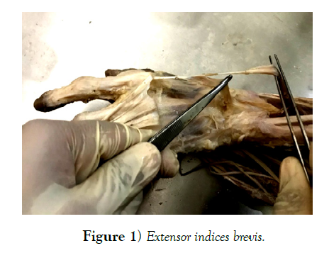
During routine dissection of the upper limb, a small elevation was noted over the dorsum of the left hand of female cadaver immediately behind the base of second metacarpal bone and on further explorative dissection, the extensor indices tendon was found interrupted by an additional small muscular belly originating from the distal end of radius and dorsum of the proximal carpal bones and the fibrous capsule of the wrist joint.
Extensor indices tendon had an additional small muscular belly originating from the lower end of the radius and the back of the fibrous capsule of the wrist joint. The 5 cm long muscular belly ended by joining the extensor indices tendon 1.5 cm distal to the base of the second metacarpal and later the single tendon joining on to the extensor expansion of the index finger (Figure 1).
Discussion
The index finger Extensor indices muscle usually arises from the posterior surface of the ulna distal to the origin of the extensor pollicis longus and passes deep to the extensor digitorum. Its tendon takes a postion on the ulnar side of the extensor digitorum tendon to the index finger and joins this tendon at the extensor expansion, at the level of the proximal phalanges. It aids in extending the metacarpophalangeal and the two interphalangeal joints of the index finger. It also adducts this finger independently.
Cauldwell, Anson and Wright in a study of the extensor indicis muscle in 263 consecutive specimens, found three cases in which the muscle had an abnormal origin. In one case the muscle had the usual origin from the ulna, became tendinous, and then again became muscular with a secondary attachment in the region of the carpal bones. In the second case a muscle with a rudimentary origin at the normal site was inserted into a second muscle arising from two heads from the proximal carpal bones. In the third case a short muscle only was present, arising from the distal end of the radius, proximal carpal bones and related ligaments [3]. They called this short muscle “extensor indices brevis manus.”
In cases of variation in extensor indices it may mimic ganglia, lipoma, tendon sheath cyst, tenosynovitis of the extensor tendons, exostosis, carpal boss or with other benign tissue tumours [4]. For example the palmaris longus with muscle fibres extending down into the palm is more likely to be misdiagnosed as a compound palmar ganglion. In such cases when the extended muscle get constricted by the normal retinacula under which they pass may cause symptoms like tenderness, numbness, weakness, restricted movements etc. This may be relieved by the division of the retinaculum.
Variations of extensor indices muscle are rare and include complete absence of the muscle and variations in its origin and insertion. During limb development the tendons originate from the lateral plate mesoderm, while the limb musculature is derived from the migrating somatic mesoderm. These observations emphasize on the role of embryological development in such variations, which has to be taken into consideration.
Extensor indices muscle allows independent extension of the index finger and it is frequently used for tendon grafts. It is also known that extensor indices are affected in extensor indices proprius syndrome. Ogura et al. have suggested that the extensor digitorum brevis manus muscle is a variant of extensor indices proprius muscle. Their anatomic investigations of 559 dissected cadavers have demonstrated that the extensor digitorum brevis manus muscle and extensor indices proprius muscle are most frequently associated together and supplied by the same neurovascular complex (posterior interosseous nerve and posterior branch of the interosseous artery) [5].
In the current variation we have observed a second muscular belly continuous with the tendon of extensor indices, seen as an elevation on the dorsum of left hand of the cadaver. Knowledge about these variations is inevitable for physicians and surgeons in making proper diagnosis and can avoid complications during surgical interventions. It would be helpful to the examiner in making proper diagnosis and might perhaps eliminate a surgical procedure. Even though the variations of extensor indices muscle are relatively rare, an awareness of this possible anatomical variation is important in the management of hand diseases or during tendon transfers. Accordingly, any surgical procedure in this particular region should be planned carefully in advance.
REFERENCES
- Singh V, Dutta S. Textbook of anatomy upper limb and thorax, 3rd Edition, Elsevier, New Delhi, India. 2018.
- Caudwell EW, Anson BJ, Wright RR. The extensor indicis proprius muscle: A study of 263 consecutive specimens, Quarterly Bulletin of Northwestern University Medical School. 1943;17:267-279.
- Tan ST, Smith PJ. Anomalous extensor muscles of the hand: A review. J Hand Surg. 1999;24:449–455.
- Ogura T, Inove H, Tanabe G. Anatomic and clinical studies of the extensor digitorum brevis manus. J Hand Surg. 1987;12:100-107.


