An anticipatory complete atrio-ventricular block and state of shock due to hyperkalemia
Received: 18-Aug-2017 Accepted Date: Sep 10, 2017; Published: 15-Sep-2017
Citation: Laachach H, Anass H, Bachrif M, et al. An anticipatory complete atrioventricular block and state of shock due to hyperkalemia. Clin Cardiol J 2017;1(1):8-9.
This open-access article is distributed under the terms of the Creative Commons Attribution Non-Commercial License (CC BY-NC) (http://creativecommons.org/licenses/by-nc/4.0/), which permits reuse, distribution and reproduction of the article, provided that the original work is properly cited and the reuse is restricted to noncommercial purposes. For commercial reuse, contact reprints@pulsus.com
Abstract
Even hyperkalemia is not a common etiology of a complete interruption of nodal conduction; it was the cause of signs of shock and complete atrioventricular bloc at a woman who was not correctly dialyzed. treatment was the early decreasing of serum potassium level. Evolution was well by restoration of normal hemodynamic values and atrial fibrillation rhythm without a need to pacing.
Keywords
Hyperkalemia; Concentration; Electrophysiologic; Renal; Symptoms
Background
The important role of potassium in the electrophysiologic regulation of myocardial function is known. Any change in extracellular potassium concentration may have a significant effect on myocyte electrophysiologic gain and bring about many electric dysfunctions [1]. Hyperkalemia occurs in 1–10% of hospitalized patients in medical department, with a mortality rate of 1 in 1,000 patients [2]. In general, High serum potassium levels results in a gradual decrease in the excitability and conduction velocity of specialized pacemaker cells and conducting tissues throughout the heart. Typical Electrocardiogram (ECG) findings in Hyperkalemia progress from tall, “peaked” T waves and a shortened QT interval to a lengthening of PR interval and loss of P waves, and then to widening of the QRS complex, culminating in a “sine wave” morphology and death if not treated [3-6]. Hyperkalemia is thought to impair the conduction in Purkinje fibers and ventricles more than that in the AV node; although complete Atrio-ventricular block (AVB) can occur, it is one of the rarest initial presentations [7].
Case Report
A 50-year-old woman who had a history of renal failure undergoing hemodialysis at 3 sessions per week, admitted into emergency department due to generalized weakness, chills, somnolence and Coldness of the extremities. The symptoms have started early the morning.
At the admission she had a respiratory rate of 24 per min, a pulse rate of 28/min, collapsed blood pressure and here blood sugar level was 120 mg/dL.
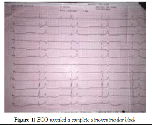
Here ECG revealed a complete atrio-ventricular block (Figure 1) Ventricular escape rate at 20/min.
The results of Laboratory examination were as the following: Creatinine=71 mg/L; urea=4.6 g/L; Na=137 mmol/L; K=8.5 mmol/L; CRP=40 mg/L; Hb=10.8 g/dL and INR=1.94. Besides, a respiratory acidosis was been objectified.
The patient was stabilized by vasoactive drugs: Dobutamine and Noradrenaline, infusion of glucose with insulin and gluconates of calcium.
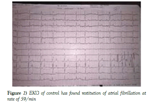
EKG of control has found restitution of atrial fibrillation at rate of 59/min (Figure 2), Blood pressure at 120/60 mmHg and potassium level at 7 mmol/L.
Figure 2: EKG of control has found restitution of atrial fibrillation at rate of 59/min
She was prepared for imperative hemodialysis simultaneously. She was admitted into the cardiology department for the rest of the treatment after hemodialysis. Transthoracic echocardiography has objectified a moderate mitral stenosis and left atrium dilation. The woman was discharged four days later, after good evolution.
Results and Discussion
Diagnosis of hyperkalemia is usually based on laboratory studies, although the electrocardiogram (ECG) may contain changes suggestive of hyperkalemia. Typical ECG findings in hyperkalemia progress from tall, “peaked” T waves and a shortened QT interval to lengthening PR interval and loss of P waves, and then to widening of the QRS complex culminating in a “sine wave” morphology and death if not treated [1,2]. Even with profound serum potassium elevations, the ECG cannot reliably be used to exclude Hyperkalemia or to monitor therapy designed to lower the Serum potassium concentration [3,4]. Many studies demonstrated that important hyperkaliemia can occur an auriculo-ventricular block [5-7]. AVB has been demonstrated experimentally in animals. Potassium, according to plasma level and infusion rate, demonstrated a quantitatively different, and sometimes opposite, effect on AV conduction, intraventricular conduction, automaticity and excitability [7]. The second or third degree BAV caused by hyperkalaemia has been rarely documented in humans [3,8,9]. Complete heart block (CHB) is the consequence of the loss of electrical transmission between the supraventricular tissues to ventricles. The lower level of block, the slower and the less reliable will be the ventricular escape rhythm. Third-degree AV block can be congenital or acquired. Three causes are known to induce an acquired CHB: Ischemic heart disease, drugs, and Hyperkalemia. However, cardiomyopathies, myocarditis, structural heart disease, hypothermia, and severe hypothyroidism are much less common causes. Acquired third-degree AV block often has a wide complex escape rhythm, with symptoms of hypo perfusion at rest or with minimal exertion. CHB may be sub-acute or chronic and relatively well tolerated providing that the block occurs at a relatively high level, (A-V nodal to His bundle region). Alternatively, the patient may be very unwell, usually in acute onset in association with myocardial ischemia or a low level block [4,9,10].
The treatment is based on two main sources: Treatment of the atrioventricular block and palliation against tissue hypoperfusion on the one hand; On the other hand decreasing the plasma potassium concentration, combating its effects on myocytes and treating the cause of hyperkalemia. When the heart block is complete, Patients with clinical hypo perfusion or not responding to medical measures may have a continuous monitoring and treated by percutaneous pacing as a temporary or conclusive solution [9,10]. Hyperkalemia should be treated as quick as possible, by antagonizing the extracellular potassium effect thus decreasing the serum potassium level by transferring on intracellular. The next line is to remove potassium from the body by ’Kayexalat‘ or ultimately by hemodialysis [11-13]. As the case of our patient, the evolution is good after early treatment.
Conclusion
Complete heart block is not a common complication of hyperkalemia, it can occur when serum potassium level is very high. Syncope and heart failure is some of clinical signs. Treatment is an imperative decreasing potassium serum level and repairing the etiology of hyperkalemia. As this case has demonstrated, the heart block is mostly regressive after kalemia normalization.
Conflict of Interest
None
Author Contribution
All the authors have contributed simultaneously to elaborate this article
REFERENCES
- Slovis C, Jenkins R. ABC of clinical electrocardiography: Conditions not primarily affecting the heart. BMJ 2002;324(7349):1320-3.
- Webster A, Brady W, Morris F. Recognizing signs of danger: EKG changes resulting from an abnormal serum potassium concentration. Emerg Med J 2002 19(1):74-7
- Kim NH, Oh SK, Jeong JW. Hyperkalemia induced complete atrioventricular block with a narrow QRS complex. Heart 2005;91(1): e5.
- Smith SC Jr, Benjamin EJ, Bonow RO, et al. AHA/ACCF secondary prevention and risk reduction therapy for patients with coronary and other atherosclerotic vascular disease: 2011 update: A guideline from the American Heart Association and American College of Cardiology Foundation Circulation. 2015;1(1);35-38
- Smith SC Jr, Benjamin EJ, Bonow RO, et al. AHA/ACCF secondary prevention and risk reduction therapy for patients with coronary and other atherosclerotic vascular disease: 2011 update: a guideline from the American Heart Association and American College of Cardiology Foundation endorsed by the World Heart Federation and the Preventive Cardiovascular Nurses Association J Am Coll Cardiol 2011;58(23):2432-46.
- Mclean FC, Bay EB, Hasting AB. Electrical changes in the isolated heart of the rabbit following changes in the potassium content of the perfusing fluid. Am J Physiol. 1933;105:72.
- Fisch C, Feigenbaum H, Bowers J. The effect of potassium on atrioventricular conduction of normal dogs. Am J Cardiol 1963;11:487-92.
- Fisch C, Knoebel S, Feigenbaum H, et al. Potassium and the monophasic action potential, electrocardiogram, conduction and arrhythmias. Prog Cardiovasc Dis 1966;8:387-418.
- Bashour T, Hsu I, Gorfinkel H, et al. Atrioventricular and intraventricular conduction in hyperkalemia. Am J Cardiol 1975;35:199-203.
- Alireza B, Pauline H, Alaleh R, et al. Hyperkalemia-induced complete heart block. Journal of Emergency Practice and Trauma, 2015;1(1);35-38.
- Yealy DM, Delbridge TR. Dysrhythmias emergency medicine concepts and clinical practice. 7th ed. Philadelphia: Mosby; 2007. p. 1000-2.
- Parham WA, Mehdirad AA, Biermann KM, et al. Hyperkalemia revisited. Tex Heart Inst J 2006;33(1):40-7.
- Diercks DB, Shumaik GM, Harrigan RA, et al. Electrocardiographic manifestations: Electrolyte abnormalities. J Emerg Med 2004;27(2):153-60.


