An unusual palmaris longus tendon: variation in the insertion and orientation at the level of wrist joint
Vasantha Kumar* and Bincy M. George
Melaka Manipal Medical College (Manipal Campus), International Centre for Health Sciences, Madhav Nagar, Manipal, Karnataka State, India
- *Corresponding Author:
- Vasantha Kumar, MSc
Lecturer in Anatomy, Melaka Manipal Medical College (Manipal Campus), International Centre for Health Sciences, Madhav Nagar, Manipal, Udupi District, Karnataka State, 576 104, India
Tel: +91 820 2922519
E-mail: vasanthui@gmail.com
Date of Received: June 29th, 2009
Date of Accepted: October 23rd, 2009
Published Online: November 4th, 2009
© IJAV. 2009; 2: 138–139.
[ft_below_content] =>Keywords
palmaris longus, unusual insertion, relation, wrist, thenar eminence
Introduction
The palmaris longus (PL) is one of the most variable muscle present in 90% of humans, since it has a short belly and a long tendon it is classified phylogenetically as a retrogressive muscle [1]. It takes origin from medial epicondyle of the humerus along with other superficial flexors. It lies between flexor carpi radialis laterally and flexor carpi ulnaris medially. Median nerve, at the level of wrist lies between its tendon and flexor carpi radialis. The long slender tendon passes anterior to the flexor retinaculum. It is attached to the distal half of flexor retinaculum and predominantly to the palmar aponeurosis. In vertebrates it is found only in mammals and is best developed in those where the forelimb is used for ambulation.
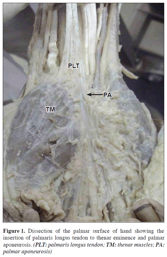
Absence of PL in humans appears to be hereditary but its genetic transmission is not clear. Studies by dissection and clinical testing show bilateral absence of PL in 8-16% of the individuals and unilateral absence in 4 to 14% [1]. Sometimes the PL may give a slip to thenar muscle group. But a variation in the insertion of PL tendon predominantly to thenar eminence and a slip to palmar aponeurosis is reported here. At wrist, the relation of median nerve is also found varied than the usual pattern, which make the variation surgically important.
Case Report
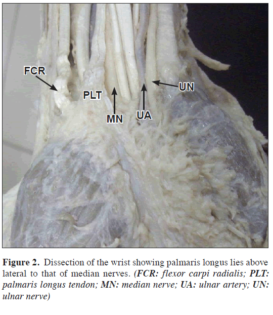
During a routine dissection in the right upper limb of a 40-year-old embalmed male cadaver in the department of Anatomy, variation in tendinous insertion of PL was noted. The presence of PL was identified on the superficial stratum of flexor compartment. Above the level of wrist, the slender tendon was running laterally to the median nerve. Before passing superficial to the flexor retinaculam, majority of the tendinous fibers were inserted to the thenar eminence. Few long medial fibers were inserted to palmar aponeurosis.
Discussion
In the development of the forelimb as a prehensile organ, its function has been taken over by the intrinsic muscles of the hand and the PL has become degenerate. The PL belly is largely replaced by tendon, and the degenerated distal tendon of the palm has become the palmar aponeurosis, which retains the five distal slip of attachment. Few case reports of hypertrophy of PL are proved to produce compression neuropathy of the median nerve.
Numerous anomalies of the origin, course and insertion of the PL have been described [1,2]. Such variants include anomalous insertion deep to the retinaculum, producing a carpal tunnel-like syndrome; compression of the median nerve by the distal belly of the PL between it and the underlying tendons. Three headed reversed PL causing compartment syndrome [2] and double PL also have been reported. We are reporting a different case with the unusual insertion of PL with the median nerve medially located to it at the wrist.
Prevalence of absence of PL has been studied clinically in few ethnic groups using variety of testing methods [3–5]. In all these cases, the examiner is looking for prominent PL tendon in the middle of the forearm. In the current case, there is a possibility that the examiner who test the PL tendon may mistake that it is absent, as he or she is unaware of such anomaly. Surgeons handling PL graft should be aware of this kind of variation in the insertion of PL tendon, so as to avoid the accidental damage of the structures like median nerve. Insertion to thenar muscles may affect its actions like abduction adduction, flexion and opposition.
References
- Pai MM, Prabhu LV, Nayak SR, Madhyastha S, Vadgaonkar R, Krishnamurthy A, Kumar A. The palmaris longus muscle: its anatomic variations and functional morphology. Rom J Morphol Embryol. 2008; 49: 215–217.
- Natsis K, Levva S, Totlis T, Anastasopoulos N, Paraskevas G. Three-headed reversed palmaris longus muscle and its clinical significance. Ann Anat. 2007; 189: 97–101.
- O’Sullivan E, Mitchell BS. Association of the absence of palmaris longus tendon with an anomalous superficial palmar arch in the human hand. J Anat. 2002; 201: 405–408.
- Sebastin SJ, Lim AY. Clinical assessment of absence of the palmaris longus and its association with other anatomical anomalies-- a Chinese population study. Ann Acad Med Singapore. 2006; 35: 249–253.
- Thompson NW, Mockford BJ, Cran GW. Absence of the palmaris longus muscle: a population study. Ulster Med J. 2001; 70: 22–24.
Vasantha Kumar* and Bincy M. George
Melaka Manipal Medical College (Manipal Campus), International Centre for Health Sciences, Madhav Nagar, Manipal, Karnataka State, India
- *Corresponding Author:
- Vasantha Kumar, MSc
Lecturer in Anatomy, Melaka Manipal Medical College (Manipal Campus), International Centre for Health Sciences, Madhav Nagar, Manipal, Udupi District, Karnataka State, 576 104, India
Tel: +91 820 2922519
E-mail: vasanthui@gmail.com
Date of Received: June 29th, 2009
Date of Accepted: October 23rd, 2009
Published Online: November 4th, 2009
© IJAV. 2009; 2: 138–139.
Abstract
Palmaris longus is a muscle often used in reconstructive plastic surgery mainly in tendon transfer procedures for replacement of long flexors of the fingers. It has also been used for many other procedures including ptosis correction, lip augmentation and management of facial paralysis. Absence of palmaris longus in humans appears to be hereditary, but its genetic transmission is not clear. We report here a variant pattern of insertion of palmaris longus, and its probable significants. Any variation in the insertion of tendon of the palmaris longus is gaining importance as it is becoming very popular amongst graft material for reconstructive surgeries.
-Keywords
palmaris longus, unusual insertion, relation, wrist, thenar eminence
Introduction
The palmaris longus (PL) is one of the most variable muscle present in 90% of humans, since it has a short belly and a long tendon it is classified phylogenetically as a retrogressive muscle [1]. It takes origin from medial epicondyle of the humerus along with other superficial flexors. It lies between flexor carpi radialis laterally and flexor carpi ulnaris medially. Median nerve, at the level of wrist lies between its tendon and flexor carpi radialis. The long slender tendon passes anterior to the flexor retinaculum. It is attached to the distal half of flexor retinaculum and predominantly to the palmar aponeurosis. In vertebrates it is found only in mammals and is best developed in those where the forelimb is used for ambulation.
Absence of PL in humans appears to be hereditary but its genetic transmission is not clear. Studies by dissection and clinical testing show bilateral absence of PL in 8-16% of the individuals and unilateral absence in 4 to 14% [1]. Sometimes the PL may give a slip to thenar muscle group. But a variation in the insertion of PL tendon predominantly to thenar eminence and a slip to palmar aponeurosis is reported here. At wrist, the relation of median nerve is also found varied than the usual pattern, which make the variation surgically important.
Case Report
During a routine dissection in the right upper limb of a 40-year-old embalmed male cadaver in the department of Anatomy, variation in tendinous insertion of PL was noted. The presence of PL was identified on the superficial stratum of flexor compartment. Above the level of wrist, the slender tendon was running laterally to the median nerve. Before passing superficial to the flexor retinaculam, majority of the tendinous fibers were inserted to the thenar eminence. Few long medial fibers were inserted to palmar aponeurosis.
Discussion
In the development of the forelimb as a prehensile organ, its function has been taken over by the intrinsic muscles of the hand and the PL has become degenerate. The PL belly is largely replaced by tendon, and the degenerated distal tendon of the palm has become the palmar aponeurosis, which retains the five distal slip of attachment. Few case reports of hypertrophy of PL are proved to produce compression neuropathy of the median nerve.
Numerous anomalies of the origin, course and insertion of the PL have been described [1,2]. Such variants include anomalous insertion deep to the retinaculum, producing a carpal tunnel-like syndrome; compression of the median nerve by the distal belly of the PL between it and the underlying tendons. Three headed reversed PL causing compartment syndrome [2] and double PL also have been reported. We are reporting a different case with the unusual insertion of PL with the median nerve medially located to it at the wrist.
Prevalence of absence of PL has been studied clinically in few ethnic groups using variety of testing methods [3–5]. In all these cases, the examiner is looking for prominent PL tendon in the middle of the forearm. In the current case, there is a possibility that the examiner who test the PL tendon may mistake that it is absent, as he or she is unaware of such anomaly. Surgeons handling PL graft should be aware of this kind of variation in the insertion of PL tendon, so as to avoid the accidental damage of the structures like median nerve. Insertion to thenar muscles may affect its actions like abduction adduction, flexion and opposition.
References
- Pai MM, Prabhu LV, Nayak SR, Madhyastha S, Vadgaonkar R, Krishnamurthy A, Kumar A. The palmaris longus muscle: its anatomic variations and functional morphology. Rom J Morphol Embryol. 2008; 49: 215–217.
- Natsis K, Levva S, Totlis T, Anastasopoulos N, Paraskevas G. Three-headed reversed palmaris longus muscle and its clinical significance. Ann Anat. 2007; 189: 97–101.
- O’Sullivan E, Mitchell BS. Association of the absence of palmaris longus tendon with an anomalous superficial palmar arch in the human hand. J Anat. 2002; 201: 405–408.
- Sebastin SJ, Lim AY. Clinical assessment of absence of the palmaris longus and its association with other anatomical anomalies-- a Chinese population study. Ann Acad Med Singapore. 2006; 35: 249–253.
- Thompson NW, Mockford BJ, Cran GW. Absence of the palmaris longus muscle: a population study. Ulster Med J. 2001; 70: 22–24.


