An unusual presentation of tibialis anterior Introduction Tibialis
Arjun burlakoti, Jacqueline Lee and Nicola Massy-Westropp*
Division of Health Sciences, University of South Australia (UniSA), School of Health Sciences, Adelaide, Australia.
- *Corresponding Author:
- Dr. Nicola Massy-Westropp
Division of Health Sciences University of South Australia School of Health Sciences City East Campus North Terrace, Adelaide SA 5000 Australia
Tel: +61 8 8302 2486
E-mail: nicola.massy-westropp@unisa.edu.au
Date of Received: November 7th, 2014
Date of Accepted: September 19th, 2015
Published Online: April 3rd, 2016
© Int J Anat Var (IJAV). 2016; 9: 1–2.
[ft_below_content] =>Keywords
variant, tibialis anterior, additional belly.
Introduction
Tibialis anterior originates from the upper, lateral shaft of the tibia and adjacent interosseus membrane. Acting as an ankle dorsiflexor and invertor of the foot, it continues medially beneath the extensor retinacula of the ankle, to insert onto the medial cuneiform and the base of the first metatarsal bone medially [1].
Past variations of the insertions of tibialis anterior have included a lateral insertion onto fifth metatarsal and cuboid bone [2], and a number of variant insertions onto the intermediate and lateral cuneiform in addition to the medial cuneiform [3]. The tendon has also been found inserting only onto the first metatarsal with no cuneiform insertion [4].
Case Report
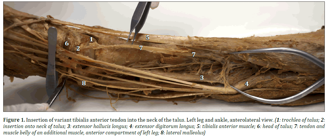
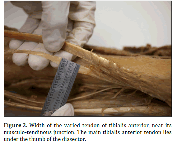
During a routine dissection of a lower limb for use in teaching, an additional slip of the tendon of tibialis anterior was found, lying deep to the main tendon and having its own smaller muscle belly, which shared a small border with the muscle of extensor digitorum longus. This tendon passed deep to the superior and inferior extensor retinacula of the ankle, to insert into the anterior, lateral neck of the talus (Figure 1). This muscle and tendon were 22 cm long, the tendon is 13.5 centimetres long and 0.8 millimetres wide at the musculo-tendinous junction, while the main tendon of this muscle was 1 centimetre wide at the same point (Figure 2). Interestingly there was a large ganglion (10 mm in length, 0.7 mm wide and up to 0.2 mm at its most thick portion) containing a clear viscous fluid (32 mm in length, 10 mm wide and up to 0.7 mm at its thickest portion) in the tarsal sinus, superficial to the cervical ligament of the subtalar joint.
Figure 1: Insertion of variant tibialis anterior tendon into the neck of the talus. Left leg and ankle, anterolateral view. (1: trochlea of talus; 2:insertion onto neck of talus; 3: extensor hallucis longus; 4: extensor digitorum longus; 5: tibialis anterior muscle; 6: head of talus; 7: tendon and muscle belly of an additional muscle, anterior compartment of left leg; 8: lateral malleolus)
Discussion
The clinical implications of this additional tendon of tibialis anterior are not known, but the dissection shows no abnormality in the neighbouring structures of the anterior tibial artery and the distal continuation of deep fibular nerve. It was not determined, whether the additional tendon produced forces that led to the development of the large ganglion superficial to the adjacent subtalar cervical ligament.
Interestingly no muscles routinely attach to the talus bone but there are many ligamentous attachments. These provide stability to the surrounding talocrural, subtalar and talocalcaneonavicular joints.
The additional muscle inserting into the lateral neck of the talus might limit the range of motion around the ankle and foot complex, such as inversion and plantar flexion. This may interfere with the normal course of nearby neurovascular structures, such as deep fibular nerve and dorsalis pedis artery and consequently give rise to the peripheral neurovascular complications distally.
With this varied attachment, which is more lateral to the usual attachment of tibialis anterior, the line of pull of this muscle may have aided in ankle dorsiflexion movement. Being a slender muscle and much shorter in length than the main belly of tibialis anterior, the functional role of this muscle is uncertain. However, it is likely that it assisted as an anterior ankle joint stabiliser in this individual.
References
- Brenner E. Insertion of the tendon of the tibialis anterior muscle in feet with and without hallux valgus. Clin Anat. 2002; 15: 217–223.
- Ikiz ZA, Ucerler H. A previously unreported variation related to the insertion of the tibialis anterior muscle and the superficial fibular (peroneal) nerve. Anat Sci Int. 2005: 80: 172–175.
- Verma P, Arora AK, Sharma RK, Lalit M, Agnihotri G. A rare incidence of abnormally broad tendon of insertion of tibialis anterior- a case report. Int J App Biol Pharm Tech. 2011; 2:359–362.
- Luchansky E, Paz Z. Variations in the insertion of tibialis anterior muscle. Anat Anz. 1986;162: 129–136.
Arjun burlakoti, Jacqueline Lee and Nicola Massy-Westropp*
Division of Health Sciences, University of South Australia (UniSA), School of Health Sciences, Adelaide, Australia.
- *Corresponding Author:
- Dr. Nicola Massy-Westropp
Division of Health Sciences University of South Australia School of Health Sciences City East Campus North Terrace, Adelaide SA 5000 Australia
Tel: +61 8 8302 2486
E-mail: nicola.massy-westropp@unisa.edu.au
Date of Received: November 7th, 2014
Date of Accepted: September 19th, 2015
Published Online: April 3rd, 2016
© Int J Anat Var (IJAV). 2016; 9: 1–2.
Abstract
During a routine dissection in the teaching laboratory of the University of South Australia, the left leg of a male donor was found to have an additional muscle belly and tendon slip of tibialis anterior in the anterior compartment of the left leg, inserting onto the talus bone. The other muscular structures in the anterior compartment, the anterior tibial artery and the distal continuation of the deep fibular nerve followed the course described in standard textbooks.
-Keywords
variant, tibialis anterior, additional belly.
Introduction
Tibialis anterior originates from the upper, lateral shaft of the tibia and adjacent interosseus membrane. Acting as an ankle dorsiflexor and invertor of the foot, it continues medially beneath the extensor retinacula of the ankle, to insert onto the medial cuneiform and the base of the first metatarsal bone medially [1].
Past variations of the insertions of tibialis anterior have included a lateral insertion onto fifth metatarsal and cuboid bone [2], and a number of variant insertions onto the intermediate and lateral cuneiform in addition to the medial cuneiform [3]. The tendon has also been found inserting only onto the first metatarsal with no cuneiform insertion [4].
Case Report
During a routine dissection of a lower limb for use in teaching, an additional slip of the tendon of tibialis anterior was found, lying deep to the main tendon and having its own smaller muscle belly, which shared a small border with the muscle of extensor digitorum longus. This tendon passed deep to the superior and inferior extensor retinacula of the ankle, to insert into the anterior, lateral neck of the talus (Figure 1). This muscle and tendon were 22 cm long, the tendon is 13.5 centimetres long and 0.8 millimetres wide at the musculo-tendinous junction, while the main tendon of this muscle was 1 centimetre wide at the same point (Figure 2). Interestingly there was a large ganglion (10 mm in length, 0.7 mm wide and up to 0.2 mm at its most thick portion) containing a clear viscous fluid (32 mm in length, 10 mm wide and up to 0.7 mm at its thickest portion) in the tarsal sinus, superficial to the cervical ligament of the subtalar joint.
Figure 1: Insertion of variant tibialis anterior tendon into the neck of the talus. Left leg and ankle, anterolateral view. (1: trochlea of talus; 2:insertion onto neck of talus; 3: extensor hallucis longus; 4: extensor digitorum longus; 5: tibialis anterior muscle; 6: head of talus; 7: tendon and muscle belly of an additional muscle, anterior compartment of left leg; 8: lateral malleolus)
Discussion
The clinical implications of this additional tendon of tibialis anterior are not known, but the dissection shows no abnormality in the neighbouring structures of the anterior tibial artery and the distal continuation of deep fibular nerve. It was not determined, whether the additional tendon produced forces that led to the development of the large ganglion superficial to the adjacent subtalar cervical ligament.
Interestingly no muscles routinely attach to the talus bone but there are many ligamentous attachments. These provide stability to the surrounding talocrural, subtalar and talocalcaneonavicular joints.
The additional muscle inserting into the lateral neck of the talus might limit the range of motion around the ankle and foot complex, such as inversion and plantar flexion. This may interfere with the normal course of nearby neurovascular structures, such as deep fibular nerve and dorsalis pedis artery and consequently give rise to the peripheral neurovascular complications distally.
With this varied attachment, which is more lateral to the usual attachment of tibialis anterior, the line of pull of this muscle may have aided in ankle dorsiflexion movement. Being a slender muscle and much shorter in length than the main belly of tibialis anterior, the functional role of this muscle is uncertain. However, it is likely that it assisted as an anterior ankle joint stabiliser in this individual.
References
- Brenner E. Insertion of the tendon of the tibialis anterior muscle in feet with and without hallux valgus. Clin Anat. 2002; 15: 217–223.
- Ikiz ZA, Ucerler H. A previously unreported variation related to the insertion of the tibialis anterior muscle and the superficial fibular (peroneal) nerve. Anat Sci Int. 2005: 80: 172–175.
- Verma P, Arora AK, Sharma RK, Lalit M, Agnihotri G. A rare incidence of abnormally broad tendon of insertion of tibialis anterior- a case report. Int J App Biol Pharm Tech. 2011; 2:359–362.
- Luchansky E, Paz Z. Variations in the insertion of tibialis anterior muscle. Anat Anz. 1986;162: 129–136.


