An unusual tunnel formation in the arm and its clinical significancewas
Vasudha Saralaya, Soubhagya R. Nayak*, Swetha Sequeira, Sampath Madhyastha, Ashwin Krishnamurthy and Sujatha D’Costa
Department of Anatomy, Centre for Basic Sciences, Kasturba Medical College, Bejai, Mangalore, Karnataka, India
- *Corresponding Author:
- Soubhagya R. Nayak, MSc
Lecturer, Department of Anatomy, Centre for Basic Sciences, Kasturba Medical College Bejai, Mangalore, Karnataka, 575004, India
Tel: +91 824 2211746
Fax: +91 824 2421283
E-mail: ranjanbhatana@gmail.com
Date of Received: November 5th, 2008
Date of Accepted: February 19th, 2009
Published Online: February 23rd, 2009
© Int J Anat Var (IJAV). IJAV. 2009; 2: 27–28.
[ft_below_content] =>Keywords
third head of biceps brachii, medial head of triceps brachii, median nerve, brachial artery, entrapment
Introduction
Third head of biceps brachii muscle (THBB) is an often-reported finding of academic interest. It becomes more significant when causing entrapment of the neurovascular bundle in the vicinity and the resultant clinical presentations. The presence of supernumerary humeral heads of biceps brachii is one of the common variations seen in the region of the front of the arm affecting an estimated population of 9-22% [1]. The most frequently occurring variation of the biceps brachii is the presence of a third head and this has been reported in 8% of the Chinese, 10% of Europeans, 12% of Africans and 18% of Japanese [2]. In the present case the THBB was forming a tunnel with the muscle fibers of the medial head of triceps (MHT); the median nerve and brachial artery were passing through the tunnel, making them vulnerable to various entrapment syndromes.
Case Report
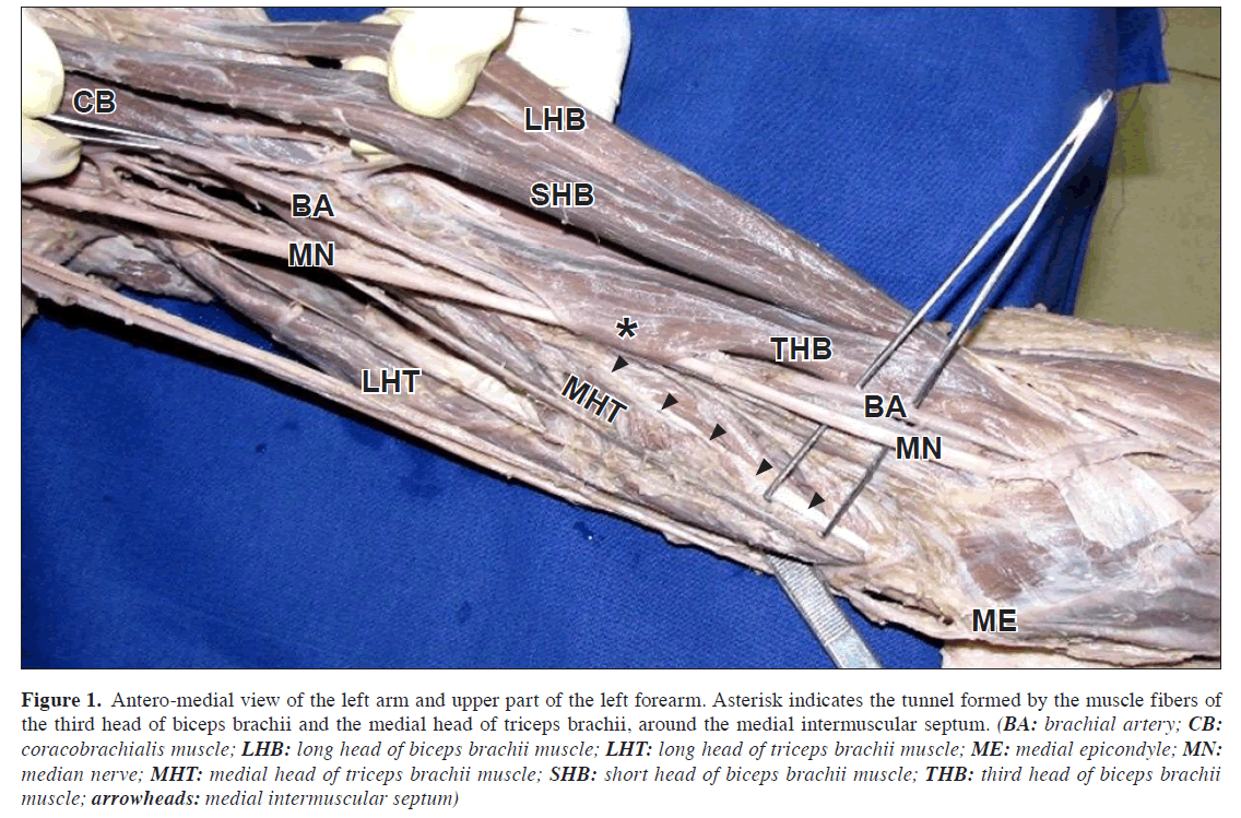
In the left arm of a 65-year-old male cadaver, the THBB (length, 21 cm; width, 6.1 cm) was originating from the humeral shaft at the site of insertion of coracobrachialis and inserted to the tendon of the biceps brachii muscle (BB). The medial fibers of the THBB were merging with the fibers of the MHT at the medial intermuscular septum. A tunnel was formed deep to the THBB and fibers of the MHT. The median nerve and brachial artery were the passing within the tunnel. Both the nerve and artery were lying on the brachialis muscle. The THBB significancewas innervated by the branch of musculocutaneous nerve as expected.
Discussion
The BB is a large fusiform muscle in the flexor compartment of the upper arm, derives its name from its two proximally attached ‘heads’. The short head arises by a thick flattened tendon from the coracoid process together with coracobrachialis. The long head arises within the capsule of the shoulder joint from the supraglenoid tubercle at the apex of the glenoid cavity. The two heads lead into elongated bellies. At the elbow joint they end in a flattened tendon attached to the posterior aspect of the radial tuberosity. The broad medial expansion, the bicipital aponeurosis descends medially across the brachial artery to fuse with the deep fascia over the origin of the flexor muscles of the forearm [3].
Figure 1: Antero-medial view of the left arm and upper part of the left forearm. Asterisk indicates the tunnel formed by the muscle fibers of the third head of biceps brachii and the medial head of triceps brachii, around the medial intermuscular septum. (BA: brachial artery; CB: coracobrachialis muscle; LHB: long head of biceps brachii muscle; LHT: long head of triceps brachii muscle; ME: medial epicondyle; MN: median nerve; MHT: medial head of triceps brachii muscle; SHB: short head of biceps brachii muscle; THB: third head of biceps brachii muscle; arrowheads: medial intermuscular septum)
A wide range of variations in BB have been reported in literature. It may arise as accessory fascicles from the coracoid process, pectoralis minor tendon or proximal end of humerus [4]. The most common variation is the muscle arising from the proximal humerus also known as humeral head or THBB [5-7]. We observed an obvious origin of the third head from the humeral shaft at the level of insertion of coracobrachialis. This variable head incidentally contributed to the formation of the bicipital aponeurosis. In 10% of cases, a third head arises from the superomedial part of the brachialis and is attached to the bicipital aponeurosis or to the medial side of tendon [3].
Pronator teres syndrome is the most common manifestation of median nerve entrapment after the carpal tunnel syndrome [8]. Other common entrapment sites include the ligament of Struther’s, Lacertus fibrosus and tendinous origin of flexor digitorum superficialis [9]. Laha et al. reported the median nerve entrapment under the bicipital aponeurosis over two decade’s ago [10]. In our case the median nerve was completely surrounded by the fleshy fibers of the THBB. The brachial artery, in close proximity to the median nerve, may also be entrapped under the muscular tunnel formed by the THBB as in our case. It has been reported in the previous studies that a possible compression of the brachial artery may result in claudication type of pain with cold intolerance and loss of radial and ulnar pulses in pronation [11,12]. As the BB muscle is a powerful supinator of the forearm and a flexor of the elbow, such movements bringing about the contraction of the muscle fibers would result in entrapment of the nerve with the resultant symptoms.
There have been plenty of reports of median nerve entrapment by the lacertus fibrosus, ligament of Struther’s and pronator teres [8-10]. This particular finding of the entrapment of the median nerve along with the brachial artery may be of great interest to the operating surgeons as well as to the physicians to use this finding as a differential diagnosis in all patients presenting with the complaints of tingling, numbness and claudication on movements of pronation and supination.
References
- Kosugi K, Shibita S, Yamashita H. Supernumerary head of biceps brachii and branching pattern of the musculocutaneus nerve in Japanese. Surg Radiol Anat. 1992; 14: 175–185.
- Bergman RA, Thompson SA, Afifi AK, Saadeh FA. Compendium of human anatomic variation. Baltimore, Urban & Schwarzenberg. 1988; 32–33.
- Williams PL. Gray’s Anatomy. 38th Ed., Edinburgh, Churchill Livingstone. 1995; 843.
- Sargon MF, Tuncali D, Celik HH. An unusual origin for the accessory head of the biceps brachii muscle. Clin Anat. 1996; 9: 160–162.
- Greig HW, Anson BJ, Budinger JM. Variations in the form and attachments of the biceps brachii muscle. Q Bull Northwest Univ Med Sch.. 1952; 26: 241–244.
- Khaledpour C. [Anomalies of the biceps muscle of the arm]. Anat Anz. 1985; 158: 79–85. German.
- Asvat R, Candler P, Sarmiento EE. High incidence of the third head of biceps brachii in South African populations. J Anat. 1993; 182: 101–104.
- Gessini L, Jandolo B, Pietrangeli A. Entrapment neuropathies of the median nerve at and above the elbow. Surg Neurol. 1983; 19: 112–116.
- Suranyi L. Median nerve compression by Struthers ligament. J Neurol Neurosurg Psychiatry. 1983; 46: 1047–1049.
- Laha RK, Lunsford D, Dujovny M. Lacertus fibrosus compression of the median nerve. J Neurosurg. 1978; 48: 838-841.
- Bassett FH 3rd, Spinner RJ, Schroeter TA. Brachial artery compression by the lacertus fibrosus. Clin Orthop Relat Res. 1994; 307: 110–116.
- Biemans RG. Brachial artery entrapment syndrome. Intermittent arterial compression as a result of muscular hypertrophy. J Cardiovasc Surg (Torino). 1977; 18: 367–371.
Vasudha Saralaya, Soubhagya R. Nayak*, Swetha Sequeira, Sampath Madhyastha, Ashwin Krishnamurthy and Sujatha D’Costa
Department of Anatomy, Centre for Basic Sciences, Kasturba Medical College, Bejai, Mangalore, Karnataka, India
- *Corresponding Author:
- Soubhagya R. Nayak, MSc
Lecturer, Department of Anatomy, Centre for Basic Sciences, Kasturba Medical College Bejai, Mangalore, Karnataka, 575004, India
Tel: +91 824 2211746
Fax: +91 824 2421283
E-mail: ranjanbhatana@gmail.com
Date of Received: November 5th, 2008
Date of Accepted: February 19th, 2009
Published Online: February 23rd, 2009
© Int J Anat Var (IJAV). IJAV. 2009; 2: 27–28.
Abstract
We present a case of third head of biceps brachii muscle forming a tunnel along with the muscle fibers of the medial head of triceps in the middle third of the left arm of a 65-year-old male cadaver found during routine dissection for the undergraduate students. The extra head of biceps brachii muscle was arising from the mid-shaft of the humerus and inserted to the tendon of the biceps brachii enclosing the median nerve and the brachial artery in a 3 cm long canal formed by it. This extra head of the biceps brachii is of particular interest as it may compress the median nerve and the brachial artery in the mid arm during the fexion at the elbow and supination of the forearm.
-Keywords
third head of biceps brachii, medial head of triceps brachii, median nerve, brachial artery, entrapment
Introduction
Third head of biceps brachii muscle (THBB) is an often-reported finding of academic interest. It becomes more significant when causing entrapment of the neurovascular bundle in the vicinity and the resultant clinical presentations. The presence of supernumerary humeral heads of biceps brachii is one of the common variations seen in the region of the front of the arm affecting an estimated population of 9-22% [1]. The most frequently occurring variation of the biceps brachii is the presence of a third head and this has been reported in 8% of the Chinese, 10% of Europeans, 12% of Africans and 18% of Japanese [2]. In the present case the THBB was forming a tunnel with the muscle fibers of the medial head of triceps (MHT); the median nerve and brachial artery were passing through the tunnel, making them vulnerable to various entrapment syndromes.
Case Report
In the left arm of a 65-year-old male cadaver, the THBB (length, 21 cm; width, 6.1 cm) was originating from the humeral shaft at the site of insertion of coracobrachialis and inserted to the tendon of the biceps brachii muscle (BB). The medial fibers of the THBB were merging with the fibers of the MHT at the medial intermuscular septum. A tunnel was formed deep to the THBB and fibers of the MHT. The median nerve and brachial artery were the passing within the tunnel. Both the nerve and artery were lying on the brachialis muscle. The THBB significancewas innervated by the branch of musculocutaneous nerve as expected.
Discussion
The BB is a large fusiform muscle in the flexor compartment of the upper arm, derives its name from its two proximally attached ‘heads’. The short head arises by a thick flattened tendon from the coracoid process together with coracobrachialis. The long head arises within the capsule of the shoulder joint from the supraglenoid tubercle at the apex of the glenoid cavity. The two heads lead into elongated bellies. At the elbow joint they end in a flattened tendon attached to the posterior aspect of the radial tuberosity. The broad medial expansion, the bicipital aponeurosis descends medially across the brachial artery to fuse with the deep fascia over the origin of the flexor muscles of the forearm [3].
Figure 1: Antero-medial view of the left arm and upper part of the left forearm. Asterisk indicates the tunnel formed by the muscle fibers of the third head of biceps brachii and the medial head of triceps brachii, around the medial intermuscular septum. (BA: brachial artery; CB: coracobrachialis muscle; LHB: long head of biceps brachii muscle; LHT: long head of triceps brachii muscle; ME: medial epicondyle; MN: median nerve; MHT: medial head of triceps brachii muscle; SHB: short head of biceps brachii muscle; THB: third head of biceps brachii muscle; arrowheads: medial intermuscular septum)
A wide range of variations in BB have been reported in literature. It may arise as accessory fascicles from the coracoid process, pectoralis minor tendon or proximal end of humerus [4]. The most common variation is the muscle arising from the proximal humerus also known as humeral head or THBB [5-7]. We observed an obvious origin of the third head from the humeral shaft at the level of insertion of coracobrachialis. This variable head incidentally contributed to the formation of the bicipital aponeurosis. In 10% of cases, a third head arises from the superomedial part of the brachialis and is attached to the bicipital aponeurosis or to the medial side of tendon [3].
Pronator teres syndrome is the most common manifestation of median nerve entrapment after the carpal tunnel syndrome [8]. Other common entrapment sites include the ligament of Struther’s, Lacertus fibrosus and tendinous origin of flexor digitorum superficialis [9]. Laha et al. reported the median nerve entrapment under the bicipital aponeurosis over two decade’s ago [10]. In our case the median nerve was completely surrounded by the fleshy fibers of the THBB. The brachial artery, in close proximity to the median nerve, may also be entrapped under the muscular tunnel formed by the THBB as in our case. It has been reported in the previous studies that a possible compression of the brachial artery may result in claudication type of pain with cold intolerance and loss of radial and ulnar pulses in pronation [11,12]. As the BB muscle is a powerful supinator of the forearm and a flexor of the elbow, such movements bringing about the contraction of the muscle fibers would result in entrapment of the nerve with the resultant symptoms.
There have been plenty of reports of median nerve entrapment by the lacertus fibrosus, ligament of Struther’s and pronator teres [8-10]. This particular finding of the entrapment of the median nerve along with the brachial artery may be of great interest to the operating surgeons as well as to the physicians to use this finding as a differential diagnosis in all patients presenting with the complaints of tingling, numbness and claudication on movements of pronation and supination.
References
- Kosugi K, Shibita S, Yamashita H. Supernumerary head of biceps brachii and branching pattern of the musculocutaneus nerve in Japanese. Surg Radiol Anat. 1992; 14: 175–185.
- Bergman RA, Thompson SA, Afifi AK, Saadeh FA. Compendium of human anatomic variation. Baltimore, Urban & Schwarzenberg. 1988; 32–33.
- Williams PL. Gray’s Anatomy. 38th Ed., Edinburgh, Churchill Livingstone. 1995; 843.
- Sargon MF, Tuncali D, Celik HH. An unusual origin for the accessory head of the biceps brachii muscle. Clin Anat. 1996; 9: 160–162.
- Greig HW, Anson BJ, Budinger JM. Variations in the form and attachments of the biceps brachii muscle. Q Bull Northwest Univ Med Sch.. 1952; 26: 241–244.
- Khaledpour C. [Anomalies of the biceps muscle of the arm]. Anat Anz. 1985; 158: 79–85. German.
- Asvat R, Candler P, Sarmiento EE. High incidence of the third head of biceps brachii in South African populations. J Anat. 1993; 182: 101–104.
- Gessini L, Jandolo B, Pietrangeli A. Entrapment neuropathies of the median nerve at and above the elbow. Surg Neurol. 1983; 19: 112–116.
- Suranyi L. Median nerve compression by Struthers ligament. J Neurol Neurosurg Psychiatry. 1983; 46: 1047–1049.
- Laha RK, Lunsford D, Dujovny M. Lacertus fibrosus compression of the median nerve. J Neurosurg. 1978; 48: 838-841.
- Bassett FH 3rd, Spinner RJ, Schroeter TA. Brachial artery compression by the lacertus fibrosus. Clin Orthop Relat Res. 1994; 307: 110–116.
- Biemans RG. Brachial artery entrapment syndrome. Intermittent arterial compression as a result of muscular hypertrophy. J Cardiovasc Surg (Torino). 1977; 18: 367–371.


